. 2007 Mar 7;26(5):1352-62.
doi: 10.1038/sj.emboj.7601582. Epub 2007 Feb 15.

Fission yeast Swi5/Sfr1 and Rhp55/Rhp57 differentially regulate Rhp51-dependent recombination outcomes

Affiliations

Fission yeast Swi5/Sfr1 and Rhp55/Rhp57 differentially regulate Rhp51-dependent recombination outcomes

Yufuko Akamatsu et al. EMBO J. .

Abstract

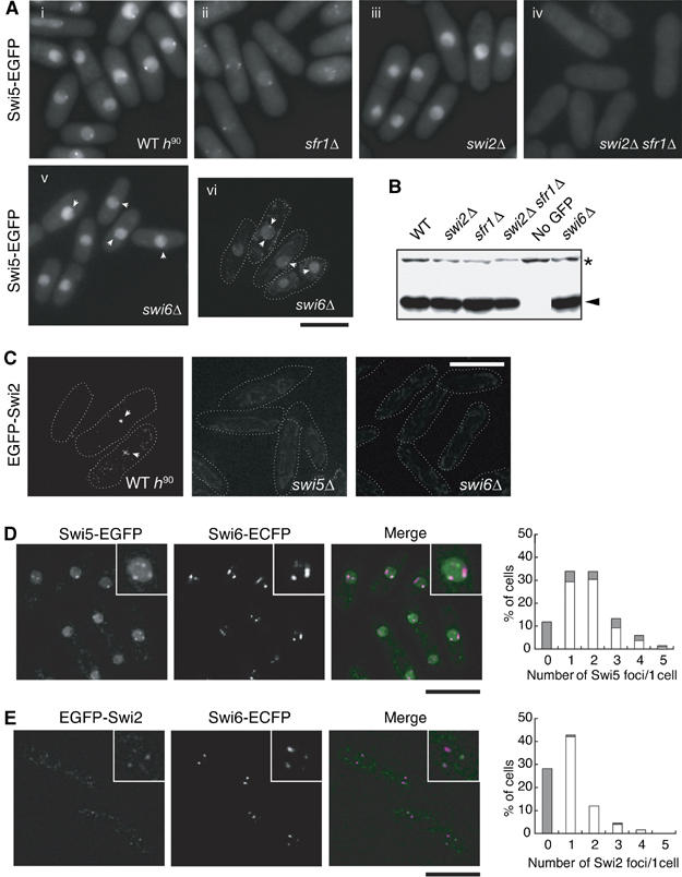
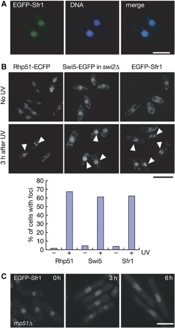
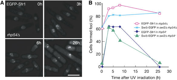
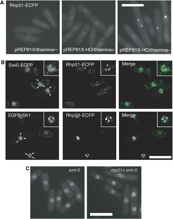
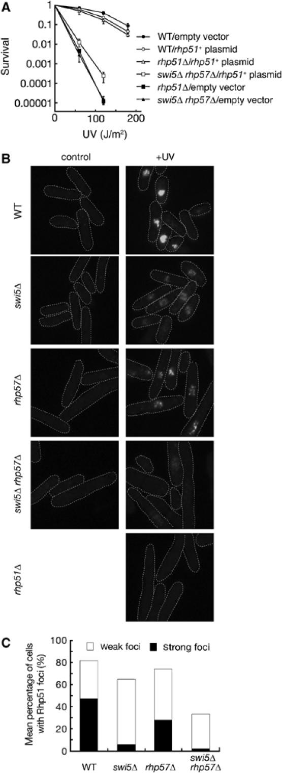
Several accessory proteins referred to as mediators are required for the full activity of the Rad51 (Rhp51 in fission yeast) recombinase. In this study, we analyzed in vivo functions of the recently discovered Swi5/Sfr1 complex from fission yeast. In normally growing cells, the Swi5-GFP protein localizes to the nucleus, where it forms a diffuse nuclear staining pattern with a few distinct foci. These spontaneous foci do not form in swi2Delta mutants. Upon UV irradiation, Swi5 focus formation is induced in swi2Delta mutants, a response that depends on Sfr1 function, and Sfr1 also forms foci that colocalize with damage-induced Rhp51 foci. The number of UV-induced Rhp51 foci is partially reduced in swi5Delta and rhp57Delta mutants and completely abolished in an swi5Delta rhp57Delta double mutant. An assay for products generated by HO endonuclease-induced DNA double-strand breaks (DSBs) reveals that Rhp51 and Rhp57, but not Swi5/Sfr1, are essential for crossover production. These results suggest that Swi5/Sfr1 functions as an Rhp51 mediator but processes DSBs in a manner different from that of the Rhp55/57 mediator.


Figures

Figure 1
Figure 1
Localization of Swi2 and Swi5 proteins. (A) Swi5 localizes to the nucleus in two patterns. The diffuse nuclear localization of Swi5 is dependent on sfr1+, whereas focal formation requires swi2+. The Swi5-EGFP signals of exponentially growing cells were observed by fluorescence microscopy. The strains used were wild-type h90 (YA598), sfr1Δ (YA763), swi2Δ (YA673), swi2Δ sfr1Δ (YA765) and swi6Δ (YA752). (i)–(v) Images were obtained by using a conventional fluorescence microscope (Nikon ECLIPSE E800). (vi) Image was obtained by using a DeltaVision microscope system as described in Materials and methods. The bar indicates 10 μm. (B) Neither the swi2Δ nor sfr1Δ mutation affects the level of Swi5-EGFP expression. Total protein extracts (10 μg) from the indicated strains were separated by SDS–polyacrylamide gel electrophoresis and subjected to immunoblot analysis with an anti-GFP antibody (JL-8, Clontech). The same strains were used as in (A) and the untagged strain was YA254. (C) Swi2 forms a few spontaneous foci in the nucleus. Swi2-EGFP signals of exponentially growing cells were observed by a DeltaVision microscope system. The strains used were wild type (YA820), swi5Δ (YA1247) and swi6Δ (YA1261). (D) Swi5 foci colocalize with Swi6 foci. A strain (YA756) with EGFP-tagged swi5+ and ECFP-tagged swi6+ was examined as in (C). In the merged image, green indicates Swi5-EGFP and magenta indicates Swi6-ECFP signals. Each inset is a magnified image of the same nucleus. The graph shows percentages of cells with unlocalized (gray bars) and colocalized (open bars) foci. X-axis gives the number of Swi5 foci in each cell. (E) Swi2 foci colocalize with Swi6 foci. A strain (YA1292) with EGFP-tagged swi2+ and ECFP-tagged swi6+ was examined as in (C). In the merged image, green indicates Swi2-EGFP and magenta indicates Swi6-ECFP signals. Each inset is a magnified image of the same nucleus. The graph shows percentages of cells with unlocalized (gray bars) and colocalized (open bars) foci. X-axis gives the number of Swi2 foci in each cell.
Figure 2
Figure 2
Analyses of damage-inducible Sfr1 and Swi5 foci. (A) The EGFP-Sfr1 signals and chromosomal DNA (stained with Hoechst 33342) of exponentially growing wild-type cells (YA848) were observed with a fluorescent microscope. (B) Rhp51 (left, YA990), Swi5 (middle, YA979) and Sfr1 (right, YA848) assemble following UV irradiation. Exponentially growing cells were irradiated with 100 J/m2 UV light. After incubation for 3 h at 30°C, cells were observed by using a DeltaVision microscope system. Note that focal signals of Swi5-EGFP were analyzed in an swi2Δ background to abolish the accumulation of spontaneous foci at heterochromatin. The graph shows percentages of cells with foci before (−) and after (+) UV irradiation. (C) Sfr1 assembly is dependent on Rhp51. EGFP-Sfr1 focal signals are not detected in cells (YA986) with the rhp51Δ mutation 3 and 6 h after UV irradiation.
Figure 3
Figure 3
(A) In rhp54Δ cells (YA1240), EGFP-Sfr1 foci form spontaneously under normal growth conditions, but much more foci of stronger intensity are observed following UV irradiation (120 J/m2). Foci persisted for at least 26 h after UV irradiation. (B) Time course of foci formations of Swi5 and Sfr1 after UV irradiation (120 J/m2). Strains used were YA848 (EGFP-Sfr1), YA979 (Swi5-EGFP in swi2Δ), YA1325 (Swi5-EGFP in swi2Δ rhp54Δ) and YA1240 (EGFP-Sfr1 in rhp54Δ).
Figure 4
Figure 4
(A) Rhp51-ECFP assembles at sites of HO-induced single DSBs (YA1083). (B) Swi5 and Sfr1 colocalize with Rhp51 following UV irradiation. Top, cells with Swi5-EGFP and Rhp51-ECFP fusion proteins (YA1263); bottom, cells with EGFP-Sfr1 and Rhp51-ECFP fusion proteins (YA1213). Each inset is a magnified image of the same nucleus. In the merged images, green indicates Swi5-EGFP or EGFP-Sfr1 and magenta indicates Rhp51-ECFP signals. Each inset is a magnified image of the same nucleus. Note that focal signals of Swi5-EGFP were analyzed in an swi2Δ background to abolish the accumulation of spontaneous foci at heterochromatin. (C) Spontaneous Swi5 foci are independent of rhp51+ and the DSB at the smt locus within the mat1 region. The strains used were rhp51+ smt-0 (YA961) and rhp51Δ smt-0 (YA984).
Figure 5
Figure 5
Rhp57- and Swi5-dependent subnuclear accumulation of Rhp51 upon UV irradiation. (A) A multicopy plasmid expressing rhp51+ suppresses the UV sensitivity of the swi5Δ rhp57Δ double mutant. Wild-type (YA119), rhp51Δ (T3) and swi5Δ rhp57Δ (YA250) cells carrying the vector alone (pSP102) or a vector expressing rhp51+ (pSP192) were examined for UV sensitivity. The data represent the average of the results from three independent experiments and standard deviations are shown by error bars. (B) Rhp51 accumulation in wild-type and mutant cells was detected by immunostaining with the anti-Rhp51 antibody. Wild-type (YA119), swi5Δ (YA177), rhp57Δ (T5), swi5Δ rhp57Δ (YA250) and rhp51Δ (T3) cells were examined without UV irradiation or 3 h after UV irradiation (200 J/m2). (C) Mean percentage of mutant cells showing Rhp51 foci at 3 h post-irradiation. Frequencies were determined as described in Materials and methods.

References

    1. Akamatsu Y, Dziadkowiec D, Ikeguchi M, Shinagawa H, Iwasaki H (2003) Two different Swi5-containing protein complexes are involved in mating-type switching and recombination repair in fission yeast. Proc Natl Acad Sci USA 100: 15770–15775 - PMC - PubMed
    1. Allers T, Lichten M (2001) Differential timing and control of noncrossover and crossover recombination during meiosis. Cell 106: 47–57 - PubMed
    1. Arcangioli B, de Lahondes R (2000) Fission yeast switches mating type by a replication–recombination coupled process. EMBO J 19: 1389–1396 - PMC - PubMed
    1. Arcangioli B, Thon G (2004) Mating-type cassettes: structure, switching and silencing. In The Molecular Biology of Schizosaccharomyces pombe, Egal R (ed) pp 129–148. Heidelberg: Springer
    1. Bishop DK, Park D, Xu L, Kleckner N (1992) DMC1: a meiosis-specific yeast homolog of E. coli recA required for recombination, synaptonemal complex formation, and cell cycle progression. Cell 69: 439–456 - PubMed

Publication types

MeSH terms