. 2007 Mar 7;26(5):1340-51.
doi: 10.1038/sj.emboj.7601574. Epub 2007 Feb 15.

Histone H2AX and Fanconi anemia FANCD2 function in the same pathway to maintain chromosome stability

Affiliations

Histone H2AX and Fanconi anemia FANCD2 function in the same pathway to maintain chromosome stability

Massimo Bogliolo et al. EMBO J. .

Abstract

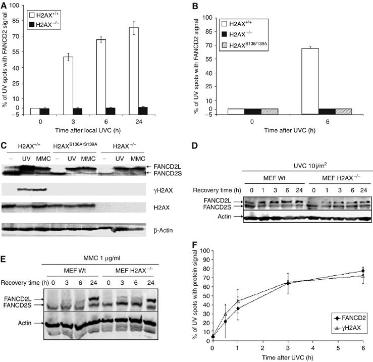
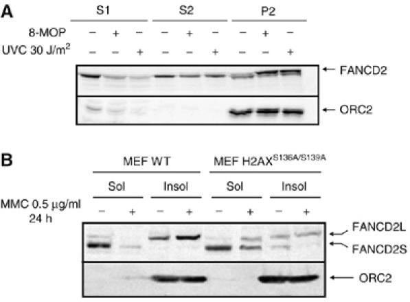
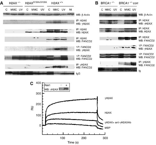
Fanconi anemia (FA) is a chromosome fragility syndrome characterized by bone marrow failure and cancer susceptibility. The central FA protein FANCD2 is known to relocate to chromatin upon DNA damage in a poorly understood process. Here, we have induced subnuclear accumulation of DNA damage to prove that histone H2AX is a novel component of the FA/BRCA pathway in response to stalled replication forks. Analyses of cells from H2AX knockout mice or expressing a nonphosphorylable H2AX (H2AX(S136A/S139A)) indicate that phosphorylated H2AX (gammaH2AX) is required for recruiting FANCD2 to chromatin at stalled replication forks. FANCD2 binding to gammaH2AX is BRCA1-dependent and cells deficient or depleted of H2AX show an FA-like phenotype, including an excess of chromatid-type chromosomal aberrations and hypersensitivity to MMC. This MMC hypersensitivity of H2AX-deficient cells is not further increased by depleting FANCD2, indicating that H2AX and FANCD2 function in the same pathway in response to DNA damage-induced replication blockage. Consequently, histone H2AX is functionally connected to the FA/BRCA pathway to resolve stalled replication forks and prevent chromosome instability.


Figures

Figure 1
Figure 1
FANCD2 relocates to the sites of UVC radiation-induced stalled replication forks in S phase: a functional assay for the FA/BRCA pathway. Cells (human wild-type MRC5 fibroblasts) irradiated through a 5-μm-pore filter with UVC (20 J/m2) present FANCD2 signal (detected with anti-FANCD2 antibodies in red) at the site of damage (detected with anti-CPD antibodies in green) 6 h after irradiation but not immediately after irradiation (A). FANCD2 relocates to UV spots following a specific dynamics and in a manner dependent on ATR, FANCA, BRCA1 and FANCD2 K561 but independent of ATM (means and s.d. of three experiments are shown in the graph) (B). PCNA and FANCD2 (red) relocate to the site of irradiation (green) following the same dynamics (C). FANCD2 (red) relocates to the irradiated site (green) only in replicating but not in G1 synchronized (>95% of the cells in G1 phase) wild-type primary fibroblasts (D); FANCD2 relocates to UV spots (right green panel) only in S-phase cells, as shown by BrdU incorporation (red middle panel). Nuclear counterstaining in DAPI (left blue panel) (E).
Figure 2
Figure 2
Relocation of FANCD2 to UVC-induced damage requires H2AX phosphorylation. FANCD2 does not relocate to locally induced stalled replication forks with UVC in H2AX−/− MEFs (A) or in H2AX−/− MEFs expressing a nonphosphorylable H2AX (H2AXS136A/S139A) (B). H2AX is not required for UVC- or MMC-induced FANCD2 monoubiquitination (C–E). H2AX-deficient MEF and their wild-type counterpart were treated with either UVC (10 J/m2; C, D) or with MMC (1 mg/ml; C, E). Cells were then harvested at the indicated recovery times and analyzed by Western blot for FANCD2 monoubiquitination. The equal loading of each slot was confirmed by probing the blot with anti-actin antibody (C–E). Local UVC irradiation results in concurrent H2AX phosphorylation and FANCD2 accumulation at the site of irradiation, with a dynamics of H2AX phosphorylation identical to the dynamics of FANCD2 relocation (F). (Means and s.d. of three experiments are shown in panels A, B and F.)
Figure 3
Figure 3
H2AX depletion by RNAi disrupts FANCD2 foci formation ability after MMC treatment but does not impair FANCD2 monoubiquitination. HeLa cells were interfered with siRNA versus H2AX or versus GFP and treated with MMC for 0, 6 and 24 h. Western blot showing inhibition of H2AX phosphorylation after siRNA and normal FANCD2 monoubiquitination (A). FANCD2 foci in HeLa cells interfered with siRNA versus GFP (B). FANCD2 foci in HeLa cells interfered with siRNA versus H2AX (C). Graph showing percentage of inhibition of FANCD2 foci formation in GFP-interfered cells versus H2AX-depleted cells (D). Mean and s.d. of two independent experiments are reported. H2AX expression was knocked down by transfection with a SMARTpool® reagent (Dharmacon, USA) designed against H2AX. SiRNA was transfected using the Oligofectamine reagent (Invitrogen, USA) according to the manufacturer's indications. As a control, an siRNA duplex directed against GFP (Dharmacon, USA) was used. All the experiments were carried out 72 h after transfection when maximal inhibition was observed as analyzed by Western blot, (A). The equal loading of each slot was confirmed by probing the blot with anti-actin antibody.
Figure 4
Figure 4
H2AX-dependent association of FANCD2 to chromatin after DNA damage. HeLa cells were left untreated or treated with 8-MOP+UVA or with 30 J/m2 of UVC, let to recover for 6 h and then fractionated and analyzed by Western blot (A). S1=cytoplasmic fraction, S2=nucleoplasmic fraction, P2=chromatin fraction. Wild type and H2AX−/− MEFs were treated with 1 μg/ml MMC for 24 h and then fractionated and analyzed by Western blot (B). S1 and S2 fractions were pooled (Sol) and 50 μg of total protein of the Sol fraction and of the chromatin fraction (Insol) were analyzed by Western blot. ORC2 was used as a marker of the chromatin fraction (A, B).
Figure 5
Figure 5
BRCA1-dependent binding of FANCD2 to γH2AX upon DNA damage. FANCD2 co-immunoprecipitates with γH2AX after DNA damage in a BRCA1-dependent manner. H2AX+/+, H2AXS136A/S139A, H2AX−/− MEF (A) or BRCA1−/− and BRCA−/− corrected cells (B) were not treated (−), MMC-treated (MMC) or UVC-irradiated (UV) as described in Materials and methods. Cells were lysed and immunoprecipitated with γH2AX, H2AX, FANCD2 or non-immune control (NI) antibodies followed by Western blot, and the protein bands were detected by probing with FANCD2, H2AX or γH2AX antibody, respectively. Loading control was monitored either by β-actin Western (A, B, upper row) or nonspecific binding to IgG (A, B, lowest row). Steady-state levels of γH2AX, FANCD2 or β-actin are maintained by Western blot of the corresponding samples before IP. In panels A and B, blots 2–4 and 5–7 represent H2AX IPs and FANCD2 IPs, respectively. Direct binding of FANCD2 to γH2AX by SPR (C). Equal aliquots of H2AX and H2AX phosphorylated by Rsk1 kinase were monitored by Western blot probed with γH2AX antibodies. Binding sensorgram of phosphorylated H2AX to the immobilized FANCD2. FANCD2 was immobilized (5 ng/mm2) on a dextran surface, as described in Materials and methods. Phosphorylated (A), nonphosphorylated (C) or phosphorylated H2AX premixed with anti-γH2AX antibody diluted at 1:500 (B) was injected over immobilized FANCD2. Control binding was performed on an unrelated (MBP) protein surface (C). The double bands of the FANCD2 WB panels in panels A and B correspond to FANCD2L (monoubiquitinated FANCD2; upper band) and FANCD2S (non-monoubiquitinated FANCD2).
Figure 6
Figure 6
H2AX deficiency leads to an excess of MMC-induced chromatid-type chromosomal aberrations and cytotoxicity. Excess of MMC-induced chromatid-type aberrations (A) and radial (B) in MEF derived from H2AX KO mice or H2AX−/− MEF expressing a nonphosphorylable H2AX (H2AXS136A/S139A). Means and s.d. of 2–4 experiments are shown. Differences between H2AX−/− and H2AXS136A/S139A are not statistically significant. Differences between wild-type and H2AX−/− or H2AXS136A/S139A cells are highly significant (P<0.001). H2AX-deficient cells are hypersensitive to the cytotoxic effect of MMC (C) (means and s.d. of 48 measures per dose and cell type are shown). Differences between wild-type and H2AX−/−cells are highly significant (P<0.001). Depletion of H2AX by RNAi in wild-type (but not in H2AX−/−) MEF increases cellular sensitivity to MMC (D). FANCD2 and H2AX function in the same pathway in response to MMC (E). FANCD2 depletion by RNAi does not further increase MMC sensibility in H2AX-deficient cells, suggesting that both FANCD2 and H2AX are in the same pathway. H2AX+/+, H2AX−/− and H2AXS136A/S139A MEFs were exposed to either FANCD2 RNAi or scramble RNAi (the same RNA sequence with five mismatches) and then exposed to MMC (200 nM for 24 h). FANCD2 immunoblot shows inhibition of FANCD2 synthesis by siRNA-FANCD2, not by scramble siRNA-FANCD2, irrespective of the H2AX genetic background (upper blot). The double bands of the FANCD2 WB panel in panel E correspond to FANCD2L (monoubiquitinated FANCD2; upper band) and FANCD2S (non-monoubiquitinated FANCD2). Percent of inhibition is calculated by TotaLab2.1 software as a ratio of FANCD2 RNAi and scRNA signals and normalized to the loading control signals (β-actin; upper blot, lowest row). The effect of FANCD2 depletion on MMC sensitivity in the three genetic backgrounds is shown in the graph. FANCD2 depletion by RNAi in wild-type cells increases MMC sensitivity. Similar and concurrent FANCD2 depletion in H2AX-deficient cells does not further increase MMC sensibility, suggesting that both FANCD2 and H2AX act in the same pathway in response to MMC.

References

    1. Abraham RT (2001) Cell cycle checkpoint signalling through the ATM and ATR kinases. Genes Dev 15: 2177–2196 - PubMed
    1. Andreassen PR, D'Andrea AD, Taniguchi T (2004) ATR couples FANCD2 monoubiquitination to the DNA-damage response. Genes Dev 18: 1958–1963 - PMC - PubMed
    1. Bassing CH, Suh H, Ferguson DO, Chua KF, Manis J, Eckersdorff M, Gleason M, Bronson R, Lee C, Alt FW (2003) Histone H2AX: a dosage-dependent suppressor of oncogenic translocations and tumors. Cell 114: 359–370 - PubMed
    1. Bassing CH, Alt FW (2004) H2AX may function as an anchor to hold broken chromosomal DNA ends in close proximity. Cell Cycle 3: 149–153 - PubMed
    1. Bogliolo M, Taylor RM, Caldecott KW, Frosina G (2000) Reduced ligation during DNA base excision repair supported by BRCA2 mutant cells. Oncogene 19: 5781–5787 - PubMed

Publication types

MeSH terms