

Review
doi: 10.1038/nrmicro1615.
Virus trafficking - learning from single-virus tracking
Affiliations
- PMID: 17304249
- PMCID: PMC2740720
- DOI: 10.1038/nrmicro1615
Item in Clipboard
Review
Virus trafficking - learning from single-virus tracking
Nat Rev Microbiol.
2007 Mar.
Abstract
What could be a better way to study virus trafficking than 'miniaturizing oneself' and 'taking a ride with the virus particle' on its journey into the cell? Single-virus tracking in living cells potentially provides us with the means to visualize the virus journey. This approach allows us to follow the fate of individual virus particles and monitor dynamic interactions between viruses and cellular structures, revealing previously unobservable infection steps. The entry, trafficking and egress mechanisms of various animal viruses have been elucidated using this method. The combination of single-virus trafficking with systems approaches and state-of-the-art imaging technologies should prove exciting in the future.
Figures
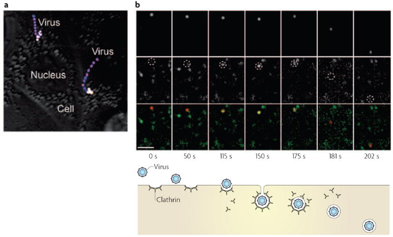
a | Stacked, time-lapse images of influenza viruses in living cells, revealing actin and microtubule-dependent transport. The virus is labelled with the lipophilic dye, DiD. The sudden colour change from blue/pink to yellow/white indicates a dramatic increase in the fluorescence signal of DiD, indicating the fusion of the virus with an endosome. A movie showing the time course of one of these virus particles can be found in the Supplementary Information S1 (movie). b | Simultaneous images of a DiD-labelled virus (upper panels and red in lower panels) and fluorescent protein-labelled clathrin (middle panels and green in lower panels) in a cell show the internalization of the virus by a clathrin-coated vesicle. The centres of dotted circles in the middle panels indicate the virus positions. Overlay of green and red signals appears yellow. The time (in seconds) after viral attachment and different stages of viral entry are shown below the images. A movie showing the time course of this virus particle is shown in Supplementary Information S2 (movie). Part (a) reproduced with permission from REF. © (2003) National Academy of Sciences, USA. Part (b) reproduced with permission from Nature Structural and Molecular Biology REF. © (2004) Macmillan Publishers Ltd.
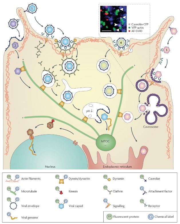
Viruses attach to the plasma membrane, surf on the cell surface or along the filopodia (1–3), and bind to specific receptors before entering the cell. Viruses can directly fuse with the plasma membrane (2). They also hijack endocytic pathways, including clathrin- dependent (1), caveolin-dependent (3) or clathrin- and caveolin-independent (4) pathways for internalization. After internalization and transport through the actin matrix, vesicles that contain virus are transported by dynein or dynactin along microtubules towards the microtubule organizing centre (MTOC). This might include trafficking of viruses through endosomes, caveosomes or the endoplasmic reticulum, prior to the release of the virus into the cytoplasm. Capsids can also be transported by dynein or dynactin along microtubules. From the MTOC, capsids can be transported by kinesin towards the replication site of the nucleus (5). Some viruses release their genetic material into the cytosol whereas others transport their genomes into the nucleus. The key shows how the different components have been labelled previously. The inset panel shows the caveolin-mediated endocytosis of Simian virus 40 (SV40). The arrowheads indicate SV40-containing caveolae co-localized with actin tails. The dye-labelled SV40 particles are shown in red and the fluorescent protein-labelled caveolin and actin are in purple and green, respectively. Scale bar represents 3μm. Inset panel reproduced with permission from REF. © (2002) American Association for the Advancement of Science.
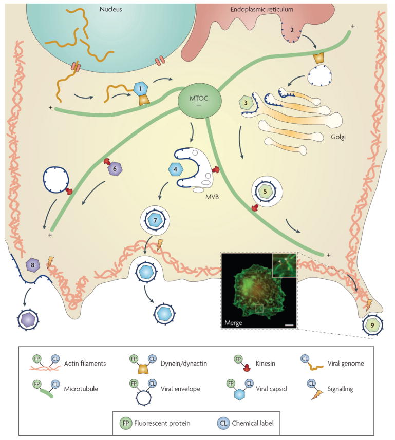
Viral genomes are packaged into capsids and are transported along microtubules (1). Viral membrane proteins are translated at the endoplasmic reticulum membrane (2) and transported along microtubules to the Golgi apparatus, where capsids can bud into an envelope (3). Viruses also bud into the multivesicular bodies (MVB) (4). Complete virions inside transport vesicles (5), or subviral particles (6), are transported by kinesin on microtubules towards the plasma membrane, and exit the cell by exocytosis (7) or budding (8) at the plasma membrane. During egress, the actin cortex might propel viruses towards neighbouring cells through a dynamic actin tail (9). The key shows how the different components have been labelled previously. The inset image shows actin-tail formation during the egress of vaccinia virus. Actin (green)and the phosphorylated viral membrane protein A36R (red) were detected by immunofluorescence. MTOC, microtubule organization centre. Scale bar represents 10 μm. Inset image reproduced with permission from REF. © (2004) American Association for the Advancement of Science.
References
-
- Pelkmans L, Helenius A. Insider information: what viruses tell us about endocytosis. Curr Opin Cell Biol. 2003;15:414–422. - PubMed
-
- Conner SD, Schmid SL. Regulated portals of entry into cells. Nature. 2003;422:37–44. - PubMed
-
- Giepmans BN, Adams SR, Ellisman MH, Tsien RY. The fluorescent toolbox for assessing protein location and function. Science. 2006;312:217–224. A review of the uses of fluorescent probes for studying proteins and protein–protein interaction in cells. - PubMed
Publication types
MeSH terms
Grants and funding
LinkOut - more resources
Full Text Sources
Other Literature Sources
Medical
