

Inactivation of LLC1 gene in nonsmall cell lung cancer
- PMID: 17304513
- PMCID: PMC1907378
- DOI: 10.1002/ijc.22577
Inactivation of LLC1 gene in nonsmall cell lung cancer
Abstract
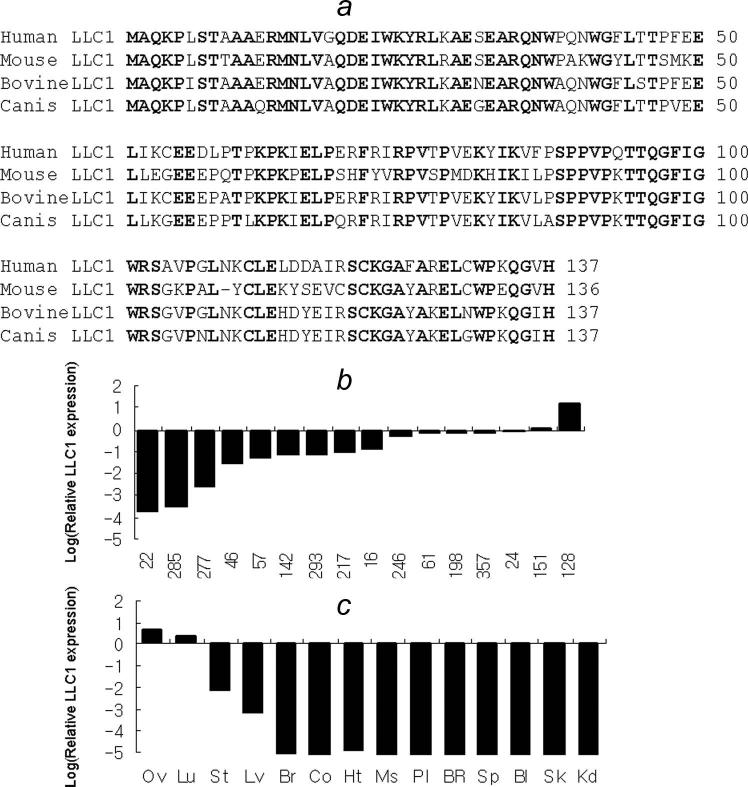
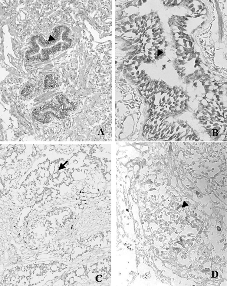
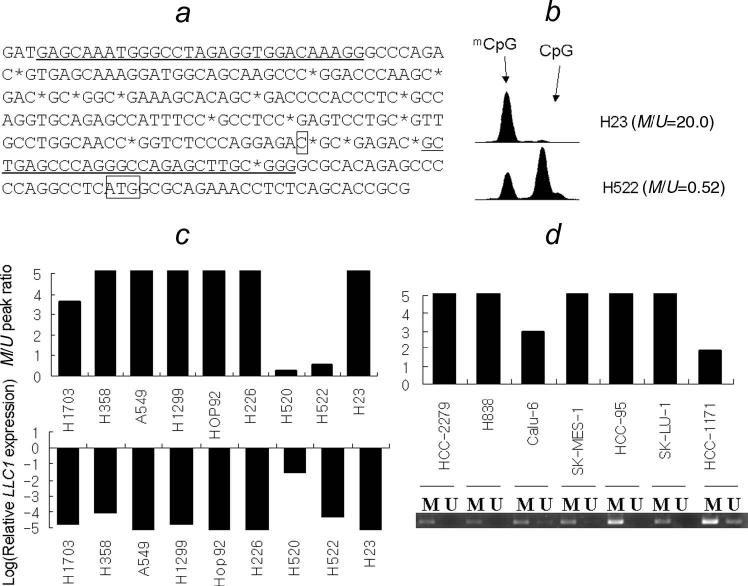
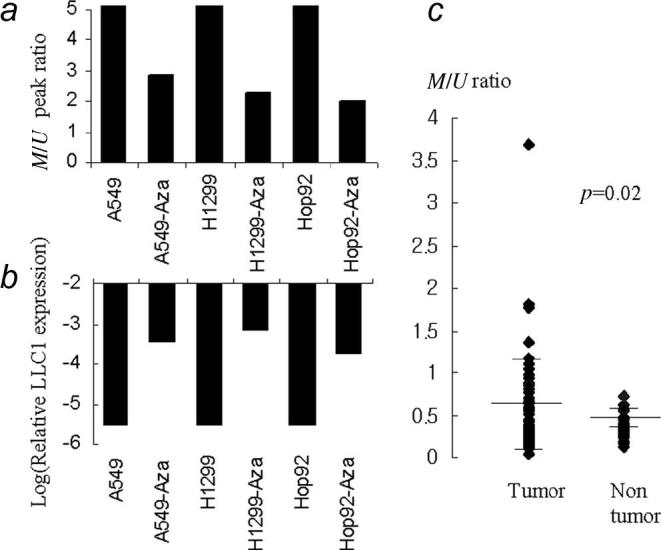
Serial analysis of gene expression studies led us to identify a previously unknown gene, c20orf85, that is present in the normal lung epithelium but absent or downregulated in most primary nonsmall cell lung cancers and lung cancer cell lines. We named this gene LLC1 for Low in Lung Cancer 1. LLC1 is located on chromosome 20q13.3 and has a 70% GC content in the promoter region. It has 4 exons and encodes a protein containing 137 amino acids. By in situ hybridization, we observed that LLC1 message is localized in normal lung bronchial epithelial cells but absent in 13 of 14 lung adenocarcinoma and 9 out of 10 lung squamous carcinoma samples. Methylation at CpG sites of the LLC1 promoter was frequently observed in lung cancer cell lines and in a fraction of primary lung cancer tissues. Treatment with 5-aza deoxycytidine resulted in a reduced methylation of the LLC1 promoter concomitant with the increase of LLC1 expression. These results suggest that inactivation of LLC1 by means of promoter methylation is a frequent event in nonsmall cell lung cancer and may play a role in lung tumorigenesis.
Figures
Similar articles
-
Aberrant methylation of TMS1 in small cell, non small cell lung cancer and breast cancer.Int J Cancer. 2003 Aug 20;106(2):198-204. doi: 10.1002/ijc.11206. Int J Cancer. 2003. PMID: 12800194
-
Epigenetic down-regulation of death-associated protein kinase in lung cancers.Clin Cancer Res. 2003 Aug 1;9(8):3034-41. Clin Cancer Res. 2003. PMID: 12912953
-
Immunohistochemical localization of LLC1 in human tissues and its limited expression in non-small cell lung cancer.Histol Histopathol. 2015 Sep;30(9):1111-20. doi: 10.14670/HH-11-608. Epub 2015 Mar 18. Histol Histopathol. 2015. PMID: 25786037
-
Genetic and epigenetic screening for gene alterations of the chromatin-remodeling factor, SMARCA4/BRG1, in lung tumors.Genes Chromosomes Cancer. 2004 Oct;41(2):170-7. doi: 10.1002/gcc.20068. Genes Chromosomes Cancer. 2004. PMID: 15287030
-
Aberrant p16 promoter methylation in smokers and former smokers with nonsmall cell lung cancer.Int J Cancer. 2003 Oct 10;106(6):913-8. doi: 10.1002/ijc.11322. Int J Cancer. 2003. PMID: 12918069
Cited by
-
Exploring the Role of Fallopian Ciliated Cells in the Pathogenesis of High-Grade Serous Ovarian Cancer.Int J Mol Sci. 2018 Aug 24;19(9):2512. doi: 10.3390/ijms19092512. Int J Mol Sci. 2018. PMID: 30149579 Free PMC article. Review.
-
Transglutaminase 2 as a cisplatin resistance marker in non-small cell lung cancer.J Cancer Res Clin Oncol. 2010 Apr;136(4):493-502. doi: 10.1007/s00432-009-0681-6. Epub 2009 Sep 18. J Cancer Res Clin Oncol. 2010. PMID: 19763620 Free PMC article.
-
Epigenetic inactivation of SOX1 promotes cell migration in lung cancer.Tumour Biol. 2015 Jun;36(6):4603-10. doi: 10.1007/s13277-015-3107-x. Epub 2015 Jan 23. Tumour Biol. 2015. PMID: 25613070
-
Comparative analysis of methods for identifying recurrent copy number alterations in cancer.PLoS One. 2012;7(12):e52516. doi: 10.1371/journal.pone.0052516. Epub 2012 Dec 20. PLoS One. 2012. PMID: 23285074 Free PMC article.
-
Schistosoma mansoni Egg, Adult Male and Female Comparative Gene Expression Analysis and Identification of Novel Genes by RNA-Seq.PLoS Negl Trop Dis. 2015 Dec 31;9(12):e0004334. doi: 10.1371/journal.pntd.0004334. eCollection 2015 Dec. PLoS Negl Trop Dis. 2015. PMID: 26719891 Free PMC article.
References
-
- Cancer Statistics. 2005. www.cancer.gov/statistics/
-
- Sanchez-Cespedes M, Ahrendt SA, Piantadosi S, Rosell R, Monzo M, Wu L, Westra WH, Yang SC, Jen J, Sidransky D. Chromosomal alterations in lung adenocarcinoma from smokers and nonsmokers. Cancer Res. 2001;61:1309–13. - PubMed
-
- Pei J, Balsara BR, Li W, Litwin S, Gabrielson E, Feder M, Jen J, Testa JR. Genomic imbalances in human lung adenocarcinomas and squamous cell carcinomas. Genes Chromosomes Cancer. 2001;31:282–7. - PubMed
-
- Hibi K, Liu Q, Beaudry GA, Madden SL, Westra WH, Wehage SL, Yang SC, Heitmiller RF, Bertelsen AH, Sidransky D, Jen J. Serial analysis of gene expression in non-small cell lung cancer. Cancer Res. 1998;58:5690–4. - PubMed
Publication types
MeSH terms
Substances
Grants and funding
LinkOut - more resources
Full Text Sources
Medical
Molecular Biology Databases
Miscellaneous
