. 2007 Mar;37(3):675-85.
doi: 10.1002/eji.200636639.

HER-2/neu antigen loss and relapse of mammary carcinoma are actively induced by T cell-mediated anti-tumor immune responses

Affiliations

HER-2/neu antigen loss and relapse of mammary carcinoma are actively induced by T cell-mediated anti-tumor immune responses

Maciej Kmieciak et al. Eur J Immunol. 2007 Mar.

Abstract

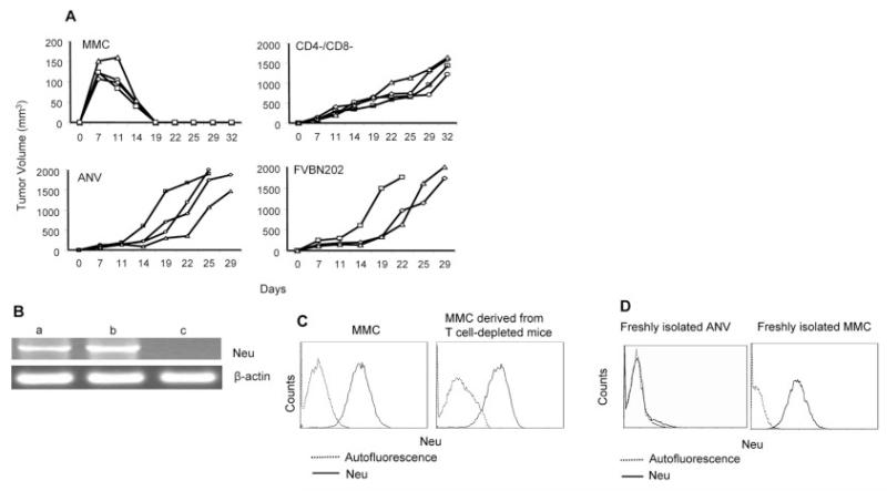
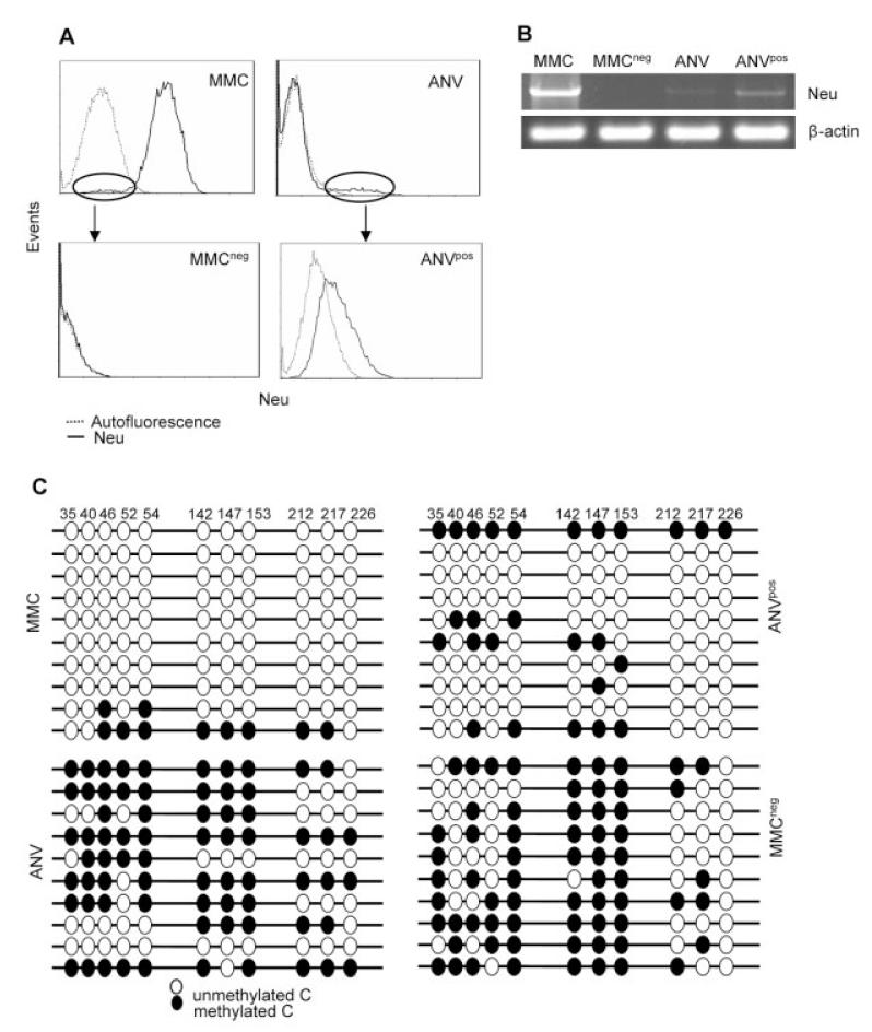
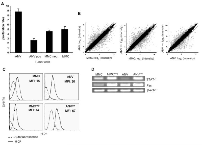
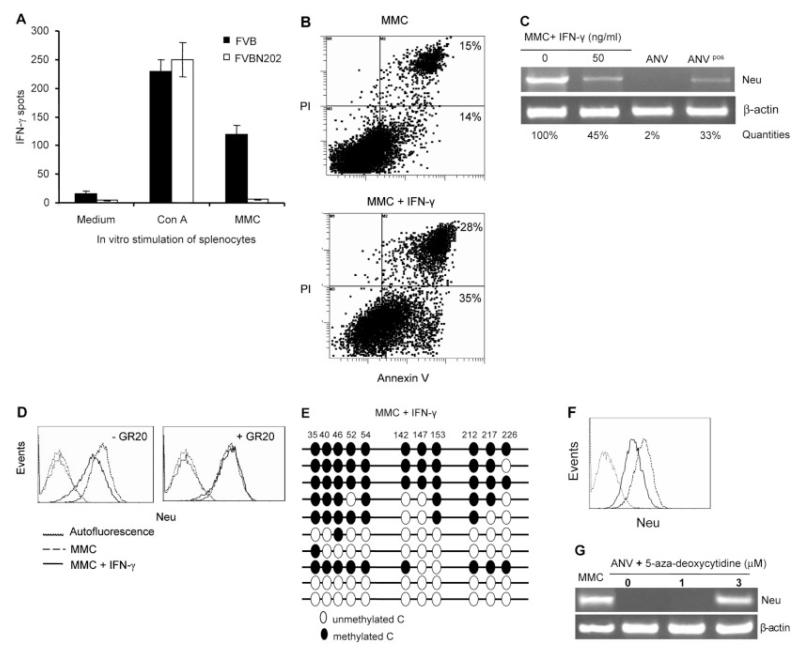
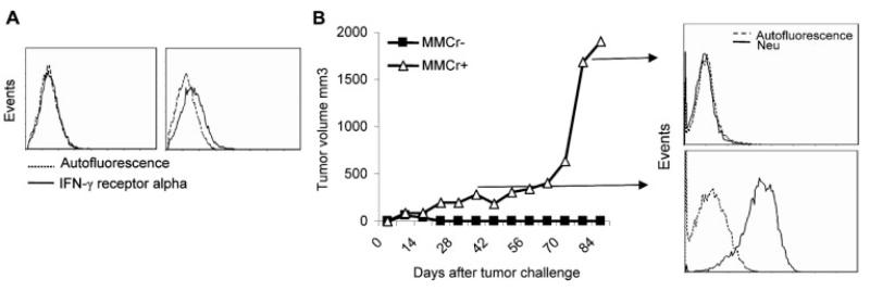
Induction of tumor-specific immune responses results in the inhibition of tumor development. However, tumors recur because of the tumor immunoediting process that facilitates development of escape mechanisms in tumors. It is not known whether tumor escape is an active process whereby anti-tumor immune responses induce loss or downregulation of the target antigen in the antigen-positive clones. To address this question, we used rat neu-overexpressing mouse mammary carcinoma (MMC) and its relapsed neu antigen-negative variant (ANV). ANV emerged from MMC under pressure from neu-specific T cell responses in vivo. We then cloned residual neu antigen-negative cells from MMC and residual neu antigen-positive cells from ANV. We found marked differences between these neu-negative clones and ANV, demonstrating that the residual neu-negative clones are probably not the origin of ANV. Since initial rejection of MMC was associated with the presence of IFN-gamma-secreting T cells, we treated MMC with IFN-gamma and showed that IFN-gamma could induce downregulation of neu expression in MMC. This appears to be due to methylation of the neu promoter. Together, these data suggest that neu antigen loss is an active process that occurs in primary tumors due to the neu-targeted anti-tumor immune responses.


Figures

Figure 1
Figure 1
Rejection of MMC in FVB mice and downregulation of neu antigen are mediated by T cell immune responses. (A) Wild-type immunocompetent (MMC) or CD4+ and CD8+ T cell-depleted (CD4/CD8) FVB mice (n=4) were inoculated with MMC. One group of FVB mice was inoculated with ANV (ANV). As a control, FVBN202-transgenic mice (n=3), which are tolerant to neu protein and fail to reject MMC, were inoculated with MMC (FVBN202). (B) RT-PCR analysis for detection of neu mRNA in tumors derived from MMC-challenged CD4/CD8 FVB mice (a) show no neu antigen loss. Expression of neu mRNA in neu-overexpressing MMC (b) and neu-negative ANV (c) was determined as positive and negative controls. (C) Detection of neu expression by flow cytometry analysis of a viable MMC line and MMC cells isolated from CD4/CD8 FVB mice. (D) Expression of the neu protein in freshly isolated ANV or MMC.
Figure 2
Figure 2
Establishment of MMCneg and ANVpos tumor lines from MMC and ANV. (A) MMC or ANV were stained with anti-neu Ab and subjected to flow cytometry analyses. Residual neu-negative clones of MMC and neu-positive clones of ANV were sorted using the Beckman Coulter EPICS Elite sorter. Sorted cells were cultured and cloned in vitro and subjected to flow cytometry for further analyses of neu expression. (B) RT-PCR analysis of the indicated tumor lines for the expression of neu mRNA using β-actin as an internal control. (C) Bisulfite genomic sequencing of the indicated cell lines. A total of ten clones per sample was sequenced.
Figure 3
Figure 3
MMC and ANV appear to have distinct morphology, proliferation rates, and gene expression profiles. (A) All the tumor lines were cultured at 2.5 × 10⁵ cells/well in triplicates using tissue culture dishes. After 3 days in culture, adherent cells were detached using 0.25% Trypsin-EDTA. Cells were then counted using trypan blue exclusion. Proliferation rates were calculated as follows: total cell numbers after 3 days in culture divided by cell numbers on day 0. (B) Microarray analysis was performed on the indicated samples. Background correction, normalization, and expression summaries were calculated. Scatter plot of log2-transformed expression summaries of the 22 690 probe sets in the Mouse430A 2.0 array are plotted for the samples indicated in the axes of the graphs. Gray dots show genes that were at least 2-fold different for MMC versus ANV, MMC versus ANVpos, or ANV versus MMCneg. Similar results were obtained in independent experiments using two different microarray analyses on biological replicates of cells. (C) The indicated tumor lines were subjected to flow cytometry-based analyses using mouse anti-H-2q and FITC-conjugated anti-mouse Ig Ab. Isotype control Ab showed MFI similar to the autofluorescence (data not shown). Representative histograms are presented, and the MFI of quadruplicate experiments are shown after subtraction of the autofluorescence. (D) RT-PCR analysis of STAT-1 and Fas mRNA isolated from MMC, MMCneg, ANV, and ANVpos. Expression of β-actin was determined as an internal control.
Figure 4
Figure 4
IFN-γ downregulates expression of neu antigen by inducing methylation of the MMTV promoter. (A) Detection of MMC-specific IFN-γ secretion by splenocytes of wild-type FVB or FVBN202-transgenic mice (n=4) 2 wk after challenge with MMC using ELISPOT assay. Splenocytes were stimulated with irradiated MMC in vitro at 6:1 ratios. Con A stimulation was used as a positive control. (B) Flow cytometry analyses of early apoptotic (Annexin V-positive) or late apoptotic (PI- and Annexin V-positive) MMC in the absence or presence of IFN-γ for 3 days. (C) Semi-quantitative RT-PCR analysis of neu expression in MMC in the absence or presence of IFN-γ. ANV and ANVpos cell lines were used as controls for loss or intermediate expression of neu mRNA. Neu expression in the PCR products was quantitated in agarose gel using Quantity One 1-D analysis Software. (D) Flow cytometry analysis of the neu expression in MMC in the absence (dashed line) or presence (solid line) of IFN-γ in vitro. Left and right panels show in vitro culture conditions in the absence or presence of IFN-γ-blocking GR20 Ab (20 lg/mL), respectively. Viable cells were gated in all the FACS analyses. Autofluorescence is shown as dotted lines. Isotype control-induced fluorescence was similar to autofluorescence. (E) Bisulfite genomic sequencing of MMC cells after 3-day treatment with IFN-γ. A total of ten clones per sample was sequenced. (F) MMC were removed from FVB mice at the time of tumor rejection (day 10–14 post-challenge). Expression of the neu protein was detected by flow cytometry either immediately after the MMC removal (solid line) or after 3–4 wk ex vivo culture (dashed line). The dotted line indicates autofluorescence. (G) RT-PCR analysis of RNA isolated from untreated MMC or ANV as well as ANV treated with different concentrations of 5-aza-deoxycytidine for 3 days. β-actin was amplified as an internal control.
Figure 5
Figure 5
IFN-γ-mediated neu antigen loss and tumor relapse in vivo. (A) Establishment of MMCr+ and MMCr clones with the presence or lack of IFN-γ receptor alpha chain obtained from freshly isolated spontaneous mammary tumors or after a number of passages of MMC in vitro, respectively. (B) FVB mice (n=3) were depleted of CD4+ T cells and inoculated with MMCr+ or MMCr. Tumor growth was monitored until 3 months after the challenge. RT-PCR analysis of neu mRNA expression in the tumors obtained from MMCr+-bearing FVB mice either at the plateau phase of tumor growth (day 40 post-challenge) or at the exponential growth phase (day 85 post-challenge).

Similar articles

Cited by

References

    1. Maeurer MJ, Gollin SM, Storkus WJ, Swaney W, Karbach J, Martin D, Castelli C, et al. Tumor escape from immune recognition: lethal recurrent melanoma in a patient associated with downregulation of the peptide transporter protein TAP-1 and loss of expression of the immunodominant MART-1/Melan-A antigen. J. Clin. Invest. 1996;98:1633–1641. - PMC - PubMed
    1. Wang Z, Marincola FM, Rivoltini L, Parmiani G, Ferrone S. Selective histocompatibility leukocyte antigen (HLA)-A2 loss caused by aberrant pre-mRNA splicing in 624MEL28 melanoma cells. J. Exp. Med. 1999;190:205–215. - PMC - PubMed
    1. Yamshchikov GV, Mullins DW, Chang CC, Ogino T, Thompson L, Presley J, Galavotti H, et al. Sequential immune escape and shifting of Tcell responses in a long-term survivor of melanoma. J. Immunol. 2005;174:6863–6871. - PubMed
    1. Facoetti A, Nano R, Zelini P, Morbini P, Benericetti E, Ceroni M, Campoli M, Ferrone S. Human leukocyte antigen and antigen processing machinery component defects in astrocytic tumors. Clin. Cancer Res. 2005;11:8304–8311. - PubMed
    1. Shankaran V, Ikeda H, Bruce AT, White JM, Swanson PE, Old LJ, Schreiber RD. IFN-γ and lymphocytes prevent primary tumour development and shape tumour immunogenicity. Nature. 2001;410:1107–1111. - PubMed

Publication types

Substances

LinkOut - more resources