. 2007 Feb;3(2):e23.
doi: 10.1371/journal.ppat.0030023.

The role of myelin in Theiler's virus persistence in the central nervous system

Affiliations

The role of myelin in Theiler's virus persistence in the central nervous system

Jean-Pierre Roussarie et al. PLoS Pathog. 2007 Feb.

Abstract

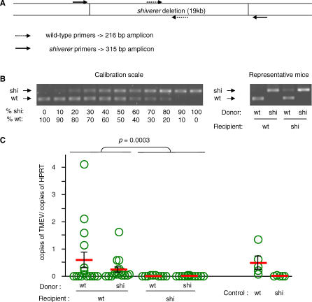
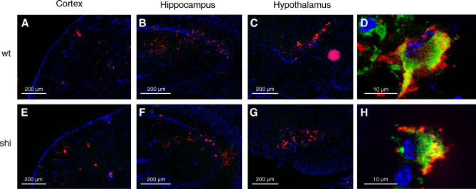
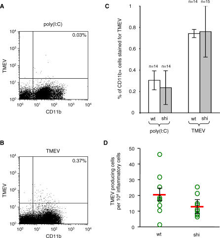
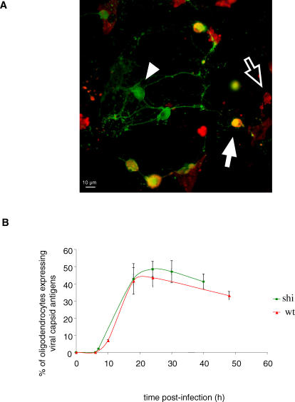
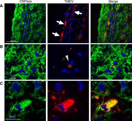
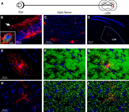
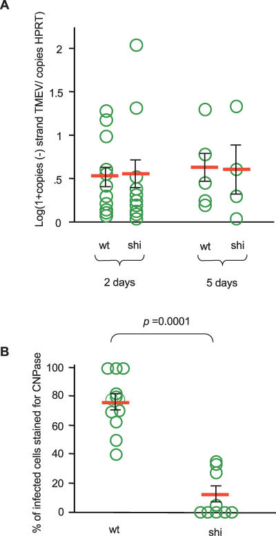
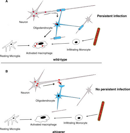
Theiler's virus, a picornavirus, persists for life in the central nervous system of mouse and causes a demyelinating disease that is a model for multiple sclerosis. The virus infects neurons first but persists in white matter glial cells, mainly oligodendrocytes and macrophages. The mechanism, by which the virus traffics from neurons to glial cells, and the respective roles of oligodendrocytes and macrophages in persistence are poorly understood. We took advantage of our previous finding that the shiverer mouse, a mutant with a deletion in the myelin basic protein gene (Mbp), is resistant to persistent infection to examine the role of myelin in persistence. Using immune chimeras, we show that resistance is not mediated by immune responses or by an efficient recruitment of inflammatory cells into the central nervous system. With both in vivo and in vitro experiments, we show that the mutation does not impair the permissiveness of neurons, oligodendrocytes, and macrophages to the virus. We demonstrate that viral antigens are present in cytoplasmic channels of myelin during persistent infection of wild-type mice. Using the optic nerve as a model, we show that the virus traffics from the axons of retinal ganglion cells to the cytoplasmic channels of myelin, and that this traffic is impaired by the shiverer mutation. These results uncover an unsuspected axon to myelin traffic of Theiler's virus and the essential role played by the infection of myelin/oligodendrocyte in persistence.


Conflict of interest statement

Competing interests. The authors have declared that no competing interests exist.

Figures

Figure 1
Figure 1. Susceptibility to Persistent Infection of Immune Chimeras between Wild-Type and shiverer Mice
Recipient mice were irradiated at 1,100 rad, reconstituted with 2.5 × 10⁶ bone marrow cells from donor mice. (A) The degree of chimerism was evaluated on peripheral blood DNA using PCR and two pairs of primers located as shown. (B) The assay was calibrated using a series of reciprocal mixtures of wild-type and shiverer DNA. The result of the assay for four representative chimeras is shown on the right. Chimerism was at least 85% for all the mice used in the experiment. (C) The immune chimeras were inoculated with 106 PFU of TMEV. The spinal cords were dissected out and the viral loads were measured by quantitative RT-PCR using HPRT mRNA as a reference. The ordinate shows the ratio (amount of viral RNA)/(amount of HPRT mRNA). Mice with a wild-type background and either a wild-type or a shiverer immune system were prone to persistent infection, whereas mice with a shiverer background and either a wild-type or a shiverer immune system were never persistently infected (Mann-Whitney test, p = 0.003). The results for age-matched wild-type and shiverer mice are shown as a control. There was no statistically significant difference between viral loads for control wild-type mice and wild-type recipients of wild-type bone marrow. Each circle corresponds to an individual mouse. wt, wild-type; shi, shiverer.
Figure 2
Figure 2. Viral Antigens in the Brain of Wild-Type and shiverer Mice
Mice were inoculated intracranially with 106 PFU of TMEV. The brain was dissected out 5 d p.i., snap-frozen, and 10-μm cryostat sections were cut. Sections were stained with an anti-NeuN antibody (green) and an anti-TMEV capsid serum (red). Nuclei were stained with DAPI. (A–D) Wild-type mice. (E–H) shiverer mice. The virus was predominantly found in cortex (A and E), hippocampus (B and F), hypothalamus (C and G), and thalamus (not shown). The distribution was the same in wild-type and in shiverer mice. (D and H) Examples of infected neurons in, respectively, wild-type and shiverer mice. Optical sections were obtained with the Zeiss ApoTome device. wt, wild-type; shi, shiverer.
Figure 3
Figure 3. Infection of Brain Macrophages during Early Disease
Wild-type and shiverer mice were inoculated intracranially with 4 × 10⁶ PFU of TMEV. The brain was dissected out 5 d p.i., mechanically and enzymatically dissociated, and inflammatory cells were purified by centrifugation on a Percoll gradient. Inflammatory cells were stained for CD11b and TMEV capsid antigens and analyzed by flow cytometry. Cells extracted from mice injected with poly(I:C) were used as negative controls for virus staining. (A) Representative analysis for a poly(I:C)-injected mouse. (B) Representative analysis for a TMEV-inoculated mouse. (C) Results obtained with 28 wild-type and 29 shiverer mice. The number of CD11b+ cells stained for capsid antigens was not statistically different between the two kinds of mice (Mann-Whitney test, p > 0.05). (D) Inflammatory cells were plated on a monolayer of BHK-21 cells and the number of viral plaques obtained was counted to measure the number of productively infected macrophages. No statistically significant difference was observed between wild-type and shiverer mice (Mann-Whitney test, p > 0.05). Each circle corresponds to an individual mouse. wt, wild-type; shi, shiverer.
Figure 4
Figure 4. Infection of Primary Cultures of Oligodendrocytes from Wild-Type or shiverer Mice
Cells were grown for 7 d with PDGF and 14 d without PDGF to induce oligodendrocyte differentiation. The cultures were infected with 500 PFU/cell of TMEV, fixed at different time points, and stained for CNPase, an oligodendrocyte marker (green) and viral capsid antigens (red). (A) Representative field of a wild-type culture 24 h p.i. White arrow, oligodendrocyte expressing viral capsid antigens; arrowhead, oligodendrocyte that did not express viral capsid; empty arrow, infected cell that was not an oligodendrocyte. (B) Percentage of oligodendrocytes that expressed viral capsid as a function of time p.i. There was no significant difference between wild-type and shiverer oligodendrocytes (Mann-Whitney test, p > 0.05, 18 and 24 h p.i.). wt, wild-type; shi, shiverer.
Figure 5
Figure 5. Viral Capsid Antigens in Myelin and Oligodendrocyte Cell Bodies during Persistent Infection
Wild-type mice were inoculated intracranially with 106 PFU of TMEV. Spinal cords were dissected out 45 d p.i., snap-frozen, and 10-μm cryostat sections (longitudinal or transverse) were cut. The sections were stained for the oligodendrocyte marker CNPase (green) and for viral capsid antigens (red). The nuclei were stained with DAPI (blue). Viral antigens were found to colocalize to a large extent with CNPase. (A) Longitudinal section. The linear pattern of capsid antigen (arrows) corresponds most probably to virus replication in myelin sheaths (see B and C). (B) Capsid antigens in myelin sheath as seen in transverse sections (arrowhead). (C) Capsid antigen in the CNPase-positive cell body of an oligodendrocyte.
Figure 6
Figure 6. TMEV Infection in the Optic Tract
Wild-type mice were inoculated intravitreously with 106 PFU of TMEV. (A) Cartoon of the optical tract. (B) Cryostat sections of the retina stained for capsid antigens (red) and with DAPI (blue), 4 d p.i. The virus replicates in the retina, in particular in the ganglion cell layer (arrow). Insert: An infected retinal ganglion cell stained for NeuN (green) and viral capsid antigens (red). (C) TMEV capsid antigens (red) in a section of the ipsilateral optic nerve, 4 d p.i. The virus is present in glial cells along the nerve. (D) Coronal section of the brain of a wild-type mouse, 5 d p.i, stained for viral capsid antigens (red). The virus reached the LGN by axonal transport. (E–J) Optic nerve 4 d p.i. (E and H); Viral capsid antigens (red); (F) CNPase (green); (I) GFAP (green); (G) Merge of (E) and (F); and (J) Merge of (H) and (I). The virus infects oligodendrocytes and astrocytes.
Figure 7
Figure 7. Infection of Retina and of Optic Nerve Oligodendrocytes in Wild-Type and shiverer Mice
Wild-type and shiverer mice were injected intravitreously with 106 PFU of TMEV. (A) Total RNA was extracted from the eye 2 d and 5 d p.i. The numbers of viral genomes of negative polarity (replicating RNA) and the number of HPRT mRNA were measured by RT-PCR. The ordinate shows the logarithm of the ratio (amount of minus-strand viral RNA)/(amount of HPRT mRNA). The replication of the virus inside the retina was the same in wild-type and shiverer mice. (B) Longitudinal sections of optic nerves obtained 4 d p.i. were scanned systematically under a fluorescence microscope equipped with the ApoTome device to assess marker colocalization. Oligodendrocytes were identified with an anti-CNPase antibody, astrocytes with an anti-GFAP antibody, and infected cells with an anti-capsid hyper-immune serum. The number of infected cells with an oligodendrocyte or an astrocyte phenotype was recorded. The ordinate shows the percentage of infected cells expressing CNPase. Each circle represents a different animal. The majority of infected cells in wild-type mice are oligodendrocytes. Conversely, most infected cells in shiverer mice are astrocytes. wt, wild-type, shi, shiverer.
Figure 8
Figure 8. Cartoon of TMEV Pathogenesis
The virus infects neurons in brain and spinal cord and is transported axonally during early disease. From the axon, it spreads to the surrounding cytoplasmic channels of myelin and from there to the cell body of oligodendrocytes. Macrophages, the main reservoir during persistent infection, are infected by virions released from infected oligodendrocytes, or by ingesting infected myelin. Macrophages can be activated infiltrating monocytes or activated microglial cells, or both.

Similar articles

Cited by

References

    1. Brahic M, Bureau JF, Michiels T. The genetics of the persistent infection and demyelinating disease caused by Theiler's virus. Annu Rev Microbiol. 2005;59:279–298. - PubMed
    1. Aubert C, Brahic M. Early infection of the central nervous system by the GDVII and DA strains of Theiler's virus. J Virol. 1995;69:3197–3200. - PMC - PubMed
    1. Lipton HL. Theiler's virus infection in mice: An unusual biphasic disease process leading to demyelination. Infect Immun. 1975;11:1147–1155. - PMC - PubMed
    1. Aubert C, Chamorro M, Brahic M. Identification of Theiler's virus infected cells in the central nervous system of the mouse during demyelinating disease. Microb Pathog. 1987;3:319–326. - PubMed
    1. Rodriguez M, Leibowitz JL, Lampert PW. Persistent infection of oligodendrocytes in Theiler's virus induced encephalomyelitis. Ann Neurol. 1983;13:426–433. - PubMed

Publication types

Substances