Comparative Study
. 2007 Apr 6;367(4):970-82.
doi: 10.1016/j.jmb.2007.01.041. Epub 2007 Jan 23.

Structural analysis of ABC-family periplasmic zinc binding protein provides new insights into mechanism of ligand uptake and release

Affiliations
Comparative Study

Structural analysis of ABC-family periplasmic zinc binding protein provides new insights into mechanism of ligand uptake and release

Beeram Ravi Chandra et al. J Mol Biol. .

Abstract

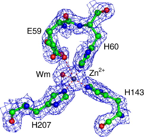
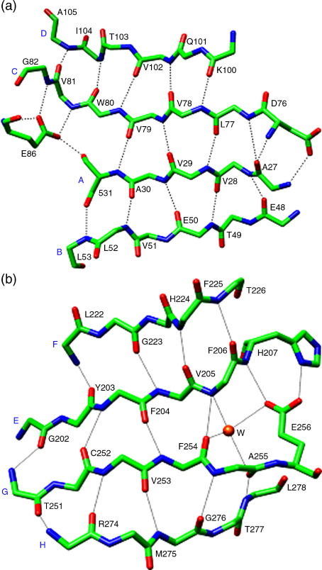
ATP-binding cassette superfamily of periplasmic metal transporters are known to be vital for maintaining ion homeostasis in several pathogenic and non-pathogenic bacteria. We have determined crystal structure of the high-affinity zinc transporter ZnuA from Escherichia coli at 1.8 A resolution. This structure represents the first native (non-recombinant) protein structure of a periplasmic metal binding protein. ZnuA reveals numerous conformational features, which occur either in Zn(2+) or in Mn(2+) transporters, and presents a unique conformational state. A comprehensive comparison of ZnuA with other periplasmic ligand binding protein structures suggests vital mechanistic differences between bound and release states of metal transporters. The key new attributes in ZnuA include a C-domain disulfide bond, an extra alpha-helix proximal to the highly charged metal chelating mobile loop region, alternate conformations of secondary shell stabilizing residues at the metal binding site, and domain movements potentially controlled by salt bridges. Based on in-depth structural analyses of five metal binding transporters, we present here a mechanistic model termed as "partial domain slippage" for binding and release of Zn(2+).


Figures

Supplementary Figure SF4
Two views of superimposed line representations of ZnuA-Ec along with two other metal binding PLBPs showing the highly conserved folds. The largest conformational differences occur in the C-domain loop region, which is proximal to interface between C-domain, N-domain and the long helix. ZnuA-Ec (red orange) TroA (light sea green), PsaA (medium blue) and MntC (purple) are superimposed along with their metal components.
Supplementary Figure SF7
(c) The conserved hydrogen bonding network at loop L7 (d) The conserved hydrogen bonding network at bottom of the metal binding cleft which links tightly for bottom part of the N- and C-domains.
Supplementary Figure SF8
Surface representation of ZnuA-Ec and vitamin B12 bound BtuF molecules and the surface is illustrated on 50% transparency. (a) In ZnuA-Ec, the bound zinc atom is shown in red sphere. (b) Vitamin B12 is shown in stick with C, O, N and P atoms in gray, red, blue and cyan, respectively. The Co and Cl ions are shown in orange and purple sphere, respectively.
Figure 1
Figure 1
A model for Zn2+ transport in bacterial systems. The three stages are as follows: (I) ZnuA binds Zn2+, (II) ZnuA and ZnuB bind each other and ZnuA transfers Zn2+ to ZnuB, and completion of the Zn2+ transport cycle (III).
Figure 2
Figure 2
Ribbon diagram of the overall structure of ZnuA from E. coli. (a) ZnuA-Ec displays a C-clamp like fold with a pseudo-2-fold axis linked by a long helix. N-domain (purple), C-domain (medium blue) and long helix (forest green) are colored differently. Bound Zn2+ is represented as an orange sphere and the protein termini are marked. (b) An overall illustration of important features of molecule A of ZnuA-Ec. The marked labels are as follows: (i) hydrogen bonding network at the metal binding arm; (ii) domain movement controlling the hydrogen bonding network and salt brides; (iii) metal binding site; (iv) unique disulfide bond in the C-domain region.
Figure 3
Figure 3
Structure-based sequence alignment of metal binding PLBPs from the ABC-type cluster 9 family. Proteins are: ZnuA-Ec (Escherichia coli), ZnuA-Syn (Synechocystis 6803), TroA (Treponema pallidum), PsaA (Streptococcus pneumoniae) and MntC-Syn (Synechocystis 6803). Metal binding residues are highlighted in red and their secondary shell stabilization residues are highlighted in blue. Twelve identical/strictly conserved residues, 37 conserved residues and 24 semi-conserved substitutions are marked with an asterisk, semicolon and dot, respectively.
Figure 4
Figure 4
Two views of superimposed line representations of ZnuA-Ec along with ZnuA-Syn and metal unbound TroA showing the highly conserved folds. The largest conformational differences occur in the C-domain loop region, which is proximal to the interface between the C-domain, N-domain and the long helix. ZnuA-Ec, ZnuA-Syn and metal unbound TroA are shown in red, orange forest green, and yellow, respectively. The bound Zn2+ is shown in sphere representation.
Figure 5
Figure 5
Metal binding residues of ZnuA-Ec. The overlayed electron density map (2Fo-Fc) is contoured at the 1.0σ level. Bound zinc is held by His60, His143, His207 and a water molecule, which is found at the apex coordination site. Two of the three metal binding residues are contributed by the N-domain and His207 comes from the C-domain. In addition, Glu59, which has a double conformation, makes a long coordination bond with zinc.
Figure 6
Figure 6
β-Strand architecture of N and C-domains. (a) Metal binding PLBPs have very similar backbone hydrogen bonding arrangements in their N-domains. Positions 86 and 82 are strictly conserved for Glu and Gly, respectively, while positions 31 and 79 are conserved for Ser/Thr and Asp/Glu, respectively. (b) Four backbone hydrogen bonds and water-mediated hydrogen bonds are present between strands E and G of ZnuA-Ec. Side-chain atoms of conserved Glu256 hydrogen bond to backbone N and side-chain Nδ1 of a metal binding residue. (c) A different architecture of the β-strands in C-domains of ZnuA-Syn, TroA, PsaA and MntC structures. In the latter group, a conserved water molecule (this is corresponding to the one involving backbone N and O atoms at positions 286 and 240) is present. In ZnuA-Syn, His243 is flipped 180° compared to ZnuA-Ec and TroA.
Figure 6
Figure 6
β-Strand architecture of N and C-domains. (a) Metal binding PLBPs have very similar backbone hydrogen bonding arrangements in their N-domains. Positions 86 and 82 are strictly conserved for Glu and Gly, respectively, while positions 31 and 79 are conserved for Ser/Thr and Asp/Glu, respectively. (b) Four backbone hydrogen bonds and water-mediated hydrogen bonds are present between strands E and G of ZnuA-Ec. Side-chain atoms of conserved Glu256 hydrogen bond to backbone N and side-chain Nδ1 of a metal binding residue. (c) A different architecture of the β-strands in C-domains of ZnuA-Syn, TroA, PsaA and MntC structures. In the latter group, a conserved water molecule (this is corresponding to the one involving backbone N and O atoms at positions 286 and 240) is present. In ZnuA-Syn, His243 is flipped 180° compared to ZnuA-Ec and TroA.
Figure 7
Figure 7
Hydrogen bonding network and inter-domain interactions in ZnuA structures. (a) Superposition of A and B monomers of ZnuA-Ec from the asymmetric unit is shown. The N-terminal metal binding arm of ZnuA has strictly conserved Tyr62 and Asp68 which hydrogen bond. The salt bridge networks between Arg65, Arg71, Glu50 and Asp68 are also shown. These interactions may restrict the movement of the metal binding arm, which holds residues His60, and Glu59. (b) The conserved hydrogen bonding networks and unique salt bridges for ZnuA-Ec and ZnuA-Syn are shown. A and B versions of ZnuA-Ec are shown in green and yellow whereas ZnuA-Syn is shown in sea blue.
Figure 8
Figure 8
(a) The Venus fly-trap mechanism proposed for capture/release of large ligands by PLBPs. The N and C-domains are held by a flexible linker which acts as a hinge. Bending of the flexible linker allows opening of the two domains so that the trapped ligand can escape. (b) Our proposed partial domain slippage mechanism for capture/release of zinc by cluster 9 family of PLBPs. The conserved hydrogen bonding networks and salt bridges restrict movement of the metal binding arm, and upon metal release a water and Glu59 will occupy the vacant metal site. The alternate conformation of Arg152 and a seesaw mechanism of C-domain helix e may cause the slippage of the bottom part of the C-domain. The breakage of backbone hydrogen bonds between strands G and E in the C-domain may trigger flexibility that leads to flipping of the active site His207, which was seen in the ZnuA-Syn (Figure 6(c)). Flipping of His207 may be the first conformational step towards release of bound metal. The marked labels are as follows: (i) hydrogen bonding network at the metal binding arm; (ii) domain movement controlling the hydrogen bonding network and salt brides; (iii) metal binding site. Wm, Wc, Ws are metal bound, conserved and structural water molecules, respectively.

References

    1. Claverys J.P. A new family of high-affinity ABC manganese and zinc permeases. Res. Microbiol. 2001;152:231–243. - PubMed
    1. Pakrasi H.B., Ogawa T., Bhattacharya-Pakarasi M. Transport of metals: a key process in oxygenic photosynthesis. In: Aro E.-M., Andersson B.A., editors. Regulatory Aspects of Photosynthesis. Kluwer Academic; Dordrecht, The Netherlands: 2001. pp. 253–264.
    1. Quiocho F.A., Ledvina P.S. Atomic structure and specificity of bacterial periplasmic receptors for active transport and chemotaxis: variation of common themes. Mol. Microbiol. 1996;20:17–25. - PubMed
    1. Bouige P., Laurent D., Piloyan L., Dassa E. Phylogenetic and functional classification of ATP-binding cassette (ABC) systems. Curr. Protein Pept. Sci. 2002;3:541–559. - PubMed
    1. Hantke K. Bacterial zinc transporters and regulators. Biometals. 2001;14:239–249. - PubMed

Publication types

MeSH terms

Associated data

LinkOut - more resources