Review
. 2007 Mar;113(3):546-93.
doi: 10.1016/j.pharmthera.2006.11.007. Epub 2006 Dec 28.

Mechanisms of action of glucagon-like peptide 1 in the pancreas

Affiliations
Review

Mechanisms of action of glucagon-like peptide 1 in the pancreas

Máire E Doyle et al. Pharmacol Ther. 2007 Mar.

Abstract

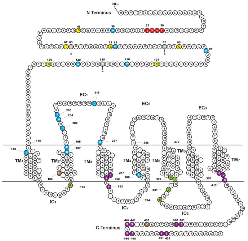
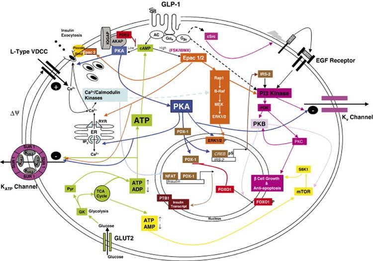
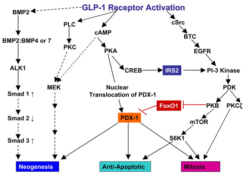
Glucagon-like peptide 1 (GLP-1) is a hormone that is encoded in the proglucagon gene. It is mainly produced in enteroendocrine L cells of the gut and is secreted into the blood stream when food containing fat, protein hydrolysate, and/or glucose enters the duodenum. Its particular effects on insulin and glucagon secretion have generated a flurry of research activity over the past 20 years culminating in a naturally occurring GLP-1 receptor (GLP-1R) agonist, exendin 4 (Ex-4), now being used to treat type 2 diabetes mellitus (T2DM). GLP-1 engages a specific guanine nucleotide-binding protein (G-protein) coupled receptor (GPCR) that is present in tissues other than the pancreas (brain, kidney, lung, heart, and major blood vessels). The most widely studied cell activated by GLP-1 is the insulin-secreting beta cell where its defining action is augmentation of glucose-induced insulin secretion. Upon GLP-1R activation, adenylyl cyclase (AC) is activated and cAMP is generated, leading, in turn, to cAMP-dependent activation of second messenger pathways, such as the protein kinase A (PKA) and Epac pathways. As well as short-term effects of enhancing glucose-induced insulin secretion, continuous GLP-1R activation also increases insulin synthesis, beta cell proliferation, and neogenesis. Although these latter effects cannot be currently monitored in humans, there are substantial improvements in glucose tolerance and increases in both first phase and plateau phase insulin secretory responses in T2DM patients treated with Ex-4. This review will focus on the effects resulting from GLP-1R activation in the pancreas.


Figures

Fig 1
Fig 1
Amino acid sequence of the rat GLP-1R showing the predicted domains, the N-terminal domain, the 7 transmembrane domains (TM1-TM7), the three extracellular domains (EC1, EC2, EC3) and the three intracellular domains (IC1, IC2, IC3). Assignment of these domains is after Thorens (Thorens, 1992). Amino acids that are critical for agonist binding are displayed in blue. The six cysteine residues highly conserved in the Class B receptor family in the N-terminal extracellular region (Thorens et al., 1993) are highlighted in yellow. Amino acids important in binding are shown in blue and are mostly located in the extracellular N-terminal region, in the TM1, TM2, and one in TM4. Glycosylation sites are shown in gray (Goke et al., 1994; Thorens, 1992). Residue H180 is shown is brown as an arginine substitution at this particular point causes both a reduction in affinity for the native ligand and in cAMP production (Heller et al., 1996). Residues known to have a functional importance in binding and/or cAMP activation are highlighted in green and those important in receptor internalization are shown in purple.
Fig 2
Fig 2
A schematic drawing outlining the main signaling pathways activated in response to ligand engagement with the GLP-1R and their major downstream effects on acute insulin secretion, insulin synthesis, preservation of β cell function and mass and regulation of proliferation. Pathways are glucose dependent hence the inclusion of glucose metabolism. GLP-1/Ex-4 bind to GLP-1R causing an increase in cAMP (Drucker et al., 1987); this leads to activation of both PKA (Wang et al., 2001) and EPAC (Holz, 2004). Localized low concentrations of cAMP lead to preferential activation of PKA. Higher cell-wide increases of cAMP by the AC stimulator forskolin (FSK) or the phosphodiesterase (PDE) inhibitor IBMX favor the EPAC pathway. cAMP is compartmentalized by PDEs most notably the PDE3B isoform as shown (Harndahl et al., 2004). PKA anchoring proteins (AKAPs) influence the specificity of cAMP response by anchoring the PKA to specific intracellular sites (Lester et al., 1997). Shown here also is the Ca2+/calmodulin binding protein IQGAP1 which co-immunoprecipitates with PKA and AKAP79 (Nauert et al., 2003). cAMP levels are increased as a consequence of ATP activation of AC consequent upon glucose metabolism. Binding of cAMP to the regulatory units of PKA results in release of the catalytic units from PKA and its activation. Sustained oscillatory increases in cAMP by GLP-1R activation lead to translocation of PKA to the nucleus (Dyachok et al., 2006; Gao et al., 2002) where it regulates PDX-1 (Wang et al., 2001) and CREB activation and subsequently insulin transcription (Chepurny et al., 2002; Hay et al., 2005; Kemp and Habener, 2001). Downstream targets of PKA and Epac in acute insulin secretion, include the KATP and Kv channels, the insulin secretory vesicles and the IP3 Ca2+ channels on the endoplasmic reticulum (ER). P38 MAPK, although activated by GLP-1R agonists (Kemp and Habener, 2001; Montrose-Rafizadeh et al., 1999) is not included as the exact mechanism of activation has not been described. Activation of the MEK/ERK pathway is coordinated through both the Epac moieties and the Ca2+/calmodulin kinases (Arnette et al., 2003; Gomez et al., 2002). The effect of PKA on CREB mediated induction of the IRS2 gene is shown, this is a prolonged effect of GLP-1R activation (Jhala et al., 2003). Acutely PI3 kinase is also stimulated by transactivation of the EGF receptor by cSrc-activated betacellulin (BTC; Buteau et al., 2003). Downstream of PI3 kinase are PKB and PKCζ both of which are implicated in β cell proliferation and PKB in prevention of β cell death (Buteau et al., 2001; Wang and Brubaker, 2002). FoxO1 is regulated by phosphorylation by PKB which results in its exclusion from the nucleus thus permitting the nuclear translocation of PDX-1 (Buteau et al., 2006). Finally enhanced ATP production due to increased mobilization of Ca2+ which in turn upregulate mitochondrial dehydrogenases leads to upregulation of mTOR activity and its downstream effector S6K1 (Kwon et al., 2004a). mTOR is implicated in increased β cell mitosis and may also be activated by PKB. GLP-1R activation also leads to stabilization of the insulin transcript by stimulating nucleocytoplasmic translocation of polypyrimidine tract binding protein (PTB) which binds to the U-rich polypyrimidine tract of insulin and insulin secretory vesicle mRNA transcripts thereby stabilizing them (Knoch et al., 2006). Mechanisms that have not been clearly demonstrated are shown by broken arrows.
Fig 3
Fig 3
Simplified schema of the human (A) and rat I (B) insulin promoters. The elements known to be regulated downstream of GLP-1R activation are shown in blue. There are four CRE sites in the insulin gene two upstream (CRE1 and CRE2) and two downstream (CRE3 and CRE4) of the transcription start site. With the exception of the first one (CRE1) all were shown to be induced when constructs containing fragments representing the individual sites were transfected into INS-1 cells (Hay et al., 2005). It is probable that the close proximity of the CRE1 site to the A3 element to which PDX-1 binds actively prohibits complex formation at this CRE site. All of the NFAT sites studied in the rat I insulin promoter are responsive to the combination of glucose and GLP-1, although NFAT2 was relatively insensitive to GLP-1 alone (Lawrence et al., 2002)
Fig 4
Fig 4
Schema outlining the signaling mechanisms reported to be involved in GLP-1R-induced differentiation/neogenesis of pancreatic precursor cells, proliferation and in the prevention of apoptosis. Dashed lines indicate mechanisms that are either not fully delineated or in the case of the cAMP activation of the MEK pathway are complex and are shown completely in Fig 2. The mechanism shown for involvement of BMP and TGFβ signaling pathways in differentiation is after Gittes and co-workers (Yew et al., 2005; Yew et al., 2004). PKB is shown as inhibiting FoxO1 (by phosphorylation). When FoxO1 is in its active and unphosphorylated state it inhibits the transcription of PDX-1 and its translocation to the nucleus (Kitamura et al., 2002).
Fig 5
Fig 5
INRI G9 cells were transfected with the rat GLP-1R. (A), (B), RT-PCR and western blot of INR1 G9 cells demonstrate the absence of GLP-1R gene and protein in native cells but its presence in transfected cells. CHO and RIN cells serve as negative and positive controls, respectively. (C), Intracellular cAMP levels in transfected cells in response to GLP-1(10 nM). (D), GLP-1-mediated glucagon (30 min) secretion into the medium of transfected cells shows no glucose-dependency.

References

    1. Abraham EJ, Leech CA, Lin JC, Zulewski H, Habener JF. Insulinotropic hormone glucagon-like peptide-1 differentiation of human pancreatic islet-derived progenitor cells into insulin-producing cells. Endocrinology. 2002;143:3152–3161. - PubMed
    1. Abrahamsen N, Nishimura E. Regulation of glucagon and glucagon-like peptide-1 receptor messenger ribonucleic acid expression in cultured rat pancreatic islets by glucose, cyclic adenosine 3′,5′-monophosphate, and glucocorticoids. Endocrinology. 1995;136:1572–1578. - PubMed
    1. Ahren B, Pacini G, Foley JE, Schweizer A. Improved meal-related beta-cell function and insulin sensitivity by the dipeptidyl peptidase-IV inhibitor vildagliptin in metformin-treated patients with type 2 diabetes over 1 year. Diabetes Care. 2005;28:1936–1940. - PubMed
    1. Ainscow EK, Rutter GA. Mitochondrial priming modifies Ca2+ oscillations and insulin secretion in pancreatic islets. Biochem J. 2001;353:175–180. - PMC - PubMed
    1. Al-Sabah S, Donnelly D. The positive charge at Lys-288 of the glucagon-like peptide-1 (GLP-1) receptor is important for binding the N-terminus of peptide agonists. FEBS Lett. 2003;553:342–346. - PubMed

Publication types

MeSH terms