. 2007 Mar 6;17(5):418-24.
doi: 10.1016/j.cub.2007.01.027. Epub 2007 Feb 15.

Ubiquitination of RIP1 regulates an NF-kappaB-independent cell-death switch in TNF signaling

Affiliations

Ubiquitination of RIP1 regulates an NF-kappaB-independent cell-death switch in TNF signaling

Marie Anne O'Donnell et al. Curr Biol. .

Abstract

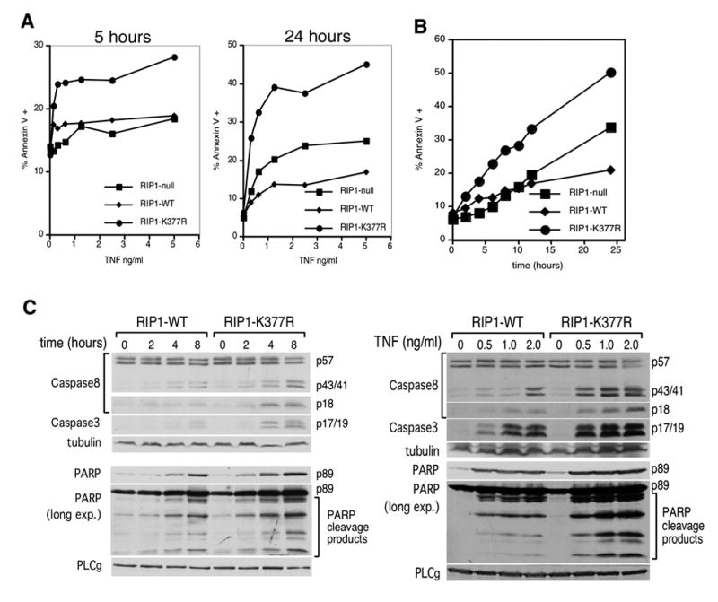
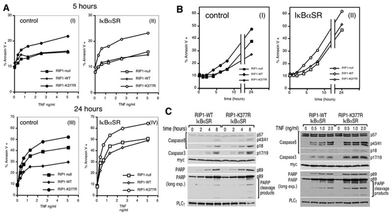
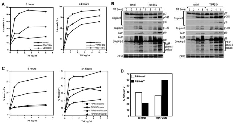
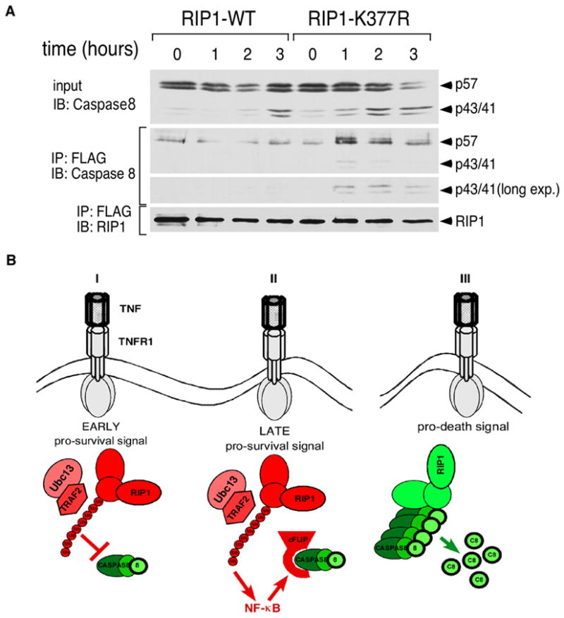
TNF receptor 1 (TNFR1) can trigger opposing responses within the same cell: a prosurvival response or a cell-death pathway [1, 2]. Cell survival requires NF-kappaB-mediated transcription of prosurvival genes [3-9]; apoptosis occurs if NF-kappaB signaling is blocked [5, 7-9]. Hence, activation of NF-kappaB acts as a cell-death switch during TNF signaling. This study demonstrates that the pathway includes another cell-death switch that is independent of NF-kappaB. We show that lysine 63-linked ubiquitination of RIP1 on lysine 377 inhibits TNF-induced apoptosis first through an NF-kappaB-independent mechanism and, subsequently, through an NF-kappaB-dependent mechanism. In contrast, in the absence of ubiquitination, RIP1 serves as a proapoptotic signaling molecule by engaging CASPASE-8. Therefore, RIP1 is a dual-function molecule that can be either prosurvival or prodeath depending on its ubiquitination state, and this serves as an NF-kappaB-independent cell-death switch early in TNF signaling. These results provide an explanation for the conflicting reports on the role of RIP1 in cell death; this role was previously suggested to be both prosurvival and prodeath [10-12]. Because TRAF2 is the E3 ligase for RIP1 [13], these observations provide an explanation for the NF-kappaB-independent antiapoptotic function previously described for TRAF2 [14-16].


Figures

Figure 1
Figure 1. Lysine 377 of RIP1 regulates cell death
(A) RIP1-null cells and RIP1-null cells reconstituted with RIP1-WT or RIP1-K377R were stimulated with the indicated doses of TNF for 5 and 24 hours. Cells were stained with annexin V-PE and analyzed by flow cytometry. The percentage of total cells that were annexin V-reactive is shown. (B) The three cell lines in (A) were stimulated with 10 ng/ml TNF for the indicated times and annexin-V levels were analyzed. (C) RIP1-WT and RIP1-K377R cells were stimulated with 10 ng/ml TNF for different periods of time (left panel) or the indicated doses of TNF for 24 hours (right panel). Cell lysates were blotted with antibodies specific for CASPASE-8, cleaved CASPASE-3 or cleaved PARP. The anti-PARP blots were also overexposed to visualize additional PARP cleavage products. Re-blotting with anti-tubulin and PLCγ was also performed to demonstrate equivalent loading. These western blots are representative of at least three similar experiments.
Figure 2
Figure 2. Lysine 377 of RIP1 provides an early NF-κB-independent and a late NF-κB-dependent anti-apoptotic signal
(A) RIP1-null, RIP1-WT or RIP1-K377R cells were transduced with retroviruses encoding either GST as a negative control (left panels) or IκBαSR (right panels). These cells were stimulated with the indicated doses of TNF for 5 hours (panels I & II) and 24 hours (panels III & IV). Apoptosis was analyzed by annexin V staining and flow cytometry as in Figure 1C. (B) The cell lines in (A) were stimulated with 10 ng/ml TNF and apoptosis analyzed at the indicated times. (C) RIP1-WT and RIP1-K377R cells stably transfected with the myc-tagged IκBαSR were stimulated with the indicated doses of TNF for 24 hours (left panel) or 10 ng/ml TNF for different periods of time (right panel). Cell lysates were blotted as in Figure 1C. The anti-myc blot serves as both a loading control and to show equivalent expression of the IκBαSR in the two cell lines. These western blots are representative of at least three similar experiments.
Figure 3
Figure 3. UBC13 and TRAF2 have a NF-κB-independent anti-apoptotic function
(A) IKKγ/NEMO-deficient Jurkat T cells stably expressing the control protein GST, UBC13DN or TRAF2DN were treated with indicated concentrations of TNF for 5 and 24 hours. Apoptosis was analyzed by annexin V staining. (B) The cell lines in (A) were treated with 2 ng/ml of TNF for the indicated times. 150 ug of cell extracts were analyzed by western blot analysis as in Figure 1C. Anti-ZAP70 blotting was performed to confirm equivalent loading. (C) RIP1-null or RIP1-WT cells were transduced with control GST or TRAF2DN-encoding retroviruses. Stably selected cells were treated with indicated concentrations of TNF for 5 or 24 hours and stained with annexin V-PE. (D) The data from (C) where the cells are stimulated with 5 ng/ml TNF for 24 hours are plotted in a bar graph to highlight the dual functional role of RIP1.
Figure 4
Figure 4. Ubiquitination of RIP1 regulates association with CASPASE-8
(A) RIP1-WT and RIP1-K377R cells were stimulated with 10 ng/ml TNF for the indicated times. FLAG-tagged RIP1 and associated proteins were immunoprecipitated with anti-FLAG beads and eluted with the FLAG peptide. The immune complexes were blotted sequentially with anti-CASPASE-8 and anti-RIP1 as a loading control. The anti-CASPASE-8 blot was also overexposed to visualize the p43/p41 cleaved form. (B) A model for the role of RIP1 in TNF-mediated apoptosis. (I) In the early phase of TNFR1 signaling, TRAF2-mediated ubiquitination of lysine 377 functions as a brake to prevent RIP1 from associating with CASPASE-8. This does not require NF-κB-mediated gene transcription. (II) In the late phase of TNFR1 signaling, a second pro-survival signal provided by the ubiquitination of lysine 377 becomes effective via the up-regulation of NF-κB-dependent anti-apoptotic genes such as c-FLIP. (III) In the absence of K63-linked poly-ubiquitination, RIP1 becomes a death-signaling molecule by forming a complex with CASPASE-8 to initiate apoptosis. Red indicates anti-apoptotic molecules and green indicates pro-apoptotic molecules.

References

    1. Micheau O, Tschopp J. Induction of TNF receptor I-mediated apoptosis via two sequential signaling complexes. Cell. 2003;114:181–190. - PubMed
    1. Aggarwal BB. Signalling pathways of the TNF superfamily: a double-edged sword. Nat Rev Immunol. 2003;3:745–756. - PubMed
    1. Wang CY, Mayo MW, Korneluk RG, Goeddel DV, Baldwin AS., Jr NF-kappaB antiapoptosis: induction of TRAF1 and TRAF2 and c-IAP1 and c-IAP2 to suppress caspase-8 activation. Science. 1998;281:1680–1683. - PubMed
    1. Wang CY, Mayo MW, Baldwin AS., Jr TNF- and cancer therapy-induced apoptosis: potentiation by inhibition of NF-kappaB. Science. 1996;274:784–787. - PubMed
    1. Van Antwerp DJ, Martin SJ, Kafri T, Green DR, Verma IM. Suppression of TNF-alpha-induced apoptosis by NF-kappaB. Science. 1996;274:787–789. - PubMed

Publication types

MeSH terms

LinkOut - more resources