. 2007 Jun 20;363(1):15-25.
doi: 10.1016/j.virol.2007.01.024. Epub 2007 Feb 15.

The equine herpesvirus-1 IR3 gene that lies antisense to the sole immediate-early (IE) gene is trans-activated by the IE protein, and is poorly expressed to a protein

Affiliations

The equine herpesvirus-1 IR3 gene that lies antisense to the sole immediate-early (IE) gene is trans-activated by the IE protein, and is poorly expressed to a protein

Byung Chul Ahn et al. Virology. .

Abstract

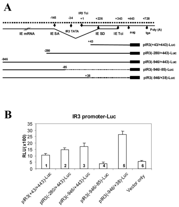
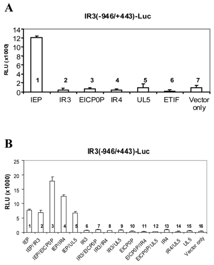
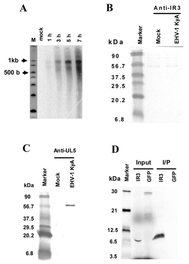
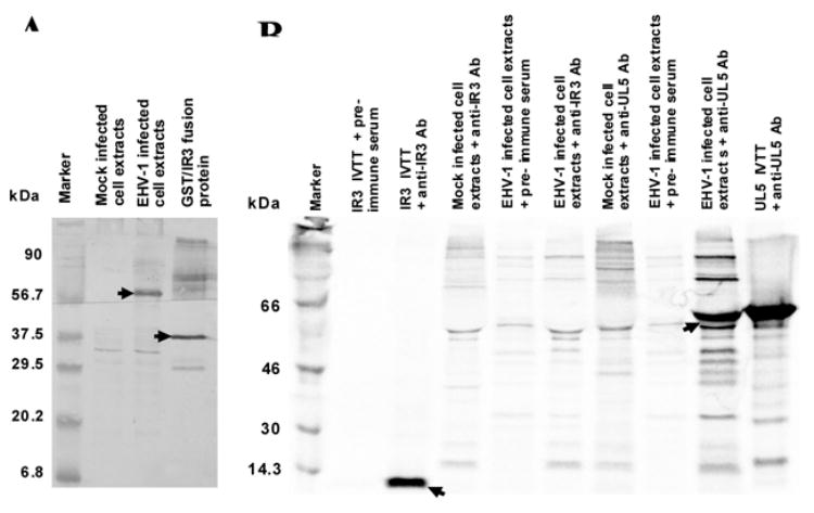
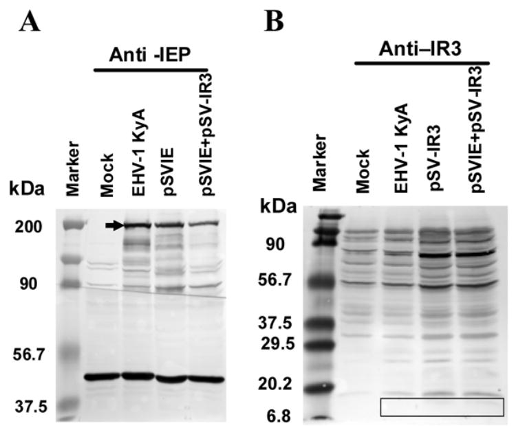
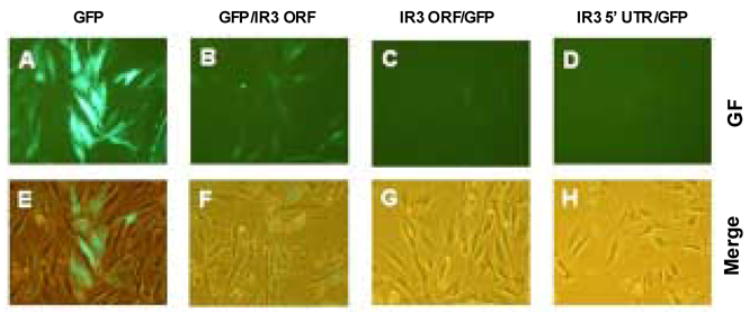
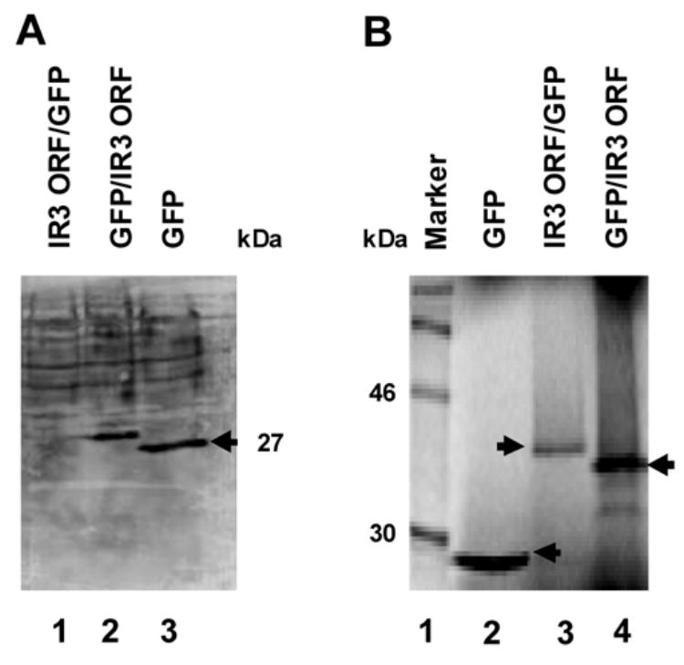
The unique IR3 gene of equine herpesvirus 1 (EHV-1) is expressed as a late 1.0-kb transcript. Previous studies confirmed the IR3 transcription initiation site and tentatively identified other cis-acting elements specific to IR3 such as a TATA box, a 443 base pair 5'untranslated region (UTR), a 285 base pair open reading frame (ORF), and a poly adenylation (A) signal [Holden, V.R., Harty, R.N., Yalamanchili, R.R., O'Callaghan, D.J., 1992. The IR3 gene of equine herpesvirus type 1: a unique gene regulated by sequences within the intron of the immediate-early gene. DNA Seq. 3, 143-152]. Transient transfection assays revealed that the IR3 promoter is strongly trans-activated by the IE protein (IEP) and that coexpression of the IEP with the early EICP0 and IR4 regulatory proteins results in maximal trans-activation of the IR3 promoter. Gel shift assays revealed that the IEP directly binds to the IR3 promoter region. Western blot analysis showed that the IR3 protein produced in E. coli was detected by antibodies to IR3 synthetic peptides; however, the IR3 protein was not detected in EHV-1 infected cell extracts by these same anti-IR3 antibodies, even though the IR3 transcript was detected by northern blot. These findings suggest that the IR3 may not be expressed to a protein. Expression of an IR3/GFP fusion gene was not observed, but expression of a GFP/IR3 fusion gene was detected by fluorescent microscopy. In further attempts to detect the IR3/GFP fusion protein using anti-GFP antibody, western blot analysis showed that the IR3/GFP fusion protein was not detected in vivo. Interestingly, a truncated form of the GFP/IR3 protein was synthesized from the GFP/IR3 fusion gene. However, GFP/IR3 and IR3/GFP fusion proteins of the predicted sizes were synthesized by in vitro coupled transcription and translation of the fusion genes, suggesting poor expression of the IR3 protein in vivo. The possible role of the IR3 transcript in EHV-1 infection is discussed.


Figures

Fig. 1
Fig. 1
The IR3 gene of EHV-1. (A) Diagram of the IR3 promoter region and IR3 promoter location. The (+/−) numbers indicate nucleotide distances from the IR3 transcription initiation site (Tci). The 117 nt of the IE/IR3 transcript overlapping sequences are located between +227 and +343. IESA, IE splicing acceptor site; IESD, IE splicing donor site. Five IR3 promoter regions were PCR-amplified and cloned into the pGL3-Basic luciferase vector as described in Materials and Methods. The IR3(−280/+443)-Luc, IR3(−946/+443)-Luc and IR3(−946/+38)-Luc include the IR3 TATA box sequences, and IR3(+43/+443)-Luc and IR3(−946/−85)-Luc do not include the IR3 TATA box sequences. (B) The IR3 promoter activity. Promoter assays of IR3 promoter sequences linked to the luciferase reporter were carried out as described in the Materials and Methods. RK13 cells were transfected with 1.0 pmol of reporter plasmid [pIR3(+43/+443)-Luc, pIR3(−280/+443)-Luc, pIR3(−946/+443)-Luc, pIR3(−946/−85)-Luc, or pIR3(−946/+38)-Luc] without effector plasmid, and luciferase activity was measured as described in Materials and Methods. RLU indicates relative luciferase units. Each experiment was carried out in triplicate. Data are averages of independent experiments, and error bars indicate standard deviations.
Fig. 2
Fig. 2
Trans-activation assays of the IR3(−946/+443)-Luc reporter construct by EHV-1 regulatory proteins. (A) 1.0 pmol of pIR3(−946/+443)-Luc reporter plasmid and 0.5 pmol of the single effector plasmid [pSVIE, pSV-IR3 ORF, pSVEICP0, pSVEICP22 (IR4), pSVEICP27 (UL5), or pSVETIF] were co-transfected into RK13 cells, and promoter activities were determined as described in Materials and Methods. (B) 1.0 pmol of pIR3(−946/+443)-Luc reporter plasmid and 0.5 pmol each of two effector plasmids [pSVIE, pSV-IR3, pSVEICP0, pSVEICP22 (IR4), or pSVEICP27 (UL5)] were co-transfected into RK13 cells, and luciferase activity was measured as described in Materials and Methods. RLU indicates relative luciferase units. Each experiment was carried out in triplicate. Data are averages of three independent experiments, and error bars indicate standard deviations.
Fig. 3
Fig. 3
The IE protein interacts with the IR3 promoter. Binding assays of the DNA sequences of the IR3 promoter region and the GST-IE protein (GST-IEP) were performed as described in Materials and Methods. Lanes 1 to 6 are the IR3 (+43/+443) DNA-IEP binding assays. Lanes 7 to 12 are IR3(−946/+38) DNA-IEP binding assays. Lanes 1 and 7 show that GST protein failed to bind to the IR3 promoter regions. Lanes 2 and 8 show DNA-GST-IEP binding. Lanes 3 and 9 show DNA-GST-IEP binding in the presence of the non-specific competitor, SP6T7 promoter sequences. Lanes 4 and 10 show that DNA-GST-IEP binding is blocked by the specific competitor, IR3 promoter DNA sequences. Lanes 5 and 11 show DNA-GST-IEP binding in the presence of control antiserum, α-GFP monoclonal antibody. Lanes 6 and 12 show that DNA-GST-IEP binding complexes were supershifted by specific α-IEP antibody.
Fig. 4
Fig. 4
Detection of the IR3 transcript and IR3 protein. Northern blot of IR3 RNA, radiolabeling of IR3 and GFP proteins, immunoprecipitation, and western blot procedures were performed as described in Materials and Methods. (A) Northern blot of the IR3 transcript. EHV-1 infected RK13 cells were harvested at 1, 3, 5, and 7 h.p.i. Total RNAs were extracted using RNA-Bee (Tel-Test, Inc., Friendwood, TX) and separated in 3% PAGE-8 M urea gels, and the IR3 transcript was detected by using a radio-labeled IR3 ORF DNA probe. M, RNA Century Marker Plus (Ambion, Austin, TX). (B) Detection of the IR3 protein. Detection of the IR3 protein in EHV-1 infected cells was performed by western blot using anti-IR3 peptide antiserum to the 20-mer peptide (see Materials and Methods). (C) Detection of UL5. UL5 in EHV-1 KyA infected cells was detected by western blot using anti-UL5 antiserum. (D) Detection of the IR3 protein by IVTT and immunoprecipitation. Samples of the IVTT reactions (Input, lanes 2 and 3) and IVTT proteins immunoprecipitated by anti-IR3 antiserum (I/P, lanes 4 and 5) were separated by SDS-PAGE. Lane 4 shows that the IR3 protein generated by IVTT was immunoprecipitated by anti-IR3 antiserum. Lane 5 shows that anti-IR3 antiserum failed to precipitate GFP generated by IVTT. The “Marker” lane shows the sizes of 14C-labeled protein molecular markers (CFA 645, Amersham Biosciences, Piscataway, NJ).
Fig. 5
Fig. 5
Detection of IR3 protein by immunological methods. (A) Detection of GST/IR3 fusion protein by western blot. GST/IR3 fusion protein and EHV-1 infected cell extracts were prepared as described in Materials and Methods. GST/IR3 fusion protein was detected using anti-IR3 antibody, and the UL5 protein was detected using anti-UL5 antibody from EHV-1 infected cell extracts by western blot. Arrows in lanes 3 and 4 indicate the UL5 protein and the GST/IR3 fusion protein, respectively. (B) Detection of the IR3 protein in extracts of EHV-1 infected radiolabeled cells. Preparation of radiolabeled IR3 and UL5 IVTT proteins, and radiolabeling of EHV-1 infected cells were performed as described in Materials and Methods. Radiolabeled IVTTs and radiolabeled cell extracts were reacted with anti-IR3 and anti-UL5 antibodies to probe for the presence of the IR3 and UL5 proteins in EHV-1 infected RK13 cells. The arrow in lane 3 indicates IVTT-synthesized IR3 protein; the arrow in lane 9 indicates the UL5 protein from EHV-1 infected cell extract.
Fig. 6
Fig. 6
Assay for the IR3 protein in EHV-1 infected cells, cells transfected with pSV-IR3, and cells cotransfected with pSVIE and pSV-IR3. Western blot procedures were performed by using anti-IR3 antibody and anti-IE antibody as described in Materials and Methods. The same amount of each sample was loaded on A and B. (A) Detection of the IEP using anti-IEP antiserum. The ~200 kDa IEP was detected in EHV-1 infected cells and in cells transfected with pSVIE and cells cotransfected with pSVIE and pSV-IR3 plasmids. As a loading control (black bands at bottom), glyceraldehyde phosphate dehydrogenase (GAPDH) was detected in cell lysates by using mouse anti-GAPDH monoclonal antibody (Chemicon international, Temecula, CA). Arrow indicates the ~200kDa IEP. (B) Assay for the IR3 protein using anti-IR3 antiserum. The ~10 kDa IR3 protein was not detected in EHV-1 infected cells, or in cells transfected with pSV-IR3, or cells cotransfected with pSVIE and pSV-IR3 plasmids. The rectangle indicates the expected location of the ~10 kDa IR3 protein.
Fig. 7
Fig. 7
Expression of GFP, IR3 5′UTR/GFP, and GFP/IR3 ORF and IR3 ORF/GFP fusion genes. Plasmid pSP6T7-EGFP-N1, pCMV-GFP/IR3 ORF, pCMV-IR3 ORF/GFP or pCMV-IR3 5′UTR/GFP was transfected into equine NBL-6 cells by using the nucleofactor kit as described in the Materials and Methods. The expression of GFP was observed using fluorescent microscopy at 48 h.p.t. Pictures in the top panels indicate green fluorescence (GF), and images in the lower panels are fluorescence and light merged images.
Fig. 8
Fig. 8
Detection of GFP, IR3 ORF/GFP and GFP/IR3 ORF proteins by western blot and IVTT. (A) Detection of GFP, IR3 ORF/GFP, and GFP/IR3 ORF proteins by western blot. Plasmids pCMV-IR3 ORF/GFP (lane 1), pCMV-GFP/IR3 ORF (lane 2), and positive control pSP6T7-EGFP-N1 (lane 3) were transfected into NBL-6 cells using the nucleofactor kit. Proteins in cellular extracts were resolved by 12% SDS-PAGE and subjected to western blot analysis using anti-GFP monoclonal antibody (Sigma, St. Louis, MO) as described in Materials and Methods. The IR3 ORF/GFP fusion protein of ~37 kDa was not detected (lane 1). A truncated form (lane 2) of the GFP/IR3 ORF fusion protein, which is smaller than the expected size of ~36 kDa, was observed. The GFP of the expected size of 27 kDa was detected (lane 3). These three vectors are under the control of the CMV promoter and identical Kozak sequences. (B) Detection of GFP, IR3 ORF/GFP, and GFP/IR3 ORF proteins by IVTT. The 35S-labeled GFP, IR3 ORF/GFP and GFP/IR3 ORF proteins generated by IVTT were separated by 12% SDS-PAGE, and assayed by autoradiography as described in the Materials and Methods. The sizes of 14C-labeled protein molecular markers (CFA626, Amersham Biosciences, Piscataway, NJ) are shown in lane 1. Arrows indicate the locations of 35S-labeled GFP (lane 2), IR3 ORF/GFP (lane 3), and GFP/IR3 ORF (lane 4) proteins.

Similar articles

Cited by

References

    1. Alam J, Cook JL. Reporter genes: application to the study of mammalian gene transcription. Anal Biochem. 1990;188:245–254. - PubMed
    1. Albrecht RA, Jang HK, Kim SK, O’Callaghan DJ. Direct interaction of TFIIB and the IE protein of equine herpesvirus 1 is required for maximal trans- activation function. Virology. 2003;316:302–312. - PubMed
    1. Albrecht RA, Kim SK, O’Callaghan DJ. The EICP27 protein of equine herpesvirus 1 is recruited to viral promoters by its interaction with the immediate-early protein. Virology. 2005;333:74–87. - PubMed
    1. Albrecht RA, Kim SK, Zhang Y, Zhao Y, O’Callaghan DJ. The equine herpesvirus 1 EICP27 protein enhances gene expression via an interaction with TATA box-binding protein. Virology. 2004;324:311–326. - PubMed
    1. Bowles DE, Holden VR, Zhao Y, O’Callaghan DJ. The ICP0 protein of equine herpesvirus type 1 is an early protein that independently transactivates expression of all classes of viral promoters. J Virol. 1997;71:4904–4914. - PMC - PubMed

MeSH terms