. 2007 May;189(9):3655-9.
doi: 10.1128/JB.00040-07. Epub 2007 Feb 16.

A riboswitch regulates expression of the coenzyme B12-independent methionine synthase in Mycobacterium tuberculosis: implications for differential methionine synthase function in strains H37Rv and CDC1551

Affiliations

A riboswitch regulates expression of the coenzyme B12-independent methionine synthase in Mycobacterium tuberculosis: implications for differential methionine synthase function in strains H37Rv and CDC1551

Digby F Warner et al. J Bacteriol. 2007 May.

Abstract

We observed vitamin B(12)-mediated growth inhibition of Mycobacterium tuberculosis strain CDC1551. The B(12) sensitivity was mapped to a polymorphism in metH, encoding a coenzyme B(12)-dependent methionine synthase. Vitamin B(12)-resistant suppressor mutants of CDC1551 containing mutations in a B(12) riboswitch upstream of the metE gene, which encodes a B(12)-independent methionine synthase, were isolated. Expression analysis confirmed that the B(12) riboswitch is a transcriptional regulator of metE in M. tuberculosis.


Figures

FIG. 1.
FIG. 1.
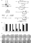
Deletion polymorphism at the PPE37-metH locus of CDC1551. Full-length MetH comprises an N-terminal homocysteine-binding domain (HCY), an N-methyltetrahydrofolate-binding domain (MTH), a CBL-binding domain, and a C-terminal SAM-binding domain (6). A large sequence polymorphism eliminates the SAM-binding domain in CDC1551 MetH (shaded box). Genomic regions deleted in the respective H37Rv metH mutants are also shown (open boxes).
FIG. 2.
FIG. 2.
Predicted M. tuberculosis metE B12 riboswitch. The secondary structure is drawn according to the scheme presented by Vitreschak et al. (23). Uppercase letters indicate residues identified by Vitreschak et al. (23) to be invariant across approximately 200 B12 riboswitches from 67 bacterial genomes; lowercase letters are M. tuberculosis specific. The conserved B12-box is highlighted. The square and circle denote sites of C→T transition mutations identified in B12 suppressor mutants of CDC1551 and H37Rv ΔmetH(B), respectively. The triangles denote single-nucleotide polymorphisms identified in the B12 riboswitch upstream of the nrdABS operon in B12 suppressor mutants of S. coelicolor (3) (see the text for details).
FIG. 3.
FIG. 3.
Construction and phenotypic characterization of M. tuberculosis methionine synthase mutants. (A) Construction and genotypic characterization of mutant strains. Genomic DNA was digested with the relevant restriction enzyme and probed with either a metE- or metH-specific probe. Restriction maps of the various strains are illustrated schematically in the line drawings adjacent to each Southern blot (not drawn to scale). For metE, Southern blots were probed with a 1,989-bp fragment (metEp, represented by a spotted box) containing 564 bp of 5′-terminal metE coding sequence obtained by PCR amplification using the primer pair metEF2/metER2 (see Table S2 in the supplemental material). The ΔmetE::hyg mutation eliminates 1,367 bp of metE coding sequence; however, an additional BamHI site is introduced by the hyg cassette (B, BamHI; WT, wild-type H37Rv). For metH, Southern blots were probed with the 644-bp MluI-BglII fragment of H37Rv metH (metHp, represented by a hatched box). The probe corresponds to the SAM-binding domain of H37Rv metH and so fails to hybridize to CDC1551 genomic DNA. The ΔmetH(B) mutation eliminates 391 bp of metH coding sequence, whereas the ΔmetH(BB) mutation eliminates 1,417 bp metH coding sequence, including an MluI site (M, MluI; WT, wild-type H37Rv). metHKin is a single-crossover homologous recombination mutant of CDC1551 containing both truncated CDC1551 and full-length H37Rv metH alleles. (B) Effect of exogenous vitamin B12 on growth of CDC1551 and H37Rv methionine synthase mutants. Cells were incubated on solid 7H10 medium containing 10 μg/ml vitamin B12 (black bars), and CFU were scored after 4 weeks. Gray bars indicate normal medium without supplement. Data represent mean CFU from two independent experiments performed in duplicate, and error bars indicate standard deviations. The H37Rv ΔmetE mutant requires B12 supplementation for growth on 7H10, ensuring an absence of CFU on unsupplemented medium; B12P2 is a representative CDC1551 B12 suppressor strain containing a mutated B12 riboswitch (see the text and Fig. 2). (C) Representative plates.
FIG. 4.
FIG. 4.
Exogenous vitamin B12 represses transcription of M. tuberculosis metE. Reverse transcription (RT)-PCR analysis of metE expression in strains H37Rv, CDC1551, and the B12 suppressor mutant B12P2 is shown. Cells were grown for 5 h in Sauton's minimal medium in the presence (+) or absence (−) of 10 μg/ml vitamin B12 before being harvested for mRNA isolation. RT-PCR methods are described in the supplemental material, and primer sequences are detailed in Table S2. The data shown are representative of two independent experiments.

References

    1. Blount, K. F., and R. R. Breaker. 2006. Riboswitches as antibacterial drug targets. Nat. Biotechnol. 24:1558-1564. - PubMed
    1. Blount, K. F., J. X. Wang, J. Lim, N. Sudarsan, and R. R. Breaker. 2006. Antibacterial lysine analogs that target lysine riboswitches. Nat. Chem. Biol. 3:44-49. - PubMed
    1. Borovok, I., B. Gorovitz, R. Schreiber, Y. Aharonowitz, and G. Cohen. 2006. Coenzyme B12 controls transcription of the Streptomyces class Ia ribonucleotide reductase nrdABS operon via a riboswitch mechanism. J. Bacteriol. 188:2512-2520. - PMC - PubMed
    1. Cole, S. T., R. Brosch, J. Parkhill, T. Garnier, C. Churcher, D. Harris, S. V. Gordon, K. Eiglmeier, S. Gas, C. E. Barry III, F. Tekaia, K. Badcock, D. Basham, D. Brown, T. Chillingworth, R. Connor, R. Davies, K. Devlin, T. Feltwell, S. Gentles, N. Hamlin, S. Holroyd, T. Hornsby, K. Jagels, A. Krogh, J. McLean, S. Moule, L. Murphy, K. Oliver, J. Osborne, M. A. Quail, M. A. Rajandream, J. Rogers, S. Rutter, K. Seeger, J. Skelton, R. Squares, S. Squares, J. E. Sulston, J. Taylor, S. Whitehead, and B. G. Barrell. 1998. Deciphering the biology of Mycobacterium tuberculosis from the complete genome sequence. Nature 393:537-544. - PubMed
    1. Dawes, S. S., D. F. Warner, L. Tsenova, J. Timm, J. D. McKinney, G. Kaplan, H. Rubin, and V. Mizrahi. 2003. Ribonucleotide reduction in Mycobacterium tuberculosis: function and expression of genes encoding class Ib and class II ribonucleotide reductases. Infect. Immun. 71:6124-6131. - PMC - PubMed

Publication types

MeSH terms