

Cytosolic proteins contribute to surface plasminogen recruitment of Neisseria meningitidis
- PMID: 17307854
- PMCID: PMC1855851
- DOI: 10.1128/JB.01966-06
Cytosolic proteins contribute to surface plasminogen recruitment of Neisseria meningitidis
Erratum in
- J Bacteriol. 2007 Jul;189(14):5404
Abstract
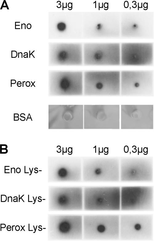
Plasminogen recruitment is a common strategy of pathogenic bacteria and results in a broad-spectrum surface-associated protease activity. Neisseria meningitidis has previously been shown to bind plasminogen. In this study, we show by several complementary approaches that endolase, DnaK, and peroxiredoxin, which are usually intracellular proteins, can also be located in the outer membrane and act as plasminogen receptors. Internal binding motifs, rather than C-terminal lysine residues, are responsible for plasminogen binding of the N. meningitidis receptors. Recombinant receptor proteins inhibit plasminogen association with N. meningitidis in a concentration-dependent manner. Besides binding purified plasminogen, N. meningitidis can also acquire plasminogen from human serum. Activation of N. meningitidis-associated plasminogen by urokinase results in functional activity and allows the bacteria to degrade fibrinogen. Furthermore, plasmin bound to N. meningitidis is protected against inactivation by alpha(2)-antiplasmin.
Figures


Similar articles
-
Analysis of plasminogen-binding M proteins of Streptococcus pyogenes.Methods. 2000 Jun;21(2):143-50. doi: 10.1006/meth.2000.0985. Methods. 2000. PMID: 10816375
-
Discriminating between cell surface and intracellular plasminogen-binding proteins: heterogeneity in profibrinolytic plasminogen-binding proteins on monocytoid cells.Thromb Haemost. 2000 Nov;84(5):882-90. Thromb Haemost. 2000. PMID: 11127872
-
Plasminogen interaction and activation on Streptococcus mutans surface.Oral Microbiol Immunol. 2004 Aug;19(4):257-61. doi: 10.1111/j.1399-302X.2004.00149.x. Oral Microbiol Immunol. 2004. PMID: 15209997
-
The analysis of Neisseria meningitidis proteomes: Reference maps and their applications.Proteomics. 2007 Aug;7(16):2933-46. doi: 10.1002/pmic.200700094. Proteomics. 2007. PMID: 17628027 Review.
-
The cell biology of the plasminogen system.FASEB J. 1995 Jul;9(10):939-45. doi: 10.1096/fasebj.9.10.7615163. FASEB J. 1995. PMID: 7615163 Review.
Cited by
-
Physical Features of Intracellular Proteins that Moonlight on the Cell Surface.PLoS One. 2015 Jun 25;10(6):e0130575. doi: 10.1371/journal.pone.0130575. eCollection 2015. PLoS One. 2015. PMID: 26110848 Free PMC article.
-
Fibrinolysis: A Primordial System Linked to the Immune Response.Int J Mol Sci. 2021 Mar 26;22(7):3406. doi: 10.3390/ijms22073406. Int J Mol Sci. 2021. PMID: 33810275 Free PMC article. Review.
-
The Complement Binding and Inhibitory Protein CbiA of Borrelia miyamotoi Degrades Extracellular Matrix Components by Interacting with Plasmin(ogen).Front Cell Infect Microbiol. 2018 Feb 2;8:23. doi: 10.3389/fcimb.2018.00023. eCollection 2018. Front Cell Infect Microbiol. 2018. PMID: 29456970 Free PMC article.
-
Distinct susceptibilities of corneal Pseudomonas aeruginosa clinical isolates to neutrophil extracellular trap-mediated immunity.Infect Immun. 2014 Oct;82(10):4135-43. doi: 10.1128/IAI.02169-14. Epub 2014 Jul 21. Infect Immun. 2014. PMID: 25047845 Free PMC article.
-
Borrelia burgdorferi enolase is a surface-exposed plasminogen binding protein.PLoS One. 2011;6(11):e27502. doi: 10.1371/journal.pone.0027502. Epub 2011 Nov 8. PLoS One. 2011. PMID: 22087329 Free PMC article.
References
-
- Ariel, N., A. Zvi, K. S. Makarova, T. Chitlaru, E. Elhanany, B. Velan, S. Cohen, A. M. Friedlander, and A. Shafferman. 2003. Genome-based bioinformatic selection of chromosomal Bacillus anthracis putative vaccine candidates coupled with proteomic identification of surface-associated antigens. Infect. Immun. 71:4563-4579. - PMC - PubMed
-
- Barlow, G. H., L. Summaria, and K. C. Robbins. 1969. Molecular weight studies on human plasminogen and plasmin at the microgram level. J. Biol. Chem. 244:1138-1141. - PubMed
-
- Berge, A., and U. Sjobring. 1993. PAM, a novel plasminogen-binding protein from Streptococcus pyogenes. J. Biol. Chem. 268:25417-25424. - PubMed
-
- Bergmann, S., M. Rohde, G. S. Chhatwal, and S. Hammerschmidt. 2001. Alpha-enolase of Streptococcus pneumoniae is a plasmin(ogen)-binding protein displayed on the bacterial cell surface. Mol. Microbiol. 40:1273-1287. - PubMed
Publication types
MeSH terms
Substances
LinkOut - more resources
Full Text Sources
Molecular Biology Databases
