. 2007 May;189(9):3525-31.
doi: 10.1128/JB.00044-07. Epub 2007 Feb 16.

Role of PBP1 in cell division of Staphylococcus aureus

Affiliations

Role of PBP1 in cell division of Staphylococcus aureus

S F F Pereira et al. J Bacteriol. 2007 May.

Abstract

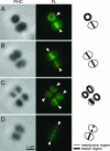
We constructed a conditional mutant of pbpA in which transcription of the gene was placed under the control of an IPTG (isopropyl-beta-D-thiogalactopyranoside)-inducible promoter in order to explore the role of PBP1 in growth, cell wall structure, and cell division. A methicillin-resistant strain and an isogenic methicillin-susceptible strain, each carrying the pbpA mutation, were unable to grow in the absence of the inducer. Conditional mutants of pbpA transferred into IPTG-free medium underwent a four- to fivefold increase in cell mass, which was not accompanied by a proportional increase in viable titer. Examination of thin sections of such cells by transmission electron microscopy or fluorescence microscopy of intact cells with Nile red-stained membranes showed a morphologically heterogeneous population of bacteria with abnormally increased sizes, distorted axial ratios, and a deficit in the number of cells with completed septa. Immunofluorescence with an antibody specific for PBP1 localized the protein to sites of cell division. No alteration in the composition of peptidoglycan was detectable in pbpA conditional mutants grown in the presence of a suboptimal concentration of IPTG, which severely restricted the rate of growth, and the essential function of PBP1 could not be replaced by PBP2A present in methicillin-resistant cells. These observations suggest that PBP1 is not a major contributor to the cross-linking of peptidoglycan and that its essential function must be intimately integrated into the mechanism of cell division.


Figures

FIG. 1.
FIG. 1.
Construction of the pbpA conditional mutant. An 850-bp fragment of pbpA containing the ribosome-binding site was cloned downstream of the Pspac promoter in the integrative vector pMGPI. The resulting plasmid, pMGPA, was introduced into S. aureus RN4220 by electroporation and was integrated into the chromosome by a Campbell-type recombination event. The only complete copy of pbpA in the resulting strain is under Pspac control. The fusion was subsequently moved to the COL and COL-S strains by transduction to yield COLspacP1 and COL-SspacP1, respectively.
FIG. 2.
FIG. 2.
Essentiality of pbpA expression for S. aureus growth and transcription of pbpA in the mutant strain COLspacP1 grown with different inducer concentrations. (A) Representative growth curves of pbpA conditional mutants COL-SspacP1 (⋄) and COLspacP1 (•) in the absence (dashed line) and presence (solid line) of 500 μM IPTG. (B) Representative growth curves of the parental MRSA strain COL (⧫) and its mutant COLspacP1. The mutant was depleted of PBP1 and grown in the absence of IPTG (○) and in the presence of 35 μM (×), 50 μM (▴), 80 μM (▵), and 500 μM (•) concentrations of inducer. (C) Representative growth curves of COL (▪), COL with pSK5632 (□), COLspacP1 in the absence of IPTG (○) and in the presence of a 500 μM concentration of inducer (•), and COLspacP1 with pSKP1 in the absence of inducer (+). (D) COLspacP1 was grown in increasing concentrations of inducer (see panel B), and the relative abundance of the pbpA transcript was expressed as an n-fold difference relative to COL. The transcription levels of pbpA were normalized to the levels of the housekeeping gene pta.
FIG. 3.
FIG. 3.
Morphology of the parental strain COL and its pbpA conditional mutant, COL-spacP1, grown in the absence of IPTG. Strain COL was grown in TSB, and COLspacP1was grown either in TSB or in TSB supplemented with 500 μM IPTG. Thin sections were processed for transmission electron microscopy to document differences in the size and morphology of the parental strain, COL (A and B), and the mutant, COLspacP1, grown in the absence of IPTG (C and D). Specimens labeled 1, 2, and 3 (in panels C and D) are representative of subpopulations 1, 2, and 3, respectively, of COLspacP1 grown in the absence of inducer (see panel F and the text for details). (E and F) Ratio between the axes defined by the equatorial (septal) and the longitudinal planes of cells of COL (black circles) and COLspacP1 grown in the absence of inducer (empty red circles) or in the presence of 500 μM IPTG (filled red circles). For the purpose of this analysis, both the longitudinal and the equatorial lengths were measured from every cell within the electron microscopy field. (G) Fluorescence images of membrane-stained representative specimens of COLspacP1 grown in the presence of IPTG and of the three subpopulations that appear when the mutant is grown in the absence of inducer. (H) Percentages of cells that show septa (completed and/or incomplete, located at the normal equatorial position) or completed septa. Black bars, strain COL; empty red bars, COLspacP1 cells grown in the absence of IPTG; filled red bars, COLspacP1 cells grown in the presence of 500 μM IPTG.
FIG. 4.
FIG. 4.
PBP1 localization by immunofluorescence. Panels show selected specimens from the same field. White arrows indicate the localization of the florescence signal in septa. Phase contrast (PHC) images are shown in the first column, and florescence (FL) images are shown in the second column, followed by a schematic representation of the cells shown in each panel.
FIG. 5.
FIG. 5.
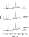
Peptidoglycan HPLC profiles for strains COL and COLspacP1 grown with optimal (500 μM) and suboptimal (50 μM) IPTG concentrations. The deficit of PBP1 did not have a significant impact on peptidoglycan composition. The only alteration observed in the HPLC profile of the mutant grown with the suboptimal IPTG concentration was a decrease in peak 1 (identified in the chromatogram), corresponding to the unsubstituted disaccharide pentapeptide monomer.

References

    1. Berger-Bachi, B., A. Strassle, and F. H. Kayser. 1986. Characterization of an isogenic set of methicillin-resistant and susceptible mutants of Staphylococcus aureus. Eur. J. Clin. Microbiol. 5:697-701. - PubMed
    1. Daniel, R. A., E. J. Harry, and J. Errington. 2000. Role of penicillin-binding protein PBP 2B in assembly and functioning of the division machinery of Bacillus subtilis. Mol. Microbiol. 35:299-311. - PubMed
    1. de Jonge, B. L., Y. S. Chang, D. Gage, and A. Tomasz. 1992. Peptidoglycan composition of a highly methicillin-resistant Staphylococcus aureus strain. The role of penicillin binding protein 2A. J. Biol. Chem. 267:11248-11254. - PubMed
    1. de Lencastre, H., S. W. Wu, M. G. Pinho, A. M. Ludovice, S. Filipe, S. Gardete, R. Sobral, S. Gill, M. Chung, and A. Tomasz. 1999. Antibiotic resistance as a stress response: complete sequencing of a large number of chromosomal loci in Staphylococcus aureus strain COL that impact on the expression of resistance to methicillin. Microb. Drug Resist. 5:163-175. - PubMed
    1. Georgopapadakou, N. H., and F. Y. Liu. 1980. Penicillin-binding proteins in bacteria. Antimicrob. Agents Chemother. 18:148-157. - PMC - PubMed

Publication types

MeSH terms

LinkOut - more resources