. 2007 Apr;143(4):1881-93.
doi: 10.1104/pp.106.093989. Epub 2007 Feb 16.

Functional genomic analysis supports conservation of function among cellulose synthase-like a gene family members and suggests diverse roles of mannans in plants

Affiliations

Functional genomic analysis supports conservation of function among cellulose synthase-like a gene family members and suggests diverse roles of mannans in plants

Aaron H Liepman et al. Plant Physiol. 2007 Apr.

Abstract

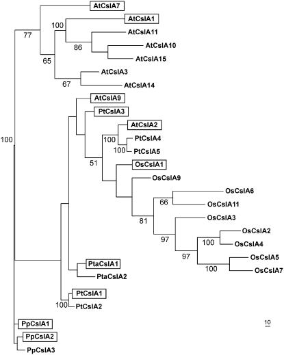
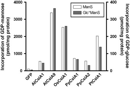
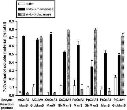
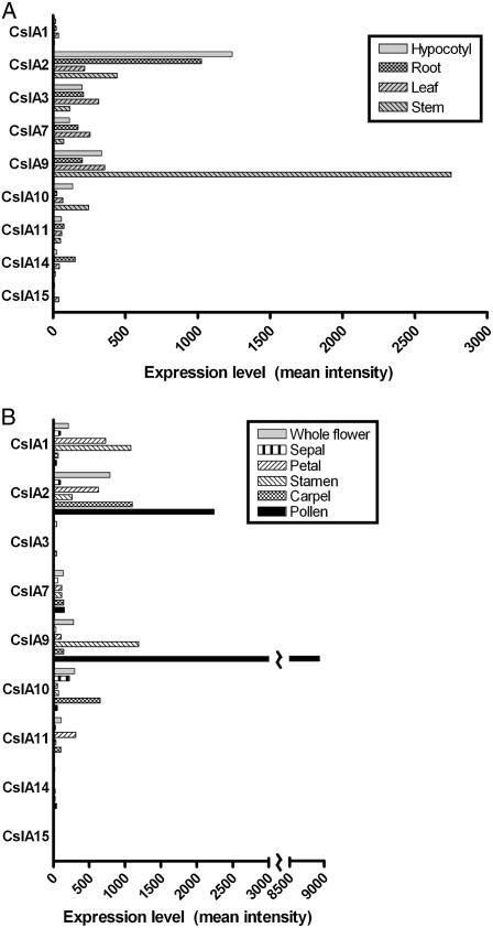
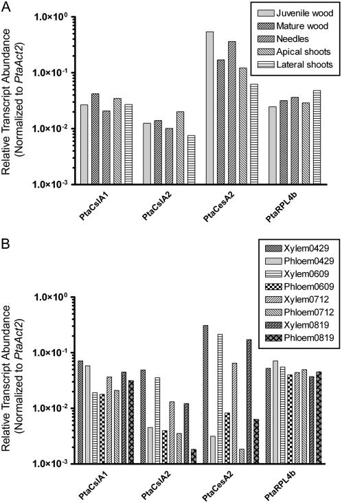
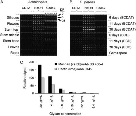
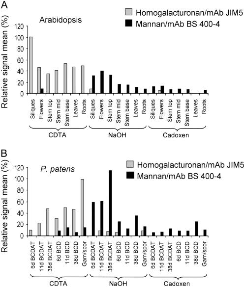
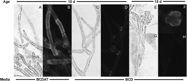
Mannan polysaccharides are widespread among plants, where they serve as structural elements in cell walls, as carbohydrate reserves, and potentially perform other important functions. Previous work has demonstrated that members of the cellulose synthase-like A (CslA) family of glycosyltransferases from Arabidopsis (Arabidopsis thaliana), guar (Cyamopsis tetragonolobus), and Populus trichocarpa catalyze beta-1,4-mannan and glucomannan synthase reactions in vitro. Mannan polysaccharides and homologs of CslA genes appear to be present in all lineages of land plants analyzed to date. In many plants, the CslA genes are members of extended multigene families; however, it is not known whether all CslA proteins are glucomannan synthases. CslA proteins from diverse land plant species, including representatives of the mono- and dicotyledonous angiosperms, gymnosperms, and bryophytes, were produced in insect cells, and each CslA protein catalyzed mannan and glucomannan synthase reactions in vitro. Microarray mining and quantitative real-time reverse transcription-polymerase chain reaction analysis demonstrated that transcripts of Arabidopsis and loblolly pine (Pinus taeda) CslA genes display tissue-specific expression patterns in vegetative and floral tissues. Glycan microarray analysis of Arabidopsis indicated that mannans are present throughout the plant and are especially abundant in flowers, siliques, and stems. Mannans are also present in chloronemal and caulonemal filaments of Physcomitrella patens, where they are prevalent at cell junctions and in buds. Taken together, these results demonstrate that members of the CslA gene family from diverse plant species encode glucomannan synthases and support the hypothesis that mannans function in metabolic networks devoted to other cellular processes in addition to cell wall structure and carbohydrate storage.


Figures

Figure 1.
Figure 1.
Phylogenetic analysis of CslA sequences. A parsimony phylogram that corresponds to the majority consensus of 1,000 bootstrap replicates (percent values above 50% shown) for full-length CslA polypeptide sequences deduced from the complete genomes of Arabidopsis (At), rice (Os), P. trichocarpa (Pt), and P. patens (Pp) and from cDNA sequences isolated from a loblolly pine (Pta) cDNA library is shown. Recombinant CslA proteins shown in boxes catalyze β-1,4-mannan and/or glucomannan synthase reactions in vitro. These activities were demonstrated in previous studies for AtCslA2, AtCslA7, and AtCslA9 by Liepman et al. (2005) and for PtCslA1 and PtCslA3 by Suzuki et al. (2006).
Figure 2.
Figure 2.
Glucomannan synthase activity assays of recombinant CslA proteins from diverse land plants produced in Drosophila S2 cells. Microsomal membranes of S2 cells producing either GFP or a CslA protein were assayed for ManS or GlcManS activity in vitro. The incorporation of GDP-Man or GDP-Glc into 70% ethanol-insoluble products by microsomal membrane fractions is graphed. The ManS product was labeled with GDP-[14C]Man, and the GlcManS product was labeled with GDP-[14C]Glc in the presence of nonradioactive GDP-Man.
Figure 3.
Figure 3.
Characterization of in vitro mannan and glucomannan synthase products. Large-scale in vitro assay products of recombinant CslA proteins from P. patens (PpCslA1), loblolly pine (PtaCslA1), rice (OsCslA1), and Arabidopsis (AtCslA9) were treated with endo-β-glucanase, endo-β-mannanase, or buffer alone. The percentage of total radioactivity released from each product into the 70% ethanol-soluble fraction after each treatment is shown.
Figure 4.
Figure 4.
Microarray expression profiling of CslA genes in vegetative and reproductive tissues of Arabidopsis. A, Mean signal intensity of Arabidopsis CslA genes in various vegetative tissues from the AtGenExpress microarray dataset (Schmid et al., 2005) is plotted. Hypocotyl, root, and leaf tissues were from 7-d-old seedlings (ATGE_2, _3, _5, respectively), and stem tissue was from 21-d-old plants (ATGE_27). B, Mean signal intensity of Arabidopsis CslA genes in flowers, flower parts, and mature pollen. Whole stage 15 flowers as well as sepals, petals, stamens, and carpels from stage 15 flowers were from 21-d-old plants (ATGE_39, _41, _42, _43, respectively); mature pollen was from 42-d-old plants (ATGE_73). All tissues were harvested from plants grown under continuous light. Low expression levels of particular CslA genes in some tissues yielded signal intensity values below the level of significance (mean intensity < 100).
Figure 5.
Figure 5.
Quantitative real-time RT-PCR analysis of PtaCslA1 and PtaCslA2 genes in tissues of loblolly pine. A, The transcript abundance of two PtaCslA genes, a cellulose synthase gene (PtaCesA2), an actin gene (PtaAct1), and a ribosomal protein L4 gene (PtaRPL4b) normalized to an actin control gene (PtaAct2) was measured in juvenile wood, mature wood, needles, apical shoot tips, and lateral shoot tips. B, Gene expression levels in xylem and phloem tissues collected at four time points, gathered from April to August, 2005.
Figure 6.
Figure 6.
Microarrays of cell wall glycan polymers. A and B, Examples of microarrays generated from polysaccharides extracted from Arabidopsis (A) and P. patens (B) probed with mannan-specific mAb (BS 400–4). Polysaccharides were sequentially extracted from the tissues/organs indicated using CDTA, followed by NaOH, then cadoxen. In the case of P. patens, cultures were grown on either BCD or BCDAT mediums as indicated. Three dilution factors (DF) of the extractions (0, 5, and 25) were printed in sextuplet (*) such that each organ/tissue is represented by 18 spots on the arrays (as indicted by the white box in A). C, Arrays were created in which samples of mannan and lime pectin were spotted at the dilutions shown and probed with antimannan or anti-HG (JIM5) mAbs. Spot signals were quantified using ImaGene 6.0 and the maximal signal for JIM5 set at 100%. Note that these values should not be used to calibrate glycan levels in Figure 7.
Figure 7.
Figure 7.
CoMPP analysis of mannan polysaccharides in Arabidopsis and P. patens. A and B, CoMPP data indicating the relative levels of HG (gray bars) and mannan (black bars) in Arabidopsis (A) and P. patens (B) organs/tissues. Mean spot signals were quantified using ImaGene 6.0 and for each species, the maximal signal obtained for HG in each plant was set at 100%. Note that signals can be used to infer difference in the relative glycan levels within, but not between, the two species.
Figure 8.
Figure 8.
Immunolocalization of mannans in P. patens. Tissue from P. patens cultured on BCDAT medium (A and B) or BCD medium (C–H) for 10 d (A–D) or 15 d (E–H), labeled with monoclonal antimannan, and imaged with brightfield (A, C, E, and G) and epifluorescence optics (B, D, F, and H). A and B, Chloronemal filaments. C and D, Chloronemal and caulonemal filaments. E and F, Bud. G and H, Gametophore leaf. No signal was observed in control samples lacking primary antibodies or incubated with antibodies that had been preadsorbed with carob galactomannan.

References

    1. Aspeborg H, Schrader J, Coutinho PM, Stam M, Kallas A, Djerbi S, Nilsson P, Denman S, Amini B, Sterky F, et al (2005) Carbohydrate-active enzymes involved in the secondary cell wall biogenesis in hybrid aspen. Plant Physiol 137 983–997 - PMC - PubMed
    1. Auxtova O, Liskova D, Kakoniova D, Kubackova M, Karacsonyi S, Bilisics L (1995) Effect of galactoglucomannan-derived oligosaccharides on elongation growth of pea and spruce stem segments stimulated by auxin. Planta 196 420–424
    1. Baldauf SL (2003) Phylogeny for the faint of heart: a tutorial. Trends Genet 19 345–351 - PubMed
    1. Benova-Kakosova A, Digonnet C, Goubet F, Ranocha P, Jauneau A, Pesquet E, Barbier O, Zhang Z, Capek P, Dupree P, et al (2006) Galactoglucomannans increase cell population density and alter the protoxylem/metaxylem tracheary element ratio in xylogenic cultures of zinnia. Plant Physiol 142 696–709 - PMC - PubMed
    1. Bilisics L, Vojtassak J, Capek P, Kollarova K, Liskova D (2004) Changes in glycosidase activities during galactoglucomannan oligosaccharide inhibition of auxin induced growth. Phytochemistry 65 1903–1909 - PubMed

Publication types

LinkOut - more resources