. 2007 Feb;19(2):582-96.
doi: 10.1105/tpc.106.049049. Epub 2007 Feb 16.

Tapetosomes in Brassica tapetum accumulate endoplasmic reticulum-derived flavonoids and alkanes for delivery to the pollen surface

Affiliations

Tapetosomes in Brassica tapetum accumulate endoplasmic reticulum-derived flavonoids and alkanes for delivery to the pollen surface

Kai Hsieh et al. Plant Cell. 2007 Feb.

Abstract

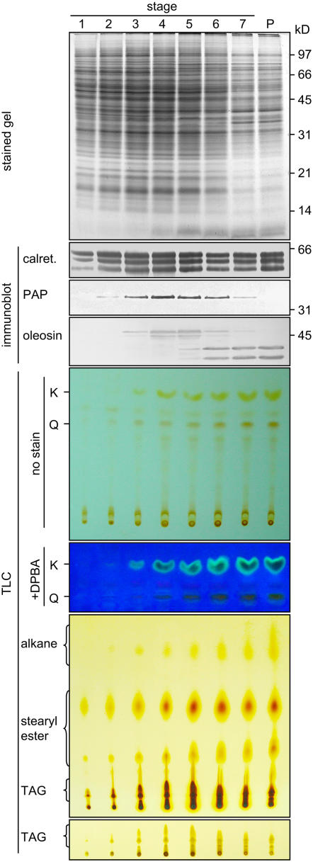
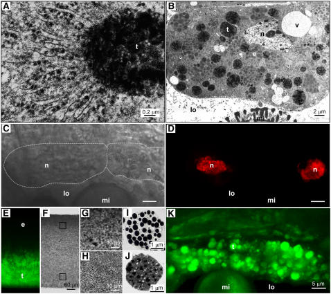
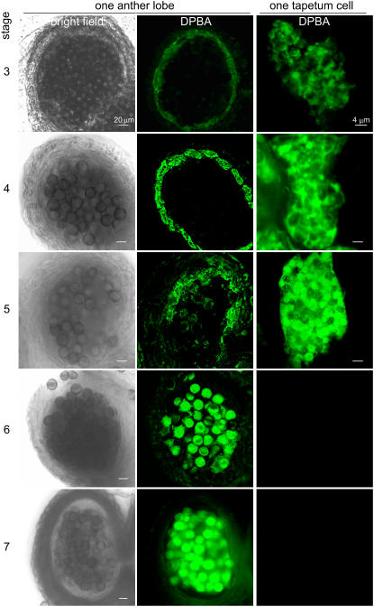
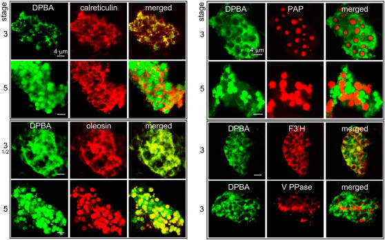
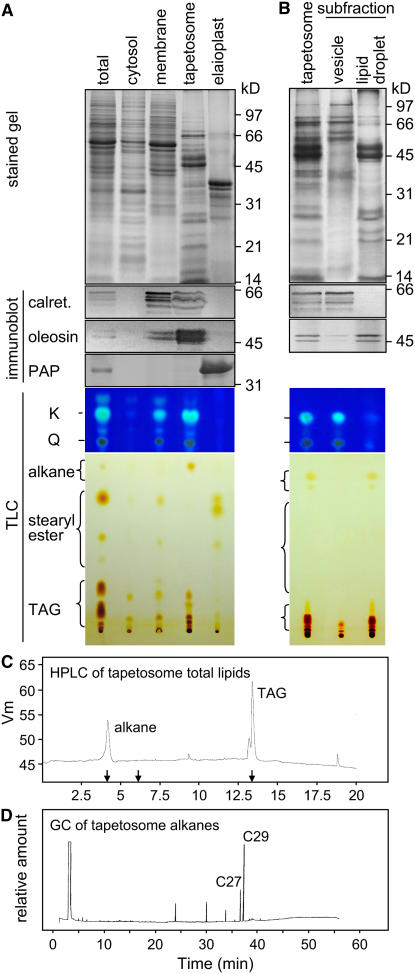
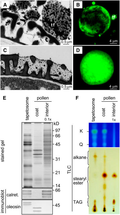
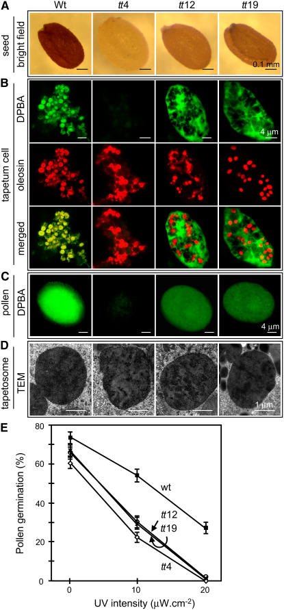
Tapetosomes are abundant organelles in tapetum cells during the active stage of pollen maturation in Brassicaceae species. They possess endoplasmic reticulum (ER)-derived vesicles and oleosin-coated lipid droplets, but their overall composition and function have not been established. In situ localization analyses of developing Brassica napus anthers revealed flavonoids present exclusively in tapetum cells, first in an ER network along with flavonoid-3'-hydroxylase and then in ER-derived tapetosomes. Flavonoids were absent in the cytosol, elaioplasts, vacuoles, and nuclei. Subcellular fractionation of developing anthers localized both flavonoids and alkanes in tapetosomes. Subtapetosome fractionation localized flavonoids in ER-derived vesicles, and alkanes and oleosins in lipid droplets. After tapetum cell death, flavonoids, alkanes, and oleosins were located on mature pollen. In the Arabidopsis thaliana mutants tt12 and tt19 devoid of a flavonoid transporter, flavonoids were present in the cytosol in reduced amounts but absent in tapetosomes and were subsequently located on mature pollen. tt4, tt12, and tt19 pollen was more susceptible than wild-type pollen to UV-B irradiation on subsequent germination. Thus, tapetosomes accumulate ER-derived flavonoids, alkanes, and oleosins for discharge to the pollen surface upon cell death. This tapetosome-originated pollen coat protects the haploidic pollen from UV light damage and water loss and aids water uptake.


Figures

Figure 1.
Figure 1.
SDS-PAGE, Immunoblot, and TLC Analyses of Extracts of Brassica Anthers of Seven Developmental Stages and Mature Pollen. The various extracts applied to the gels or TLC plates represented those from an equal number of anthers. Gels were stained for proteins or subjected to immunoblot analyses with antibodies against calreticulin, elaioplast PAP, or tapetosome oleosin. The two major oleosins of 48 and 45 kD were converted to fragments of 37 and 35 kD, respectively, during anther development; the fragments were more immunoreactive than the originals (Ting et al., 1998). Positions of markers for polypeptide molecular masses are shown at right. The TLC plate after separation of deglycosylated flavonoids was photographed directly (full plate shown) or after being treated with DPBA and placed on top of a UV irradiation source (portion of the plate shown). K and Q denote kaempferol and quercetin, respectively. The TLC plate for lipid analyses was stained with iodine for 24 h to reveal all lipids, including the less reactive alkanes (full plate shown), or for 0.5 h to reveal TAGs more clearly (portion of the plate shown).
Figure 2.
Figure 2.
Microscopy of Brassica Tapetum Cells and Isolated Tapetosomes and Elaioplasts. (A) Enlarged electron microscopy image of a maturing tapetosome (t) connected to numerous rough ER cisternae in a tapetum cell (stage 3). (B) Electron microscopy image of a stage 5 tapetum cell, in which most tapetosomes had become solitary organelles. Nucleus (n), vacuoles (v), tapetosomes (t), and elaioplasts (e) in the tapetum cells, as well as portion of a microspore (mi) in the locule (lo), were present. (C) and (D) Two tapetum cells stained for nuclei with ethidium bromide and viewed by CLSM with a bright-field (the cell circumference was marked with white dotted lines [C]) or a fluorescence (nuclei in red [D]) setting. (E) to (H) CLSM images of a lipid pad obtained from a stage 5 anther extract by centrifugation. The lipid pad contained elaioplasts (lighter density) at the top half and tapetosomes at the bottom half. It was treated with DPBA for flavonoid staining, cut perpendicularly along the direction of the original centrifugal force, and viewed by CLSM with a fluorescence (E) or a bright-field ([F] to [H]) setting. Tapetosomes at the bottom half but not elaioplasts at the top half of the lipid pad showed DPBA fluorescence (shown in green). Portions (squares in [F]) of the lipid pad were enlarged to reveal packed particles ([G] and [H] for the top and bottom portions, respectively). (I) and (J) Electron microscopy images of an isolated elaioplast (I) and tapetosome (J). (K) Two tapetum cells adjacent to the anther locule (lo) in a stage 5 anther observed by CLSM after DPBA staining. Numerous spherical particles (to be shown as tapetosomes) exhibited DPBA fluorescence (in green).
Figure 3.
Figure 3.
CLSM of Brassica Anthers and Tapetum Cells of Different Developmental Stages after DPBA Staining for Flavonoids. The left and middle columns show CLSM images of one anther lobe of developmental stages 3 to 7 taken with a bright-field or fluorescence setting. Each lobe contained microspores in the locule enclosed by the tapetum and several outer wall layers. At stages 3 to 4, the tapetum cells were intact and possessed most of the DPBA fluorescence (shown in green) in the lobe. At stage 5, the tapetum cells were fully mature, with a few already broken; the microspores began to show DPBA fluorescence. At stages 6 to 7, the tapetum had broken, and fluorescence was present on the microspores. The right column shows CLSM images of one tapetum cell of each developmental stage taken with a fluorescence setting. Fluorescence was present first in a subcellular network and then in spherical particles.
Figure 4.
Figure 4.
CLSM of Brassica Tapetum Cells of Developmental Stages 3 or 5 to Test for Colocalization of Flavonoids and Organelle-Specific Proteins. Anthers were treated with DPBA for flavonoid fluorescence (shown in green) and then antibodies against calreticulin (markers of ER), oleosin (tapetosomes), F3′H (a flavonoid-synthesizing enzyme presumed to be located on the ER), V-PPase (vacuoles), or PAP (elaioplasts) for immunodetection (shown in red). Yellow/orange color in the merged images indicates colocalization.
Figure 5.
Figure 5.
SDS-PAGE, Immunoblot, and Chromatography Analyses of Subcellular Fractions of Stage 5 Brassica Anthers and Subfractions of Isolated Tapetosomes. (A) An anther extract (labeled as total), representing materials mostly from tapetum cells (see Methods), was subfractionated by centrifugation into fractions of cytosol, membranes (densities of 1.10 to 1.12 g/cm), tapetosomes (∼1.04 g/cm), and elaioplasts (∼1.02 g/cm). For clarity in analyses, samples containing approximately equal amounts of proteins were applied to the gel or TLC lanes. (B) Isolated tapetosomes were treated with 0.1 M Na2CO3 and then subfractionated into vesicle and lipid droplet subfractions. Samples of the two subfractions applied to the gel or TLC lanes represented those from an equal amount of the tapetosome fraction. For (A) and (B), the analysis methodology and labels are as described for Figure 1. (C) Lipids in the total tapetosome fraction were separated by HPLC. Arrows on the x axis indicate the elution times of the lipid standards C-29 alkane, cholesteryl palmitate, and triolein (from left to right). (D) Tapetosome alkanes obtained by TLC were separated by gas chromatography. Alkanes of C-29 and C-27, identified by MS or comigration with C-29 alkane standard, and three unknown alkanes were resolved.
Figure 6.
Figure 6.
Microscopy of Brassica Microspores in Anther Locule after Tapetum Lysis, and SDS-PAGE, Immunoblot, and TLC Analyses of Isolated Tapetosomes, Pollen Coat, and Pollen Interior. (A) Electron microscopy of a stage 6 anther shows tapetosomes or their fragments (tf) adjacent to the surface of a microspore, whose exine cavities had not acquired coat materials. (C) Electron microscopy of a stage 7 anther shows the exine cavities of a microspore after acquisition of coat materials, which had a semitranslucent matrix embedded with electron-dense droplets. (B) and (D) Microspores in stage 5/6 (B) and 7 (D) anthers, which had been stained with DPBA for flavonoid fluorescence (shown in green) and analyzed by CLSM with the same CLSM settings (laser power and detection gain). (E) and (F) SDS-PAGE, immunoblot, and TLC analyses of isolated tapetosomes, pollen coat, and pollen interior. Analysis methodology and labels are as described for Figure 1. The interior fraction applied to the gels for protein analyses and to TLC plates for flavonoid and lipid analyses represented 0.1× and 1×, respectively, the coat fraction from the same amount of mature pollen.
Figure 7.
Figure 7.
Microscopy of Seeds, Tapetum Cells, and Pollen, and Germination of Pollen in the Absence and Presence of UV-B Irradiation of Arabidopsis Wild Type and Homozygous Mutants. Mutant tt4 has no chalcone synthase, and mutants tt12 and tt19 are deficient in a flavonoid membrane transporter. (A) Seeds were photographed with a light microscope under an identical bright-field setting. (B) Tapetum cells in anthers of a late developmental stage, when most of the tapetosomes had become solitary organelles, were stained with DPBA for flavonoids (shown in green) and then treated with antibodies against oleosin for immunodetection (in red) and analyzed by CLSM. Yellow color in the merged images indicates colocalization. Fluorescence imaging of the mutant and wild-type samples for immunodetection of oleosins used the same CLSM settings (laser power and detection gain). Fluorescence imaging of the mutant samples (which had no or reduced flavonoids) for DPBA stain used higher CLSM settings than those of the wild-type samples. (C) Mature pollen was stained with DPBA (shown in green) and analyzed by CLSM. Fluorescence imaging of the mutant pollen (which had no or reduced flavonoids) for DPBA stain used higher CLSM settings than those of the wild-type pollen. (D) Stage 5 tapetum cells were observed by transmission electron microscopy (TEM). Each image shows a tapetosome with patches of electron-dense materials. (E) Pollen was allowed to germinate on agar plates in dim visible light (0.43 μW/cm) without or with supplemented UV-B irradiation of 10 or 20 μW/cm. The Petri dishes were covered with a 0.13-mm-thick cellulose diacetate film. After 2 h of exposure, the agar plates were placed in the dark for 5 h, and germination was observed by light microscopy. The se of each data point (n ≥ 500) is shown.

Similar articles

Cited by

References

    1. Bewley, J.D., Hempel, F.D., McCormick, S., and Zambryski, P. (2000). Reproductive development. In Biochemistry and Molecular Biology of Plants, B.B. Buchanan, W. Gruissem, and R.L. Jones, eds (Rockville, MD: American Society of Plant Physiologists), pp. 988–1043.
    1. Bieza, K., and Lois, R. (2001). An Arabidopsis mutant tolerant to lethal UV-B levels shows constitutively elevated accumulation of flavonoids and other phenolics. Plant Physiol. 126 1105–1115. - PMC - PubMed
    1. Bih, F.Y., Wu, S.S.H., Ratnayake, C., Walling, L.L., Nothnagel, E.A., and Huang, A.H.C. (1999). The predominant protein on the surface of maize pollen is an endoxylanase synthesized by a tapetum mRNA with a long 5′ leader. J. Biol. Chem. 32 22884–22894. - PubMed
    1. Buer, C.S., and Muday, G.K. (2004). The transparent testa4 mutation prevents flavonoid synthesis and alters auxin transport and the response of Arabidopsis root to gravity and light. Plant Cell 16 1191–1205. - PMC - PubMed
    1. Burbulis, I.E., Iacobucci, M., and Shirley, B.W. (1996). A null mutation in the first enzyme of flavonoid biosynthesis does not affect male fertility in Arabidopsis. Plant Cell 8 1013–1025. - PMC - PubMed

MeSH terms

Associated data