. 2007 May;75(5):2374-80.
doi: 10.1128/IAI.01465-06. Epub 2007 Feb 16.

Chlamydial Hsp60-2 is iron responsive in Chlamydia trachomatis serovar E-infected human endometrial epithelial cells in vitro

Affiliations

Chlamydial Hsp60-2 is iron responsive in Chlamydia trachomatis serovar E-infected human endometrial epithelial cells in vitro

Richard W LaRue et al. Infect Immun. 2007 May.

Abstract

Chlamydial 60-kDa heat shock proteins (cHsp60s) are known to play a prominent role in the immunopathogenesis of disease. It is also known that several stress-inducing growth conditions, such as heat, iron deprivation, or exposure to gamma interferon, result in the development of persistent chlamydial forms that often exhibit enhanced expression of cHsp60. We have shown previously that the expression of cHsp60 is greatly enhanced in Chlamydia trachomatis serovar E propagated in an iron-deficient medium. The objective of this work was to determine which single cHsp60 or combination of the three cHsp60 homologs encoded by this organism responds to iron limitation. Using monospecific polyclonal peptide antisera that recognize only cHsp60-1, cHsp60-2, or cHsp60-3, we found that expression of cHsp60-2 is responsive to iron deprivation. Overall, our studies suggest that the expression of cHsp60 homologs differs among the mechanisms currently known to induce persistence.


Figures

FIG. 1.
FIG. 1.
PCR amplification of C. trachomatis serovar E groEL. (A) Initial attempt to amplify groEL-1, groEL-2, and groEL-3 (lanes 1, lanes 2, and lanes 3, respectively) using a C. trachomatis serovar E DNA template and primers based on the sequence of C. trachomatis serovar D. (B) Strategy used to amplify groEL-3 and flanking sequences. (C) Result of amplification of groEL-3 and flanking sequences. Lanes 1 through 5 contained areas indicated in panel B, and amplification of groEL-1 was used as a control. (D) Difference in the starting sequences of C. trachomatis serovars D and E. (E) Amplification of C. trachomatis serovar E groEL-1, groEL-2, and groEL-3 with redesigned primers for groEL-3.
FIG. 2.
FIG. 2.
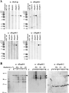
Specificity of peptide antisera and response of cHsp60-2 to iron deprivation. (A) Samples used for Western blotting included uninfected HEC-1B cells, E. coli LMG194 alone, and arabinose-induced recombinants E. coli LMG194 (pJER516), LMG194 (pJER517), and LMG194 (pJER518), representing cHsp60-1, cHsp60-2 and cHsp60-3, respectively. An anti-His tag monoclonal antibody was used as a control (upper left panel). (B) Samples included uninfected HEC-1B cells (control) (lanes C), cells mock exposed for 30 min and 1 and 2 h, and cells exposed to Desferal for 30 min and 1 and 2 h. One milligram of protein was loaded onto preparative gels (A), whereas 15 μg was loaded into each lane in panel B. Arrowheads indicate the position of cHsp60. The asterisk indicates the position of a major proteolytic product of cHsp60-2, and the circle indicates the position of a cross-reactive protein in HEC-1B cells.
FIG. 3.
FIG. 3.
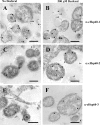
Immunolabeling transmission electron microscopy showing the response of cHsp60-2 to iron limitation. Chlamydia-infected HEC-1B cells at 36 hpi were either not exposed to Desferal (A, C, and E) or exposed to 500 μM Desferal (B, D, and F) for 1 h and labeled using a 1:100 (vol/vol) dilution of anti-cHsp60-1 (A and B), a 1:20 (vol/vol) dilution of anti-cHsp60-2 (C and D), or a 1:40 (vol/vol) dilution of anti-cHsp60-3 (E and F). A 15-nm gold-conjugated anti-rabbit serum (Amersham Biosciences) was used at a 1:200 (vol/vol) dilution for visualization. Bars = 0.5 μm. The arrowheads indicate gold particles.

Similar articles

Cited by

References

    1. Andrews, N. C. 2000. Iron homeostasis: insights from genetics and animal models. Nat. Rev. 1:208-216. - PubMed
    1. Beatty, W. L., G. I. Byrne, and R. P. Morrison. 1993. Morphologic and antigenic characterization of interferon gamma-mediated persistent Chlamydia trachomatis infection in vitro. Proc. Natl. Acad. Sci. USA 90:3998-4002. - PMC - PubMed
    1. Beatty, W. L., G. I. Byrne, and R. P. Morrison. 1994. Repeated and persistent infection with Chlamydia and the development of chronic inflammation and disease. Trends Microbiol. 2:94-98. - PubMed
    1. Belland, R. J., D. E. Nelson, D. Virok, D. D. Crane, D. Hogan, D. Sturdevant, W. L. Beatty, and H. D. Caldwell. 2003. Transcriptome analysis of chlamydial growth during IFN-γ-mediated persistence and reactivation. Proc. Natl. Acad. Sci. USA 100:15971-15976. - PMC - PubMed
    1. Belland, R. J., L. Rose, and Y. AbdelRahman. 2006. Promoter mapping of coding and non-coding RNAs using an intergenic microarray for Chlamydia trachomatis, p. 17-20. In M. Chernesky, H. Caldwell, G. Christiansen, I. N. Clarke, B. Kaltenboek, C. Knirsch, C.-C. Kuo, J. Mahony, R. G. Rank, P. Saikku, J. Schachter, W. E. Stamm, R. S. Stephens, J. T. Summersgill, P. Timms, and P. B. Wyrick (ed.), Proceedings of the 11th International Symposium on Human Chlamydial Infections, Niagara-on-the-Lake, Ontario, Canada. International Chlamydia Symposium, San Francisco, CA.

Publication types

Associated data