. 2007;35(5):1660-72.
doi: 10.1093/nar/gkm065. Epub 2007 Feb 18.

Interaction between trichosanthin, a ribosome-inactivating protein, and the ribosomal stalk protein P2 by chemical shift perturbation and mutagenesis analyses

Affiliations

Interaction between trichosanthin, a ribosome-inactivating protein, and the ribosomal stalk protein P2 by chemical shift perturbation and mutagenesis analyses

Denise S B Chan et al. Nucleic Acids Res. 2007.

Abstract

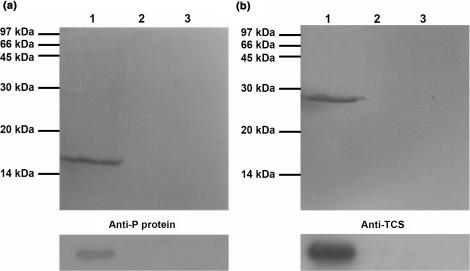
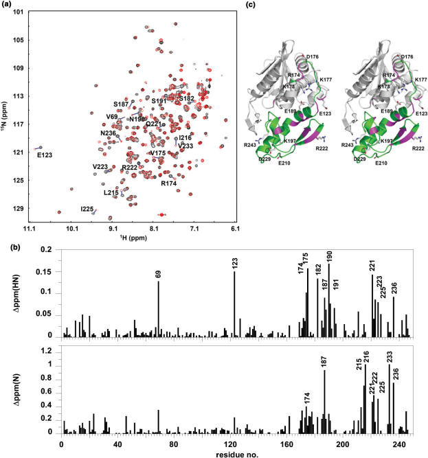
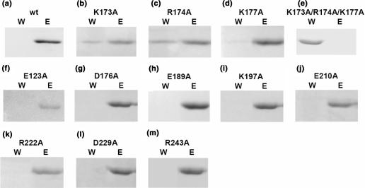
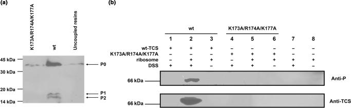
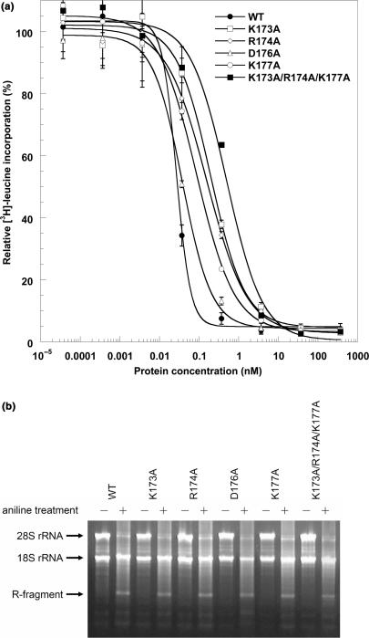
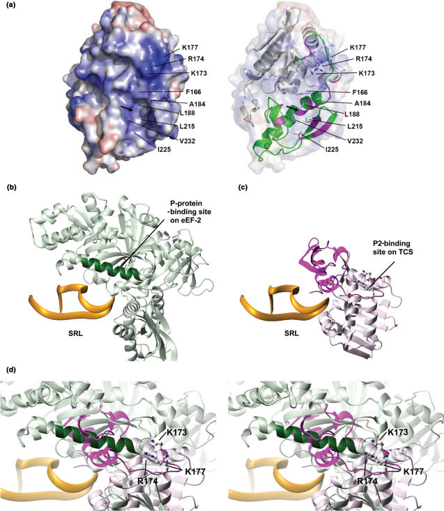
Trichosanthin (TCS) is a type I ribosome-inactivating protein that inactivates ribosome by enzymatically depurinating the A(4324) at the alpha-sarcin/ricin loop of 28S rRNA. We have shown in this and previous studies that TCS interacts with human acidic ribosomal proteins P0, P1 and P2, which constitute the lateral stalk of eukaryotic ribosome. Deletion mutagenesis showed that TCS interacts with the C-terminal tail of P2, the sequences of which are conserved in P0, P1 and P2. The P2-binding site on TCS was mapped to the C-terminal domain by chemical shift perturbation experiments. Scanning charge-to-alanine mutagenesis has shown that K173, R174 and K177 in the C-terminal domain of TCS are involved in interacting with the P2, presumably through forming charge-charge interactions to the conserved DDD motif at the C-terminal tail of P2. A triple-alanine variant K173A/R174A/K177A of TCS, which fails to bind P2 and ribosomal stalk in vitro, was found to be 18-fold less active in inhibiting translation in rabbit reticulocyte lysate, suggesting that interaction with P-proteins is required for full activity of TCS. In an analogy to the role of stalk proteins in binding elongation factors, we propose that interaction with acidic ribosomal stalk proteins help TCS to locate its RNA substrate.


Figures

Figure 1.
Figure 1.
TCS interacts with P2 in vitro. (a) P2 was loaded to TCS-coupled NHS-activated column pre-equilibrated with binding buffer. After extensive washing, P2 was eluted with 1 M NaCl (lane 1). (b) TCS was loaded to P2-coupled NHS-activated column pre-equilibrated with binding buffer. After extensive washing, TCS was eluted with 1 M NaCl (lane 1). The eluted proteins were analysed by SDS-PAGE with Coomassie blue staining (upper panel), and the identity of P2 and TCS was confirmed by western blot (lower panel) using anti-P and anti-TCS antibody, respectively. In both cases, two negative controls were carried out. First, BSA was loaded to the TCS- or P2- coupled column and no BSA was found in the elution (lane 2). Second, P2 or TCS were loaded to uncoupled NHS-Sepharose, and no P2 or TCS were found in the elution (lane 3).
Figure 2.
Figure 2.
TCS interacts with the conserved C-terminal region of P2. (a) The primary sequence of human P2 is aligned with the C-terminal residues of other acidic ribosomal stalk proteins P0 and P1. The last 17 residues of P0, P1 and P2 are highly conserved and are highlighted in the figure. (b) The last 11 residues of P-proteins are highly conserved. The sequences of the C-terminal residues of P-proteins from the SWISS-PROT database were aligned by the program CLUSTAL W (41), and are shown in a sequence logo representation (42) generated by a web-based program WebLogo (43). Sequences are numbered according to the human P2 sequence. (c and d) The interaction between C-terminal deletion mutants of P2 and TCS was checked by in vitro pull-down assay. In (c), deletion mutants of P2 were loaded to TCS-coupled NHS-Sepharose; while in (d), TCS was loaded to NHS-Sepharose coupled with P2 deletion mutants. The elution fractions from the pull-down assay were analysed by 15% SDS-PAGE stained with Coomassie blue. The presence of P2 mutants in (c) or TCS in (d) in the elution fractions indicates positive interaction between TCS and the P2 deletion mutants. In both (c and d), wild-type P2 was included as a positive control. (e) In vitro pull-down assay on the interaction between TCS and C-terminal tail of P2. The last 7, 11, 14, 17, 29, 36 residues of P2 were fused to MBP to create MBP-C7, MBP-C11, MBP-C14, MBP-C17, MBP-C29 and MBP-C36 fusion proteins. Bacterial lysates containing these fusion proteins were loaded to a TCS-coupled NHS-Sepharose column. The elution fractions from the pull-down assay were analysed by 15% SDS-PAGE stained with Coomassie blue. Our data indicate that MBP-C11, MBP-C14, MBP-C17, MBP-C29, MBP-C36 fusion proteins, but not the MBP control, MBP-C7 and MBP-C11 DDD(106–108)AAA, were retained by the TCS-coupled column. The results of the in vitro pull-down assay are summarized in (f).
Figure 3.
Figure 3.
P2-binding site on TCS was mapped to the C-terminal domain by chemical shift perturbation. (a) 1H–15N correlation spectra of TCS in the absence (black contours) and in the presence (red contours) of equal molar ratio of P2 were compared, and (b) changes in chemical shifts, ▵ppm(HN) and ▵ppm(N), of amide resonances of TCS were measured. Residues with ▵ppm(HN) >0.075 ppm or ▵ppm(N) >0.5 ppm are indicated in (a) and (b), and colour-coded magenta in the stereo diagram of TCS in (c). These residues are localized in or near the C-terminal domain (173–247, colour-coded green) of TCS. Scanning alanine mutagenesis was performed on all charge residues in the C-terminal domain and E123, which are indicated in (c).
Figure 4.
Figure 4.
In vitro pull-down assay on TCS variants suggests that K173, R174 and K177 are involved in binding P2. TCS (a) or its variants (bm) were loaded to a P2-coupled NHS-Sepharose pre-equilibrated with binding buffer. Bound protein was eluted with 1 M NaCl in 20 mM Tris/HCl buffer pH 8.0. Fractions containing unbound protein collected during washing (W) and bound protein collected during elution (E) were analysed in 15% SDS-PAGE stained with Coomassie blue. As indicated by the presence of TCS in the wash fraction, substitution of alanine at K173, R174 and K177 positions decreases the binding of TCS on P2-coupled column (b–d). Triple-alanine substitutions in these residue positions resulted in a TCS variant (K173A/R174A/K177A) that was unable to bind P2 (e).
Figure 5.
Figure 5.
Interaction between TCS and ribosome was compromised by K173A/R174A/K177A triple-alanine substitutions. (a) Pull-down assay. Rat ribosome was loaded to NHS-Sepharose coupled with TCS or its triple-alanine (K173A/R174A/K177A) variants. After extensive washing, the bound proteins were eluted with 1 M NaCl, and detected by western blot using anti-P antibody. Ribosomal proteins P0, P1 and P2 were pull-down by wild-type TCS (lane 2), while the interaction between ribosome and the triple-alanine variants (lane 1) was greatly reduced to that similar to the control (lane 3), in which the faint band of P0 was due to non-specific interactions between ribosome and the uncoupled resins. (b) Cross-linking experiments. After rat ribosome was incubated with TCS or the triple-alanine variants in room temperature for 20 min, DSS was added to induce cross-linking between TCS and ribosomal proteins, and cross-linking product was detected by western blot using anti-P or anti-TCS antibodies. A protein band at ∼66 kDa, corresponding to the size of TCS–P0 complex, was detected by both anti-P and anti-TCS antibodies when ribosome was cross-linked with wild-type TCS (lane 2), but not with the triple-alanine variants (lane 5) and in other negative controls (lanes 1 and 4: without addition of ribosome; lanes 3, 6 and 8: without addition of DSS; lanes 7 and 8: without addition of TCS or its variants).
Figure 6.
Figure 6.
(a) Translation inhibition assay. The potency of TCS variants to inhibit in vitro translation was measured by adding 3.7 pM to 370 nM of protein samples to nuclease-untreated rabbit reticulocyte lysate as described in the Materials and methods section. IC50, the concentration of TCS required to achieve 50% inhibition, was determined by fitting the data to a four-parameter logistic equation. The values of IC50 for WT (filled circle), K173A (open square), R174A (open diamond), D176A (open triangle), K177A (open circle), and the triple-alanine variant (filled square) were 0.027 ± 0.001, 0.20 ± 0.04, 0.16 ± 0.02, 0.038 ± 0.004, 0.09 ± 0.01, 0.5 ± 0.1nM, respectively. (b) Depurination assay of TCS and its variants. After incubation with 10 nM of TCS or its variants, RNA from the rabbit reticulocyte lysate was extracted, treated with aniline and analysed by electrophoresis as described in the Materials and methods section. Depurination of 28S rRNA at A4324 was detected by the ∼450 bp R-fragments (indicated by an arrow). Control lanes, in which RNA samples were not treated with aniline, are labelled with the ‘−’ marks.
Figure 7.
Figure 7.
Comparison of P-protein-binding sites on eEF2 and TCS. (a) Putative P2-binding surface of TCS. The electrostatic calculation was performed using the program APBS (44) and visualized by PyMOL (45), where positive and negative potential surface is colour-coded blue and red, respectively, at ±10 kT. K173, R174 and K177 contribute to a strongly positive-charged surface which may interact with the DDD motif of P2. The arrow indicates the location of the hydrophobic pocket constituted by F166, A184, L188, L215, I225 and V232. A ribbon representation of TCS, with the C-terminal domain colour-coded green, is shown on the right panel. Residues with large changes in amide chemical shifts (as in Figure 3c) are colour-coded magenta. (b) The structure of eEF2–SRL complex was derived from an 11.7-Å cryo-electron microscopy map of the 80S ribosome complexed with eEF2 and sordarin (38) (PDB code 1S1H and 1S1I). Residues Q176-T191 (colour-coded dark green) of eEF2 were found to be in contact with P-proteins in the cryo-electron microscopy map (38). (c) The model of TCS–SRL complex was obtained as described in the Materials and methods section. The C-terminal domain of TCS is colour-coded magenta. (d) The model of TCS–SRL complex is superimposed onto the structure of eEF2–SRL complex. The three basic residues (K173, R174 and K177) that were found to be involved in binding P2 are shown in ball-and-stick representation in (c) and (d). Noteworthy, the P-protein-binding site (dark green) on eEF2 is in close proximity to the P-protein-binding site (magenta) of TCS.

References

    1. Shaw PC, Lee KM, Wong KB. Recent advances in trichosanthin, a ribosome-inactivating protein with multiple pharmacological properties. Toxicon. 2005;45:683–689. - PubMed
    1. Endo Y, Tsurugi K. RNA N-glycosidase activity of ricin A-chain. Mechanism of action of the toxic lectin ricin on eukaryotic ribosomes. J. Biol. Chem. 1987;262:8128–8130. - PubMed
    1. Endo Y, Mitsui K, Motizuki M, Tsurugi K. The mechanism of action of ricin and related toxic lectins on eukaryotic ribosomes. The site and the characteristics of the modification in 28 S ribosomal RNA caused by the toxins. J. Biol. Chem. 1987;262:5908–5912. - PubMed
    1. Stirpe F, Bailey S, Miller SP, Bodley JW. Modification of ribosomal RNA by ribosome-inactivating proteins from plants. Nucleic Acids Res. 1988;16:1349–1357. - PMC - PubMed
    1. Zhang JS, Liu WY. The mechanism of action of trichosanthin on eukaryotic ribosomes – RNA N-glycosidase activity of the cytotoxin. Nucleic Acids Res. 1992;20:1271–1275. - PMC - PubMed

Publication types

MeSH terms