. 2007 Feb 21;2(2):e242.
doi: 10.1371/journal.pone.0000242.

Epigenetic activation of a subset of mRNAs by eIF4E explains its effects on cell proliferation

Affiliations

Epigenetic activation of a subset of mRNAs by eIF4E explains its effects on cell proliferation

Yaël Mamane et al. PLoS One. .

Abstract

Background: Translation deregulation is an important mechanism that causes aberrant cell growth, proliferation and survival. eIF4E, the mRNA 5' cap-binding protein, plays a major role in translational control. To understand how eIF4E affects cell proliferation and survival, we studied mRNA targets that are translationally responsive to eIF4E.

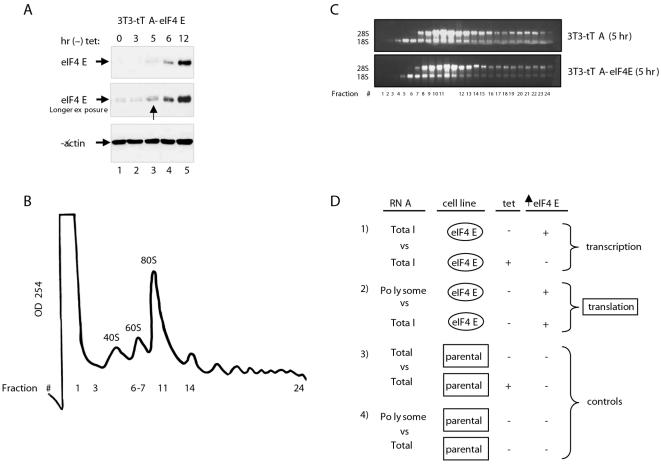
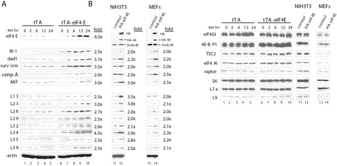
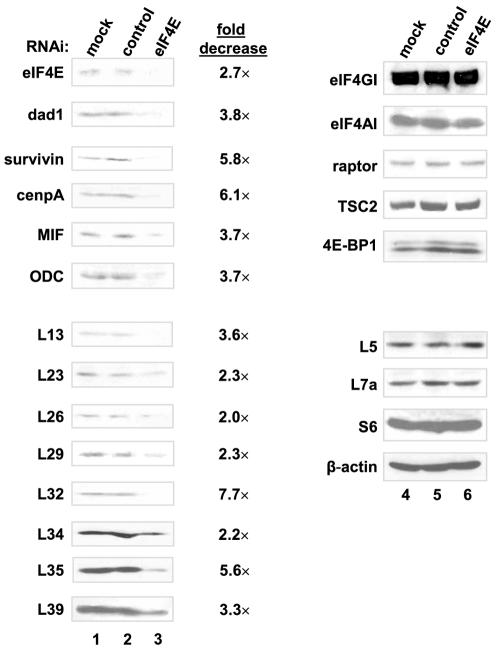
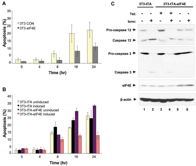
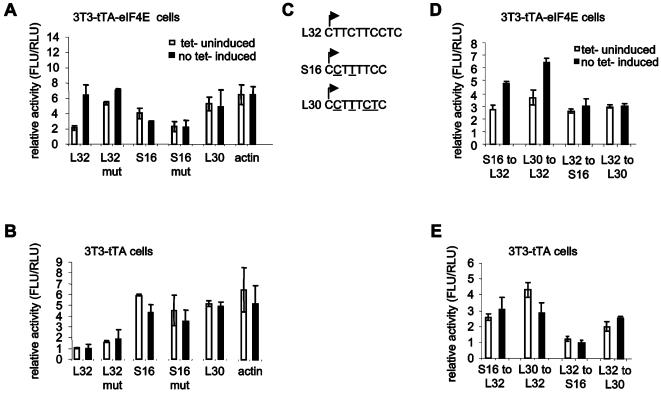
Methodology/principal findings: Microarray analysis of polysomal mRNA from an eIF4E-inducible NIH 3T3 cell line was performed. Inducible expression of eIF4E resulted in increased translation of defined sets of mRNAs. Many of the mRNAs are novel targets, including those that encode large- and small-subunit ribosomal proteins and cell growth-related factors. In addition, there was augmented translation of mRNAs encoding anti-apoptotic proteins, which conferred resistance to endoplasmic reticulum-mediated apoptosis.

Conclusions/significance: Our results shed new light on the mechanisms by which eIF4E prevents apoptosis and transforms cells. Downregulation of eIF4E and its downstream targets is a potential therapeutic option for the development of novel anti-cancer drugs.


Conflict of interest statement

Competing Interests: The authors have declared that no competing interests exist.

Figures

Figure 1
Figure 1. Induction of eIF4E in NIH 3T3 fibroblast cells and microarray analysis.
A) eIF4E overexpression was induced by culturing 3T3-tTA-eIF4E cells in a tetracycline free medium. Immunoblots for eIF4E and β-actin were performed. B) A characteristic fractionation profile of 3T3-tTA and 3T3-tTA-eIF4E cells is depicted. Absorbance at 254 nm was monitored. C) Fractions from 3T3-tTA and 3T3-tTA-eIF4E (induced) cells were analyzed on a denaturing agarose gel to visualize the 18S and 28S rRNAs. D) The experimental design used for microarray analysis is shown.
Figure 2
Figure 2. eIF4E induction stimulates the translation of a subset of mRNAs.
A) Western blotting of extracts from 3T3-tTA versus 3T3-tTA-eIF4E cells after eIF4E induction (0 to 24 hr) and from NIH 3T3 cells and MEFs that constitutively express HA-eIF4E or vector alone. eIF4E induction was determined by using anti-HA and anti-eIF4E antibodies. Fold increase at the 24 hr time point was determined using NIH Image. B) Western blotting experiments were performed as described in (A). These experiments were repeated three times using three different sets of whole-cell extracts.
Figure 3
Figure 3. eIF4E induction causes an increase in the recruitment of a subset of mRNAs to polysomes.
A) Total and polysomal (24 fractions) RNA from induced (−tet for 5 hr) and uninduced (+tet for 5 hr) 3T3-tTA-eIF4E cells was reverse transcribed into cDNA. Primers for BI-1, survivin, MIF, L23, L34, L9, S17 and actin were used to assess mRNA levels. Amplified PCR bands from the polysomal fractions were quantified using NIH Image, and absolute values were plotted. B) The effect of eIF4E induction on L34 mRNA distribution was assessed by northern blotting. Polysomal RNA was isolated from induced (−tet for 5 hr) and uninduced (+tet for 5 hr) 3T3-tTA-eIF4E cells and fractionated into 12 fractions (for purpose of detection). The RNA was loaded on an agarose denaturing gel and transferred to a nitrocellulose membrane. Membranes were probed with radiolabeled murine L34 and actin probes. Bands were quantified using NIH Image, and absolute values were plotted.
Figure 4
Figure 4. siRNA mediated knockdown of eIF4E in NIH 3T3 cells.
NIH 3T3 cells were transiently transfected with an siRNA against murine eIF4E or with a control siRNA, 4E-T-inv (scrambled sequence of human 4E-T), for 48 hr. Cells were lysed, and protein extracts were subjected to SDS-PAGE, followed by western blot analysis. The RNAi-mediated knockdown was repeated three times.
Figure 5
Figure 5. eIF4E induction protects cells against ER-mediated apoptosis.
A) 3T3 cells that stably express HA-eIF4E were treated with 5 µM ionomycin for different periods, fixed and stained with propidium iodide. The percentage of apoptosis was quantified by flow cytometry (triplicates were pooled to generate the s.d.) B) 3T3-tTA and 3T3-tTA-eIF4E cells were seeded at 75% confluency and cultured for 8 hr. Tetracycline containing medium was then replaced by a tetracycline free medium for 16 hr to induce eIF4E expression. Uninduced cells were cultured for the same period without removal of tetracycline. Cells were treated with ionomycin and processed as described in (A). C) 3T3-tTA and 3T3-tTA-eIF4E cells were seeded and cultured with or without tetracycline as described in (B). Cells were then cultured for 24 hr in complete medium±5 µM ionomycin. Protein extracts were resolved by SDS-PAGE and transferred to membranes, which were immunoblotted with anti–caspase 3 and anti–caspase 12. eIF4E expression was also examined by immunoblot; elevated eIF4E levels were only detected in 3T3-tTA-eIF4E induced cells (−tet).
Figure 6
Figure 6. eIF4E and TOP mRNA translation.
A) DNA segments encompassing the promoters and 5′ UTRs of L32, L32 mut (non-TOP), S16, S16 mut (non-TOP), L30 and β-actin were subcloned upstream of the firefly luciferase gene. 3T3-tTA-eIF4E cells were transfected with the various firefly luciferase reporters and a renilla luciferase reporter, which was used for transfection efficiency, and were cultured for 32 hr. Tetracycline containing medium was then replaced by a tetracycline free medium for 16 hr; control (non-induced) cells were cultured in parallel with tetracycline. Firefly luciferase activity (FLU) was measured and normalized against renilla luciferase activity (RLU). B) Luciferase activity of the reporters was measured in the parental cell line 3T3-tTA as described in (A). C) TOP sequences of L32, S16 and L30 are depicted. The arrows indicate the transcriptional start site. The nucleotide changes between L32 and S16 and L32 and L30 are underlined. D) Mutated reporters were generated by exchanging the TOP sequences of L32, S16 and L30. Luciferase assays were performed as described in (A). E) Luciferase activity of the reporters was measured in the parental cell line 3T3-tTA as described in (A). Assays were carried out in triplicate. Luciferase activities represent an average obtained from three independent experiments.

Similar articles

Cited by

References

    1. Thompson B, Wickens M, Kimble J. Translational Control in Development. In: Mathews M, Sonenberg N, Hershey J, editors. Translational Control in Biology and Medicine. Cold Spring Harbor, NY: Cold Spring Harbor Laboratory Press; 2007.
    1. Schneider R, Sonenberg N. Translational Control in Cancer Development and Progression. In: Mathews M, Sonenberg N, Hershey J, editors. Translational Control in Biology and Medicine. Cold Spring Harbor, NY: Cold Spring Harbor Laboratory Press; 2007.
    1. Tee AR, Blenis J. mTOR, translational control and human disease. Semin Cell Dev Biol. 2005;16:29–37. - PubMed
    1. Dua K, Williams TM, Beretta L. Translational control of the proteome: relevance to cancer. Proteomics. 2001;1:1191–1199. - PubMed
    1. Holcik M, Sonenberg N. Translational control in stress and apoptosis. Nat Rev Mol Cell Biol. 2005;6:318–327. - PubMed

Publication types

MeSH terms

Substances