. 2007 Mar;5(3):e59.
doi: 10.1371/journal.pbio.0050059.

Experimental test of connector rotation during DNA packaging into bacteriophage phi29 capsids

Affiliations

Experimental test of connector rotation during DNA packaging into bacteriophage phi29 capsids

Thorsten Hugel et al. PLoS Biol. 2007 Mar.

Abstract

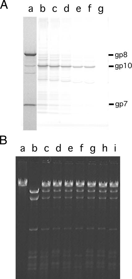
The bacteriophage phi29 generates large forces to compact its double-stranded DNA genome into a protein capsid by means of a portal motor complex. Several mechanical models for the generation of these high forces by the motor complex predict coupling of DNA translocation to rotation of the head-tail connector dodecamer. Putative connector rotation is investigated here by combining the methods of single-molecule force spectroscopy with polarization-sensitive single-molecule fluorescence. In our experiment, we observe motor function in several packaging complexes in parallel using video microscopy of bead position in a magnetic trap. At the same time, we follow the orientation of single fluorophores attached to the portal motor connector. From our data, we can exclude connector rotation with greater than 99% probability and therefore answer a long-standing mechanistic question.


Conflict of interest statement

Competing interests. The authors have declared that no competing interests exist.

Figures

Figure 1
Figure 1. Schematic of the Experimental Geometry (Not to Scale)
Stalled-packaging complexes are attached to the surface of the flow-chamber via biotinylated antibodies to the prohead major capsid protein, gp8. The dye molecule, attached to the connector, is excited via an evanescent wave using total internal reflection. The biotinylated free end of the DNA is attached to streptavidin-coated superparamagnetic beads that are pulled away from the fluorescence excitation by a magnetic field gradient that is created by a pair of magnets next to the objective. The magnetic beads are illuminated for video microscopy using a red LED; both the signals for bright-field and fluorescence images are collected by a high NA microscope objective.
Figure 2
Figure 2. Connector Structure with Position of Dye Labels
A schematic of the structure of the connector, based on the crystal structure by Simpson et al. [5] is shown. The positions of the residues that were mutated to cysteines and investigated with single-molecule fluorescence are indicated.
Figure 3
Figure 3. Dye Labeling and DNA Packaging of ϕ29 Proheads
(A) SDS-PAGE of 170C-connector-mutant proheads. Protein stain of proheads shows the structural components gp8 (capsid), gp8.5 (fiber), gp10 (connector), and gp7 (scaffold) in lane (a). Fluorescence scan of the gel showing labeled proheads with various amounts of dye per gp10 monomer used in labeling reaction: 1 dye per gp10, lane (b); 0.5 dyes per gp10, lane (c); 0.25 dyes per gp10, lane (d); 0.125 dyes per gp10, lane (e); 0.0625 dyes per gp10, lane (f); and no dye, lane (g). The bands in the fluorescence scan with no match in the protein stain originate from highly reactive but quantitatively minor E. coli proteins. (B) DNA packaging tested by nuclease (EcoRI) protection assay using the labeled proheads from (A). Lane (a) shows input DNA-gp3; lane (b) shows a negative (no ATP) control. Packaged DNA is protected from nuclease digestion. Packaging activity is unaffected by dye labeling when compared to a 193C packaging control, lane (c). (B) Shows labeled proheads from (A), ranging from 1 dye per gp10, lane (d); 0.5 dyes per gp10, lane (e); through to no dye, lane (i).
Figure 4
Figure 4. Schematic of the Experimental Setup
Combined SMFP and magnetic tweezers setup. M, mirror; P, pinhole; λ/2, λ/2 plate; PBS, polarizing beam splitter; EOM, electro optical modulator; S, shutter; F, filter; Pol, polarizer; DC, dichroic mirror. Dashed components can be removed and are solely used for alignment purpose.
Figure 5
Figure 5. Fluorescence Polarization Studies of Dye-Labeled, Stalled-Packaging Complexes
Dye-labeled, stalled-packaging complexes were attached to the surface of a flow chamber and excited using the total internal reflection microscope. The excitation polarization was rotated between s- and p-polarization with a frequency of 0.7 Hz. The emitted fluorescence was separated into s- and p-polarization, respectively, and simultaneously detected (black and red). The dye bleached after 22 s. The integration time per data point was 75 ms. a.u., arbitrary units.
Figure 6
Figure 6. Fluorescence Signal from Packaging Complexes
The graphs show the fluorescence intensity of vertical (black) and horizontal (red) polarization detected simultaneously. One example for each of the six investigated mutants is shown. The data were recorded with an integration time of 75 ms and three-points smoothing was applied. The camera background (closed shutter) was subtracted. The signal was normalized using the fluorescence intensity of the bead after packaging was completed. The traces are vertically shifted for clarity. The inset shows a zoom of the data. Here, the unfiltered data is displayed (scatter) together with the three-point sliding average. (A) Fluorescence recorded for a single complex of mutant 170C. The shutter is opened after about half a second. After t = 4 s the first dye bleaches and after 19 s the second. After about t = 100 s the fluorescence signal again starts to increase, since the magnetic bead is pulled into the evanescent field as the prohead reels in the DNA. The signal increases in an exponential fashion as the fluorescent bead samples the intensity profile of the evanescent wave. Similar behavior was observed for (B) 97C; (C) 168C; (D) 189C; (E) 190C; and (F) 260C.

Comment in

References

    1. Grimes S, Jardine PJ, Anderson D. Bacteriophage phi29 DNA packaging. Adv Virus Res. 2002;58:255–294. - PubMed
    1. Smith DE, Tans SJ, Smith SB, Grimes S, Anderson DL, et al. The bacteriophage straight phi29 portal motor can package DNA against a large internal force. Nature. 2001;413:748–752. - PubMed
    1. Iyer LM, Makarova KS, Koonin EV, Aravind L. Comparative genomics of the FtsK-HerA superfamily of pumping ATPases: Implications for the origins of chromosome segregation, cell division, and viral capsid packaging. Nucleic Acids Res. 2004;32:5260–5279. - PMC - PubMed
    1. Kainov DE, Tuma R, Mancini EJ. Hexameric molecular motors: P4 packaging ATPase unravels the mechanism. Cell Mol Life Sci. 2006;63:1095–1105. - PMC - PubMed
    1. Simpson AA, Tao Y, Leiman PG, Badasso MO, He Y, et al. Structure of the bacteriophage phi29 DNA packaging motor. Nature. 2000;408:745–750. - PMC - PubMed

Publication types