. 2007;9(1):R19.
doi: 10.1186/bcr1656.

Induction of epithelial to mesenchymal transition in PMC42-LA human breast carcinoma cells by carcinoma-associated fibroblast secreted factors

Affiliations

Induction of epithelial to mesenchymal transition in PMC42-LA human breast carcinoma cells by carcinoma-associated fibroblast secreted factors

Stephanie C Lebret et al. Breast Cancer Res. 2007.

Abstract

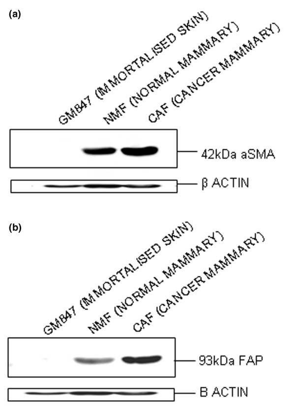
Background: Breast carcinoma is accompanied by changes in the acellular and cellular components of the microenvironment, the latter typified by a switch from fibroblasts to myofibroblasts.

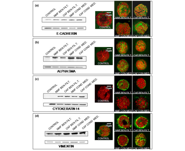
Methods: We utilised conditioned media cultures, Western blot analysis and immunocytochemistry to investigate the differential effects of normal mammary fibroblasts (NMFs) and mammary cancer-associated fibroblasts (CAFs) on the phenotype and behaviour of PMC42-LA breast cancer cells. NMFs were obtained from a mammary gland at reduction mammoplasty, and CAFs from a mammary carcinoma after resection.

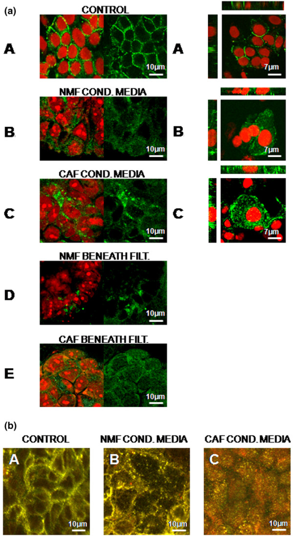
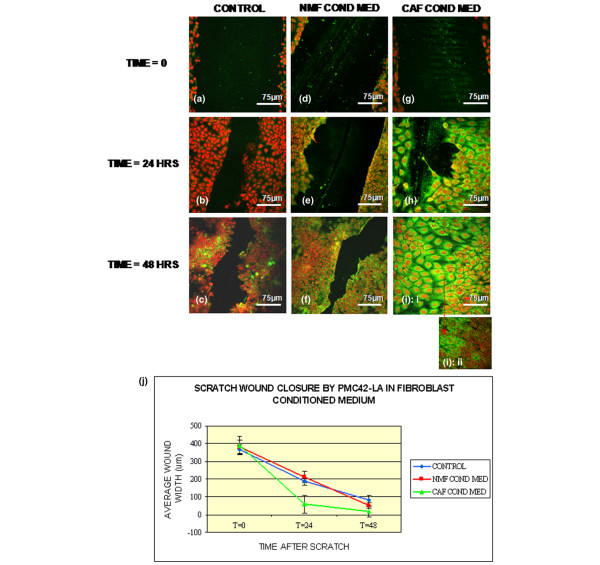
Results: We found greater expression of myofibroblastic markers in CAFs than in NMFs. Medium from both CAFs and NMFs induced novel expression of alpha-smooth muscle actin and cytokeratin-14 in PMC42-LA organoids. However, although conditioned media from NMFs resulted in distribution of vimentin-positive cells to the periphery of PMC42-LA organoids, this was not seen with CAF-conditioned medium. Upregulation of vimentin was accompanied by a mis-localization of E-cadherin, suggesting a loss of adhesive function. This was confirmed by visualizing the change in active beta-catenin, localized to the cell junctions in control cells/cells in NMF-conditioned medium, to inactive beta-catenin, localized to nuclei and cytoplasm in cells in CAF-conditioned medium.

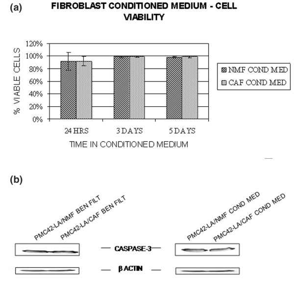
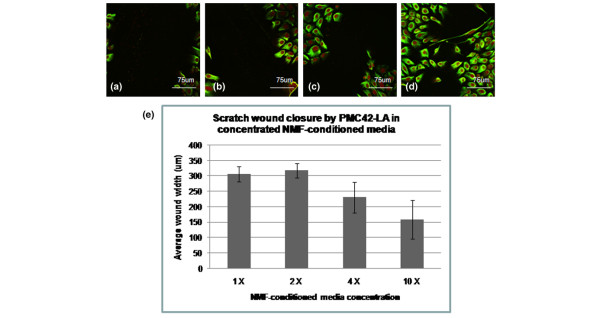
Conclusion: We found no significant difference between the influences of NMFs and CAFs on PMC42-LA cell proliferation, viability, or apoptosis; significantly, we demonstrated a role for CAFs, but not for NMFs, in increasing the migratory ability of PMC42-LA cells. By concentrating NMF-conditioned media, we demonstrated the presence of factor(s) that induce epithelial-mesenchymal transition in NMF-conditioned media that are present at higher levels in CAF-conditioned media. Our in vitro results are consistent with observations in vivo showing that alterations in stroma influence the phenotype and behaviour of surrounding cells and provide evidence for a role for CAFs in stimulating cancer progression via an epithelial-mesenchymal transition. These findings have implications for our understanding of the roles of signalling between epithelial and stromal cells in the development and progression of mammary carcinoma.


Figures

Figure 1
Figure 1
Characterisation of cancer and normal mammary fibroblast lines. (a) No α-SMA detected in control GM847 skin fibroblasts (lane 1), but α-SMA was detected in NMFs (lane 2) and in CAFs (lane 3), with an average 2.7-fold increase in band densities for CAFs compared to NMFs. (b) FAP was expressed by both NMFs and CAFs (lanes 2 and 3, respectively), but it was not detected in control GM847 skin fibroblasts (lane 1). An average 2-fold increase in band density for CAFs, compared with NMFs, was observed. CAF, cancer-associated fibroblast; FAP, fibroblast activation protein; NMF, normal mammary fibroblast; SMA, smooth muscle actin.
Figure 2
Figure 2
Effect of fibroblasts on major protein expression by PMC42-LA. Western blot analysis for a range of markers on control PMC42-LA organoids (control), and organoids cultured with NMFs beneath the filter (lane 1), with CAFs beneath the filter (lane 2), in medium conditioned by NMFs (lane 3), or in medium conditioned by CAFs (lane 4). Controls had no fibroblasts or fibroblast conditioned-medium. Immunocytochemistry was performed to view organization within organoid structures, and ethidium bromide was used to stain nuclei (red). (a) E-cadherin was not significantly upregulated in any of the fibroblast conditions, with no obvious changes in organization. (b) α-SMA expression was found to be significantly upregulated by all fibroblast conditions, with no obvious changes in organization. (c) Cytokeratin 14 expression was found to be significantly induced by all fibroblast conditions, with no obvious changes in organization. (d) Vimentin expression was also found to be significantly upregulated in all fibroblast conditions, with slight changes in organoid organization detected by immunocytochemistry. In control PMC42-LA cells (control lane in panel d), intermediate filament protein vimentin is localized in the cytoplasm of cells throughout organoids. With NMFs beneath the filter (panel d lane 1), vimentin remains localized in the cytoplasm of PMC42-LA cells and vimentin-positive cells are more organized, visible only on outer layer of organoids. With CAFs beneath the filter (panel d lane 2), the same organizational changes are observed. With NMF-conditioned media (panel d lane 3), the same organizational changes are observed. With CAF-conditioned medium (panel d lane 4), no change in organization is observed, with vimentin expression throughout organoids. CAF, cancer-associated fibroblast; NMF, normal mammary fibroblast; SMA, smooth muscle actin.
Figure 3
Figure 3
Effect of fibroblasts and conditioned media on E-cadherin and β-catenin (co)localization in PMC42-LA organoids. (a) Using immunocytochemistry and confocal microscopy, localization of E-cadherin was analyzed. Nuclei were visualized using ethidium bromide (red). (Part A) In control PMC42-LA organoids (no fibroblasts or conditioned media), E-cadherin staining was observed at cell junctions, which was confirmed by confocal microscopy sectioning of PMC42-LA in 2-dimensional culture (right panel). (Part B) In NMF-conditioned medium E-cadherin label was detected as both junctional and cytoplasmic in PMC42-LA organoids, as confirmed by confocal microscopy sectioning of PMC42-LA in 2-dimensional culture containing NMF-conditioned media (right panel). (Part C) In CAF-conditioned media E-cadherin was also detected as junctional and cytoplasmic in PMC42-LA organoids, with more predominant cytoplasmic localization. This was confirmed in 2-dimensional PMC42-LA culture containing CAF-conditioned media by confocal microscopy sectioning (right panel). With (part D) NMFs beneath filter or (part E) CAFs beneath the filter, E-cadherin was again detected at cell junctions and within cytoplasm. (b) E-cadherin and β-catenin are indicated by green and red label, respectively. Areas of colocalization appear yellow. (Part A) In control PMC42-LA cells (no fibroblasts or conditioned media), E-cadherin and β-catenin colocalized at cell junctions with some areas of non-colocalization. (Part B) When in NMF-conditioned medium, colocalization was detected at cell junctions with some independent localization. (Part C) In CAF-conditioned medium, E-cadherin localized to cytoplasm and β-catenin to cytoplasm and nuclei, with some overlap. CAF, cancer-associated fibroblast; NMF, normal mammary fibroblast.
Figure 4
Figure 4
Effect of fibroblast conditioned on apoptosis and cell viability. For PMC42-LA cells cultured in fibroblast-conditioned medium, trypan blue dye exclusion was used to analyze cell viability, and total number of cells were counted and averaged to determine the average percentage of viable cells. (a) There was a slight variation in cell viability at 24 hours (75–100% viable cells [average 91.67 ± 14.43% in NMF-conditioned medium] and 85–100% [91.90 ± 7.33%] in CAF-conditioned medium). Similar cell viability was observed at 3 days (97–100% [99.21 ± 1.37%] in NMF-conditioned medium and 98–100% [average 99.40 ± 1.05%] in CAF-conditioned medium). There was no difference at 5 days (97–98% [98.48 ± 0.69%] in NMF-conditioned medium and 97–100% [98.93 ± 1.13%] in CAF-conditioned medium). (b) There was no significant difference in active caspase-3 expression between cultures. CAF, cancer-associated fibroblast; NMF, normal mammary fibroblast.
Figure 5
Figure 5
Effect of different fibroblast-conditioned media on cell migration in two-dimensional cultures. PMC42-LA cells were cultured on glass in either normal medium (control), NMF-conditioned medium, or CAF-conditioned medium. Using scratch tests, over a period of 48 hours, PMC42-LA cells were analyzed to determine the rate and extent of wound closure, and vimentin expression. Four scratches averaging between 367 μm (minimum) and 392 μm (maximum) in width were made at time 0. Scratches were measured and averaged, and the extent of wound closure calculated and averaged. (a-c) Cells in normal medium (control) exhibited some wound closure at 24 hours (179 ± 21.4 μm), and this had progressed by 48 hours (106 ± 29.0 μm) with small, sparing vimentin expression appearing at 48 hours. (d-f) Cells in NMF-conditioned medium exhibited similar rates of wound closure at 24 hours (171 ± 32.9 μm) and 48 hours (160 ± 15.6 μm), with vimentin expression appearing at wound edges at 24 hours. (g-i) Cells in CAF-conditioned medium exhibited an accelerated wound closure rate, with the width of scratches reduced by 24 hours (333 ± 52.0 μm) and almost full wound closure by 48 hours (panel i part i: 41.63 ± 28.8 μm). Vimentin expression in these cells was abundant around wound area. (Panel i part ii) Closed wounds were identified at 48 hours by visualizing areas of elongated, vimentin-profuse cells in apparently normal areas of lesser vimentin expression. (j) Graphical representation of wound closure by these cultures. CAF, cancer-associated fibroblast; NMF, normal mammary fibroblast.
Figure 6
Figure 6
The effect of concentrated NMF-conditioned media on two-dimensional scratch wound closure. PMC42-LA cells were cultured on glass in 1×, 2×, 4×, or 10× NMF-conditioned medium. Using scratch tests, PMC42-LA cells were analyzed for the extent of wound closure 24 hours after scratch. Scratches were measured and averaged, and the extent of wound closure calculated and averaged. (a) Cells in 1× NMF-conditioned media (control) had average wound sizes of 307 ± 24.3 μm 24 hours post-scratch. (b) Cells in 2× NMF-conditioned medium had average wound sizes of 319 ± 23.2 μm 24 hours post-scratch. (c) Cells in 4× NMF-conditioned medium had average wound widths of 232 ± 49.7 μm 24 hours post-scratch, and (d) cells in 10× NMF-conditioned medium had average wound widths of 160 ± 62.4 μm 24 hours post-scratch. Vimentin localization appeared unchanged in all cultures; some change in cell morphology was noted in 10× NMF-conditioned media cultures. CAF, cancer-associated fibroblast; NMF, normal mammary fibroblast.
Figure 7
Figure 7
The effect of fibroblast-conditioned media on PMC42-LA organoid morphology in three-dimensional cultures. CAF-conditioned medium was added to or below PMC42-LA filter cultures, and the cultures analysed for changes in organoid morphology possibly representative of increased invasiveness. Controls had no fibroblast conditioned-medium. (a,b) In control cultures, organoids appeared spherical with little, if any, single cells present. (c,d) With CAF-conditioned media on the filter/culture, organoids remained predominantly spherical, with some budding edges and the presence of single cells and clusters of single cells. (e,f) With CAF-conditioned medium below the filter/culture, organoids appeared less spherical, with uneven budding edges and many single cells and clusters of single cells. CAF, cancer-associated fibroblast.

Similar articles

Cited by

References

    1. Noel A, Foidart JM. The role of stroma in breast carcinoma growth in vivo. J Mammary Gland Biol Neoplasia. 1998;3:215–225. doi: 10.1023/A:1018703208453. - DOI - PubMed
    1. Dvorak HF. Tumors: wounds that do not heal. Similarities between tumor stroma generation and wound healing. N Engl J Med. 1986;315:1650–1659. - PubMed
    1. Gache C, Berthois Y, Martin PM, Saez S. Positive regulation of normal and tumoral mammary epithelial cell proliferation by fibroblasts in coculture. In Vitro Cell Dev Biol Anim. 1998;34:347–351. - PubMed
    1. Barcellos-Hoff MH, Medina D. New highlights on stroma-epithelial interactions in breast cancer. Breast Cancer Res. 2005;7:33–36. doi: 10.1186/bcr972. - DOI - PMC - PubMed
    1. Farber E. The multistep nature of cancer development. Cancer Res. 1984;44:4217–4223. - PubMed