Review
. 2008 Jan 12;363(1489):23-38.
doi: 10.1098/rstb.2006.2010.

Temporal and epigenetic regulation of neurodevelopmental plasticity

Affiliations
Review

Temporal and epigenetic regulation of neurodevelopmental plasticity

Nicholas D Allen. Philos Trans R Soc Lond B Biol Sci. .

Abstract

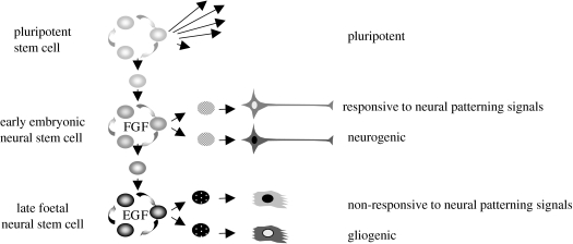
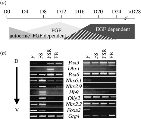
The anticipated therapeutic uses of neural stem cells depend on their ability to retain a certain level of developmental plasticity. In particular, cells must respond to developmental manipulations designed to specify precise neural fates. Studies in vivo and in vitro have shown that the developmental potential of neural progenitor cells changes and becomes progressively restricted with time. For in vitro cultured neural progenitors, it is those derived from embryonic stem cells that exhibit the greatest developmental potential. It is clear that both extrinsic and intrinsic mechanisms determine the developmental potential of neural progenitors and that epigenetic, or chromatin structural, changes regulate and coordinate hierarchical changes in fate-determining gene expression. Here, we review the temporal changes in developmental plasticity of neural progenitor cells and discuss the epigenetic mechanisms that underpin these changes. We propose that understanding the processes of epigenetic programming within the neural lineage is likely to lead to the development of more rationale strategies for cell reprogramming that may be used to expand the developmental potential of otherwise restricted progenitor populations.


Figures

Figure 1
Figure 1
Developmental potential of neural progenitor cells is temporally regulated. Early-stage embryonic and ES cell-derived neural progenitor cells proliferate in response to FGF2. They are neurogenic and responsive to morphogen patterning cues to generate a diversity of neural subtypes. Later-stage neural progenitors that are EGF responsive are non-responsive to patterning cues and undergo a switch from neurogenic to gliogenic differentiation.
Figure 2
Figure 2
Epigenetic modifications regulate the accessibility of genes for transcription by modifying chromatin structure. Expressed genes are present in chromatin with an open configuration and are characterized by histones that are acetylated (Ac) and have activity-related patterns of histone methylation, such as methylated lysine-4 on histone H3 (K4m). Transition to more repressed and silent chromatin states involves dynamic changes in the histone code, including histone deacetylation by HDACs, and changes in the histone methylation status, including demethylation of H3-K4 (e.g. by the demethylase LSD1) and gain in methylation of H3-K9 (K9m) by histone methyltransferases (HMTs). Repression and silencing are also mediated by DNA methylation (m) catalysed by DNA methyltransferases (DNMTS). Modified DNA and histones provide the substrate for the recruitment of further chromatin-modifying proteins (MBDs, HMTs), and non-histone chromatin proteins involved in chromatin compaction and long-term gene repression (e.g. HP1, PcGs, HMGs).
Figure 3
Figure 3
Responsiveness to morphogen and neural patterning involves repressive functions of fate-determining genes. (a) The graded activity of morphogens, such as SHH expressed in the floor plate (FP) of the neural tube, establishes progenitor domains of cells committed to differentiate with restricted neural fates (e.g. motor neurons derive from the pMN domain and domains p0–p3 derive four distinct classes of interneuron). In the ventral spinal cord, progenitor domains are established through the cross-repressive actions of transcription factors (TCFs) that are either activated (class II genes, e.g. Nkx2.2, Nkx6.1) or repressed (class I genes, e.g. Pax6, Dbx1) by SHH. (b) Repressive effects of TCFs possessing eh1 domains are mediated by Groucho family co-repressors (TLE). TLEs recruit repressor complexes, including HDAC activity, and may promote a spread of repressor activity in cis through auto-oligomerization.
Figure 4
Figure 4
Activation of the glial marker gene Gfap is regulated at multiple levels. (a) Gfap expression involves activation of STAT transcription factors and recruitment of SMAD and CBP/p300 coactivator complexes; at early time points, the availability of the SMAD and CBP/p300 coactivators are limiting due to competitive binding by neurogenic transcription factors. (b) The binding of activated STAT and coactivator complexes to the Gfap promoter is regulated by its epigenetic status. In neurogenic cells, the Gfap promoter is repressed by histone H3-lysine-9 methylation and CpG methylation within the STAT-binding site. In later-stage gliogenic cultures, there is an FGF-dependent chromatin remodelling that results in CpG demethylation, histone H3-lysine-9 demethylation and histone K4 methylation.
Figure 5
Figure 5
Temporal development of ES cell-derived neural progenitors in serum-free conditions. (a) Neural differentiation initially occurs under autocrine FGF control. Passage and proliferation of early-born neural progenitors is dependent on exogenous FGF2, while later-stage cultures acquire responsiveness to EGF and eventually become dependent on exogenous EGF for long-term propagation. (b) Only early-stage cultures (D8) are responsive to instructive patterning cues; the figure shows a semi-quantitative RT-PCR analysis of a panel of genes expressed in progressively dorsal to ventral progenitor domains of the neural tube being regulated in response to FGF2 alone (F), FGF2+SHH (FS), FGF2+SHH+retinoic acid (FSR) or FGF2+BMP4 (FB). Later-stage cultures (D20) become non-responsive and instead exhibit constitutive patterns of gene expression.

References

    1. Agarwala S, Sanders T.A, Ragsdale C.W. Sonic hedgehog control of size and shape in midbrain pattern formation. Science. 2001;291:2147–2150. - PubMed
    1. Aiba K, Sharov A.A, Carter M.G, Foroni C, Vescovi A.L, Ko M.S. Defining a developmental path to neural fate by global expression profiling of mouse embryonic stem cells and adult neural stem/progenitor cells. Stem Cells. 2005;4:889–895. - PubMed
    1. Ajamian F, Suuronen T, Salminen A, Reeben M. Upregulation of class II histone deacetylases mRNA during neural differentiation of cultured rat hippocampal progenitor cells. Neurosci. Lett. 2003;346:57–60. - PubMed
    1. Arney K.L, Fisher A.G. Epigenetic aspects of differentiation. J. Cell Sci. 2004;117:4355–4363. - PubMed
    1. Aubert J, et al. Screening for mammalian neural genes via fluorescence-activated cell sorter purification of neural precursors from Sox1-gfp knock-in mice. Proc. Natl Acad. Sci. USA. 2003;100(Suppl. 1):11 836–11 841. - PMC - PubMed

Publication types