. 2007 Apr 13;282(15):11110-21.
doi: 10.1074/jbc.M609040200. Epub 2007 Feb 20.

Basic fibroblast growth factor stimulates matrix metalloproteinase-13 via the molecular cross-talk between the mitogen-activated protein kinases and protein kinase Cdelta pathways in human adult articular chondrocytes

Affiliations

Basic fibroblast growth factor stimulates matrix metalloproteinase-13 via the molecular cross-talk between the mitogen-activated protein kinases and protein kinase Cdelta pathways in human adult articular chondrocytes

Hee-Jeong Im et al. J Biol Chem. .

Abstract

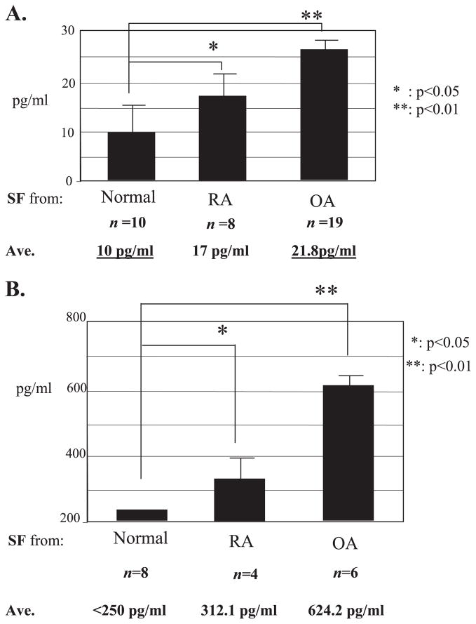
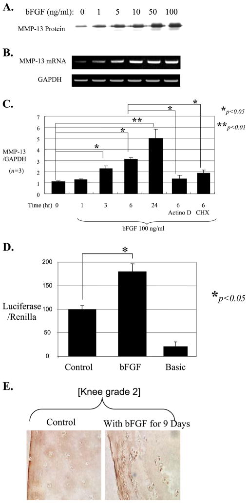
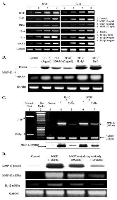
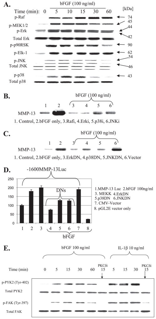
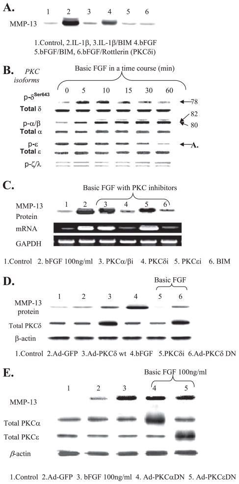
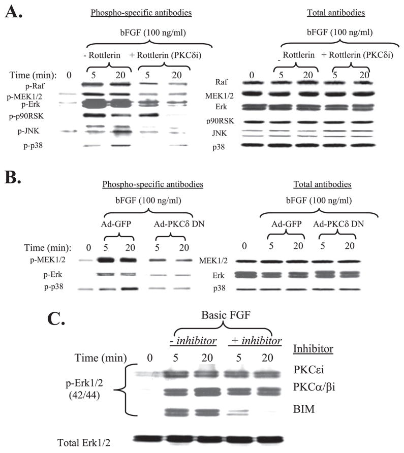
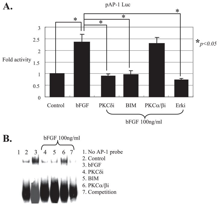
Excessive release of basic fibroblast growth factor (bFGF) during loading and/or injury of the cartilage matrix may contribute to the onset or progression of osteoarthritis. This pathological role may be related to the ability of bFGF to decrease proteoglycan synthesis and to antagonize the activity of anabolic growth factors in cartilage such as insulin-like growth factor-1 and bone morphogenetic protein 7 (BMP7 or OP-1). Matrix metalloproteinase-13 (MMP-13), a catabolic cartilage-degrading enzyme, is dramatically up-regulated by inflammatory cytokines or by fibronectin fragments in articular chondrocytes. In this study, we investigated MMP-13 production by bFGF using human articular chondrocytes. Endogenous concentration of bFGF in synovial fluids collected from arthritis patients and asymptomatic subjects showed a good linear correlation with the endogenous levels of MMP-13. bFGF stimulation of MMP-13 was mediated at the transcriptional level and, at least in part, by stimulation of interleukin-1 production. Also, our findings suggest that bFGF stimulation of MMP-13 required the activation of multiple MAPKs (ERK, p38, and JNK) by bFGF, and more importantly, bFGF activation of protein kinase C (PKC) delta played a key role in the MMP-13 stimulation. Indeed, PKCdelta is the only isoform associated with MMP-13 stimulation among the PKC isoforms tested. PKCdelta controls the bFGF response by regulating multiple MAPK pathways. Our results suggest that PKCdelta activation is a principal rate-limiting event in the bFGF-dependent stimulation of MMP-13 in human adult articular chondrocytes. We propose that deregulation of cross-talk between MAPK and PKCdelta signaling may contribute to the etiology of osteoarthritis in human patients.


Figures

FIGURE 1
FIGURE 1. Endogenous concentrations of bFGF (A) and MMP-13 (B) in human synovial fluids
The concentrations of bFGF and MMP-13 were measured by ELISA in synovial fluid samples collected from asymptomatic donors (Normal), from osteoarthritis (OA), and from rheumatoid arthritis (RA) patients. The data are means ± S.E., and statistical significance was determined by analysis of variance.
FIGURE 2
FIGURE 2. Basic FGF stimulates MMP-13 expression
Human adult articular chondrocytes cultured in monolayer were serum-starved and then treated with bFGF at gradually increasing concentrations (0 –100 ng/ml). The conditioned media were collected after 24 h and used for immunoblotting for MMP-13 protein (A), and the cells were used for total RNA extraction to perform semi-quantitative RT-PCR (B). C, real time RT-PCR was performed using total RNA extracted from cells treated with bFGF (100 ng/ml) in a time course (1–24 h). Cells were also preincubated for 30 min with either the transcriptional inhibitor actinomycin D (Actino D, 5 μM) or the protein synthesis inhibitor cycloheximide (CHX, 10 μM) prior to stimulation with bFGF for 6 h. The mRNA level of GAPDH was evaluated as an internal control for the mRNA level of MMP-13. D, −1600MMP-13 promoter-reporter construct was transiently transfected into human adult articular chondrocytes. The transfected cells were incubated in the presence or absence of bFGF (100 ng/ml) for an additional 24 h, and the luciferase activity representing MMP-13 promoter activity was measured. A Renilla vector was co-transfected as an internal control for normalization. The data represent four different donors measured in triplicate for each experiment. E, human cartilage explants were incubated in mini-ITS in the presence or absence of bFGF (100 ng/ml) for 9 days. Tissue sections were stained with an anti-MMP-13 antibody. The picture is representative of experiments performed with tissue from five different donors.
FIGURE 3
FIGURE 3. IL-1β-dependent stimulation of MMP-13 by bFGF
A, cells in monolayer were serum-starved overnight and treated with varied concentrations of bFGF or IL-1β as indicated for 24 h. The cells were then harvested and subjected to total RNA extraction. Modulation of various pro-inflammatory cytokines, including IL-1β, tumor necrosis factor-α, IL-6, IL-8, and MCP-1, were analyzed by semi-quantitative RT-PCR. B, cells in monolayers are incubated with cytokines, growth factor, and/or Fn-f for 36 h as indicated. The conditioned media were used to analyze MMP-13 protein production, and the cells were harvested and subjected to total RNA extraction for mRNA levels of MMP-13. Lower concentrations of bFGF (2 ng/ml instead of 100 ng/ml), IL-1β (2 ng/ml instead of 5–10 ng/ml), and Fn-f (100 nM instead of 250 nM to 1 μM) were used to observe additive and/or synergistic effects after combination of these factors. C, modulation of MMP-13 levels of mRNA and protein secretion by bFGF or IL-1β in the presence of IL-1ra was analyzed by semi-quantitative RT-PCR and immunoblotting, respectively. Cells stimulated with bFGF (10 ng/ml) or IL-1β (5 ng/ml) were treated with IL-1ra (100 ng/ml) for 36 h. The cells were harvested, and the total RNA was extracted for RT-PCR analysis. Lane1, genomic DNA;lane2, 100-bp DNA size marker; lane 3, control (untreated cells); lane 4, IL-1β treatment; lane 5, IL-1β with IL-1ra; lane 6, bFGF treatment; lane 7, bFGF with IL-1ra. D, cells were preincubated with FGF-neutralizing antibody (10 and 100 μg/ml concentrations) prior to stimulation with bFGF (10 ng/ml) for 36 h. Basic FGF-mediated stimulation of MMP-13 (protein and mRNA levels) and IL-1β was determined by immunoblot and RT-PCR. The picture represents two individual donors with duplicates.
FIGURE 4
FIGURE 4. Basic FGF activates multiple MAPK pathways
A, cells in monolayer were serum-starved overnight and treated with a 100 ng/ml concentration of bFGF in a time course (0 – 60 min). Cell lysates were prepared and analyzed by immunoblotting with phospho-specific antibodies for Raf, MEK1/2, ERK, p90 RSK, Elk-1, JNK, and p38. After stripping of the membranes, immunoreactivity using total antibodies for each molecule was detected for normalization. B, co-incubation of bFGF with the chemical inhibitors of Raf (Rafi, Raf1 kinase peptide inhibitor, 20 μM), ERK activation by MEK (ERKi, PD098059, 20 μM), p38 (p38i, SB203580, 10 μM), and JNK (JNKi, SP600125, 20 μM) for 24 h significantly reduced bFGF-induced MMP-13 production. C, human articular chondrocytes were transfected to overexpress dominant negative forms of MAPK subgroups (ERKDN, p38DN, and JNKDN). After incubation for 48 h, the conditioned media were analyzed for MMP-13 production by immunoblotting. Cytomegalovirus (CMV) vector was overexpressed as a control. D, −1600MMP-13 promoter-reporter construct was transiently co-expressed with MEKK and DN forms of MAPK subgroups (ERKDN, p38DN, and JNKDN). Cytomegalovirus vector (backbone of the DN constructs) and pGL2E vector (MMP-13 construct backbone) were used for controls. E, monolayer cultures of chondrocytes were serum-starved overnight and treated with bFGF (100 ng/ml) or IL-1β (10 ng/ml) in a time course (0 – 60 min) in the presence or absence of selective inhibitor of PKCδ (rottlerin). Cell lysates were then prepared and analyzed by immunoblotting with phospho-specific antibodies for FAK and PYK-2. The membranes were stripped and reprobed with total antibodies for normalization purpose.
FIGURE 5
FIGURE 5. Basic FGF activation of PKCδ is associated with the stimulation of MMP-13 expression
A, cells were serum-starved overnight and treated with bFGF (100 ng/ml) or IL-1β (10 ng/ml) in the presence or absence of a broad spectrum PKC inhibitor (BIM, 10 μM) and PKCδ-specific inhibitor, rottlerin (4 μM). The conditioned media were collected and immunoblotted for the production of MMP-13. B, serum-starved cells were treated with bFGF (100 ng/ml) in a time course. Cell lysates were immunoblotted using phospho-specific anti-PKCδ, -α/β, -ε, and -ξ/λ. The membrane was re-stripped and blotted with anti-total PKC antibodies for each isoform. C, specific chemical inhibitors of PKCα/β (α/βi, GÖ6976, 5 nM), PKCδ (δi, rottlerin, 5 μM), PKCε (εi, 5 μM), and BIM were co-incubated with bFGF (100 ng/ml) for 24 h. Conditioned media and cells were used for immunoblotting and RT-PCR for protein and mRNA levels of MMP-13, respectively. D, cells were infected with adenovirus encoding PKCδ-WT (Ad-PKCδ-WT, lane 3) or Ad-PKCδ DN (lane 6). Infected cells were treated with bFGF (100 ng/ml). Conditioned media were analyzed for MMP-13 protein production, and the cells were extracted and immunoblotted using anti-total PKCδ to validate the overexpression of PKCδ-WT and DN via adenovirus infection. In parallel, the addition of PKCδi (rottlerin, 5 μM, lane 5) and infection with adenovirus encoding GFP (Ad-GFP, lane 2) were included as controls. E, adenoviruses encoding DN forms of PKCα (α DN) and -ε (ε DN) were infected. These infected cells were stimulated with bFGF (100 ng/ml) for 24 h. The conditioned media were analyzed for immunoblotting for MMP-13 production by using an anti-MMP-13 antibody.
FIGURE 6
FIGURE 6. PKCδ activation by bFGF is required for MAPK signaling
A, serum-starved cells were co-incubated with a specific chemical inhibitor of PKCδ for 30 min prior to the stimulation with bFGF (100 ng/ml) in two time intervals (5 and 20 min). The cell extracts were analyzed by immunoblotting for the activation of Raf, MEK1/2, ERK1/2, p90 RSK, JNK, and p38 using phospho-specific antibodies. For loading normalization, corresponding total antibodies were used, and the resulting immunoblots are shown in the right panel. B, cells were infected with adenovirus encoding PKCδ DN in the presence of bFGF (100 ng/ml) at two time intervals (5 and 20 min). Cells were infected with Ad-GFP as control. Cell lysates were used for immunoblotting for the activation of MEK1/2, ERK, and p38. For loading normalization, corresponding total antibodies were used, and the resulting immunoblots are shown in the right panel. C, cells were incubated with selective PKC isoforms of ε (PKCεi), α/β (PKCα/βi), or a broad spectrum PKC inhibitor (BIM) for 30 min prior to the stimulation with bFGF in two time intervals (5 and 20 min). and the cell extracts were immunoblotted with a phospho-specific ERK1/2 antibody. Representative immunoblotting results using anti-total ERK antibody for loading normalization is shown in the lower panel.
FIGURE 7
FIGURE 7. PKCδ activation by bFGF is required for the activation of AP-1, a direct downstream transcriptional regulatory factor of MAPK signaling
A, cis-acting control plasmid, pAP-1 Luc, which contains seven tandem repeats of AP-1 consensus sequences was transiently transfected (2 μg per reaction) into human primary adult articular chondrocytes with and without bFGF (100 ng/ml) and in the presence or absence of a general inhibitor of PKC, BIM, or selective inhibitors of PKCδ or PKCα/β. The luciferase activity was calculated by comparing with the luciferase activity of a control plasmid, pAP-1 Luc (assigned as 1). A Renilla vector (100 ng per reaction) was co-transfected as an internal control for normalization. The data represent the means of three independent experiments in duplicate. B, serum-starved cells (>16 h) in monolayer that were stimulated with and without bFGF (100 ng/ml) for 45 min were subjected to nuclear preparation and incubated with biotin-labeled double-stranded AP-1 consensus sequences. Shifted AP-1 protein-DNA complex band was visualized by chemiluminescence imaging. Unlabeled double-stranded AP-1 oligonucleotides were used for competition assays to determine the binding specificity of the assay.

Similar articles

Cited by

References

    1. Middleton JF, Tyler JA. Ann Rheum Dis. 1992;51:440–447. - PMC - PubMed
    1. Keyszer GM, Heer AH, Kriegsmann J, Geiler T, Keysser C, Gay RE, Gay S. J Rheumatol. 1995;22:275–281. - PubMed
    1. Salminen HJ, Saamanen AM, Vankemmelbeke MN, Auho PK, Perala MP, Vuorio EI. Ann Rheum Dis. 2002;61:591–597. - PMC - PubMed
    1. Smeets TJ, Barg EC, Kraan MC, Smith MD, Breedveld FC, Tak PP. Ann Rheum Dis. 2003;62:635–638. - PMC - PubMed
    1. Pendas AM, Uria JA, Jimenez MG, Balbin M, Freije JP, Lopez-Otin C. Clin Chim Acta. 2000;291:137–155. - PubMed

Publication types

MeSH terms

Substances