. 2007 Feb 26;176(5):719-27.
doi: 10.1083/jcb.200609008. Epub 2007 Feb 20.

Matrix-specific p21-activated kinase activation regulates vascular permeability in atherogenesis

Affiliations

Matrix-specific p21-activated kinase activation regulates vascular permeability in atherogenesis

A Wayne Orr et al. J Cell Biol. .

Abstract

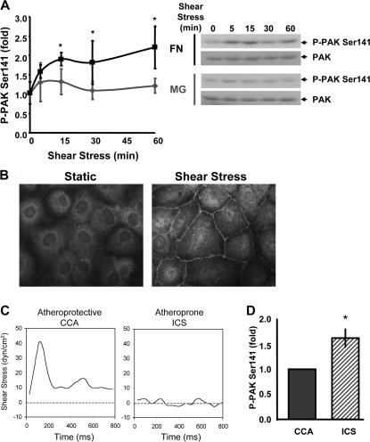
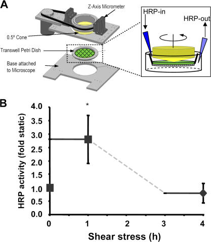
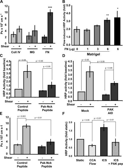
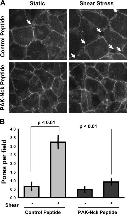
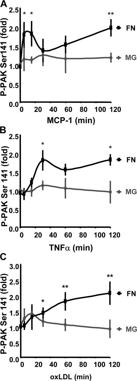
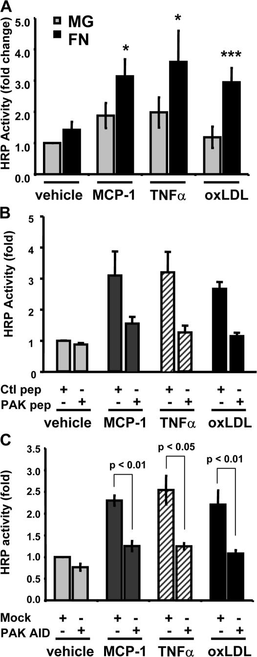
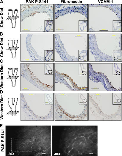
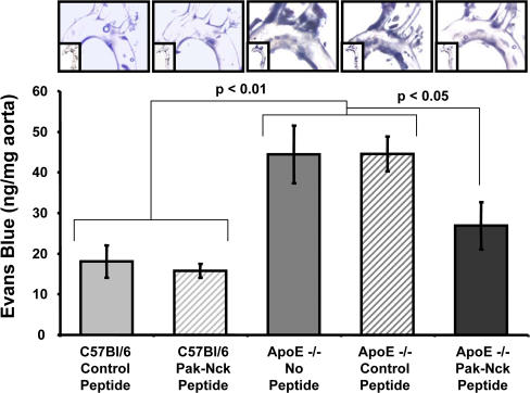
Elevated permeability of the endothelium is thought to be crucial in atherogenesis because it allows circulating lipoproteins to access subendothelial monocytes. Both local hemodynamics and cytokines may govern endothelial permeability in atherosclerotic plaque. We recently found that p21-activated kinase (PAK) regulates endothelial permeability. We now report that onset of fluid flow, atherogenic flow profiles, oxidized LDL, and proatherosclerotic cytokines all stimulate PAK phosphorylation and recruitment to cell-cell junctions. Activation of PAK is higher in cells plated on fibronectin (FN) compared to basement membrane proteins in all cases. In vivo, PAK is activated in atherosclerosis-prone regions of arteries and correlates with FN in the subendothelium. Inhibiting PAK in vivo reduces permeability in atherosclerosis-prone regions. Matrix-specific PAK activation therefore mediates elevated vascular permeability in atherogenesis.


Figures

Figure 1.
Figure 1.
Flow stimulates matrix-specific PAK phosphorylation. (A) BAE cells plated on MG or FN for 4 h were sheared for the indicated times. Phosphorylation of PAK on Ser141 was assessed by immunoblotting total cell lysates with a phosphorylation site–specific antibody. Values are means ± SD normalized for total PAK (n = 3–4). Representative blots are shown. (B) Endothelial cells plated on FN were sheared for 15 min or kept under static conditions, and PAK phospho-Ser141 localization was assessed by immunocytochemistry. Representative images are shown. (C) Shear stress flow profiles for the CCA and the ICS were determined using MRI-generated near-wall velocity gradient profiles of normal carotid arteries. (D) Endothelial cells on FN were stimulated for 24 h with either CCA or ICS flow. Phosphorylation of PAK on Ser141 normalized to total PAK was assessed as described in A. Values are means ± SD after normalization for total protein (n = 3). *, P < 0.05.
Figure 2.
Figure 2.
Onset of flow stimulates endothelial leak. (A) To monitor flow-induced permeability, we used large transwell membranes mounted into a modified cone and plate viscometer and subjected to shear stress. HRP was infused into the top well for 1 h, and permeability was assessed by measuring final HRP activity in the bottom well media. (B) BAE cell monolayers were stimulated with acute onset of flow, and the permeability response was measured as described in A during both the first and fourth hour after application of flow. Values are means ± SD (n = 3). *, P < 0.05.
Figure 3.
Figure 3.
Flow-induced endothelial permeability is matrix and PAK dependent. (A) BAE cells plated on transwell membranes coated with collagen IV, MG, or FN were stimulated with laminar flow at 12 dynes/cm2. HRP leak across the membrane was assessed during the first hour of flow; data are presented as absolute solute permeability as described in Materials and methods. Values are means ± SD (n = 3). (B) Endothelial cells were plated on transwells coated with MG and increasing concentrations of FN, stimulated with 12 dynes/cm2 laminar flow for 1 h, and HRP leak across the membrane was assessed. Values are means ± SD (n = 3). (C) Endothelial cells plated on FN-coated transwell membranes were treated with either control peptide or PAK-Nck inhibitory peptide (20 μg/ml for 1 h). HRP leak across the membrane was assessed during the first hour of steady laminar flow at 12 dynes/cm2. Values are means ± SD (n = 4). (D) Endothelial cells plated on FN-coated transwell membranes were transfected with HA-tagged PAK AID. At 24 h after transfection, monolayers were exposed to flow, and HRP leak across the membrane was assessed during the first hour. Values are means ± SD (n = 3). Transfection efficiency ranged from ∼35 to ∼50% as determined by immunocytochemistry. (E) Endothelial cells plated on FN-coated transwell membranes were treated with either control or PAK-Nck inhibitory peptide (20 μg/ml for 1 h). Alexa 488–conjugated BSA leak across the membrane was assessed during the first hour of steady laminar flow at 12 dynes/cm2. Data (means ± SD; n = 3) are shown as absolute solute permeability. (F) Endothelial cells plated on FN-coated transwell membranes were stimulated for 4 h with CCA or ICS flow. HRP leak across the membrane was assessed during the last 1-h period. Some cells were pretreated with the PAK-Nck inhibitory peptide (20 μg/ml for 1 h) and stimulated with ICS flow in the continued presence of the peptide. Values are means ± SD (n = 3).
Figure 4.
Figure 4.
Flow stimulates PAK-dependent formation of paracellular pores. BAE cells plated on FN were treated with either control or PAK-Nck inhibitory peptide (20 μg/ml for 1 h) and stimulated with flow for 30 min, and cell–cell junctions were visualized by staining for β-catenin. (A) Representative β-catenin stains are shown. Arrows indicate the presence of a paracellular pore. (B) The total number of paracellular pores per high power field was determined. 20 fields were counted per experiment (n = 3).
Figure 5.
Figure 5.
PAK activation by multiple atherogenic stimuli is matrix dependent. BAE cells plated on MG or FN for 4 h were treated with MCP-1 at 50 ng/ml (A), TNFα at 10 ng/ml (B), or oxLDL at 100 μg/ml (C) for the indicated times. Phosphorylation of PAK on Ser141 was assessed by immunoblotting total cell lysates with a phosphorylation site–specific antibody. Values are means ± SD after normalization for total PAK (n = 3–4). *, P < 0.05; **, P < 0.01.
Figure 6.
Figure 6.
Matrix-specific PAK activation stimulates permeability in response to multiple atherogenic stimuli. (A) Endothelial cells plated on transwell membranes coated with either MG or FN were stimulated with 50 ng/ml MCP-1, 10 ng/ml TNFα, or 100 μg/ml oxLDL for 2 h, and the HRP leak across the membrane was assessed. Values are means ± SD (n = 3–4). (B) BAE cells plated on FN-coated transwell membranes were treated with either a control or a PAK-Nck inhibitory peptide (20 μg/ml for 1 h). Monolayers were stimulated with MCP-1, TNFα, or oxLDL for 2 h, and the HRP leak across the membrane was assessed. Values are means ± SD (n = 3). (C) Endothelial cells plated on FN-coated transwell membranes were transfected with HA-tagged PAK AID. After 24 h, monolayers were stimulated with MCP-1, TNFα, or oxLDL for 2 h, and the HRP leak across the membrane was assessed. Values are means ± SD (n = 3). Transfection efficiency ranged from ∼35 to ∼50% as determined by immunocytochemistry.
Figure 7.
Figure 7.
PAK activation and FN deposition in vivo. Male ApoE−/− mice fed a chow diet (A and B) or a Western diet (C and D) for 10 wk were killed, and the carotid arteries were removed and embedded in paraffin. Nearby sections were stained for PAK phospho-Ser141, FN, and VCAM-1, and shown at high magnification (40×) with lower magnification (10×) views of the entire vessels shown as insets. A and B show different areas of the same carotid sinus, and C and D show the carotid sinus and CCA, respectively, from the same animal. A 3D representation of the artery indicates the location of the sections. Bars: 50 μm; (insets) 200 μm. (E) Male ApoE−/− mice fed a chow diet for 10 wk were killed, the aortic arch was excised, and en face staining was performed for PAK phospho-Ser141. Representative images are shown at both 20× and 40× magnifications. Bars: (left) 200 μm; (right) 100 μm.
Figure 8.
Figure 8.
PAK inhibition reduces permeability in vivo. C57Bl/6 and ApoE−/− mice fed a chow diet were treated with PAK-Nck inhibitory or control peptides, and leakage of Evans blue dye into the aortas was assessed as described in Materials and methods. Images were recorded by bright field microscopy. Results were quantified by extracting the dye and measuring absorbance at 620 nm. Values were normalized to the dry weight of the aorta (n = 3). *, P < 0.05. Representative images are shown.

Similar articles

Cited by

References

    1. Alahari, S.K., P.J. Reddig, and R.L. Juliano. 2004. The integrin-binding protein Nischarin regulates cell migration by inhibiting PAK. EMBO J. 23:2777–2788. - PMC - PubMed
    1. Blackman, B.R., G. Garcia-Cardena, and M.A. Gimbrone Jr. 2002. A new in vitro model to evaluate differential responses of endothelial cells to simulated arterial shear stress waveforms. J. Biomech. Eng. 124:397–407. - PubMed
    1. Bokoch, G.M. 2003. Biology of the p21-activated kinases. Annu. Rev. Biochem. 72:743–781. - PubMed
    1. Brett, J., H. Gerlach, P. Nawroth, S. Steinberg, G. Godman, and D. Stern. 1989. Tumor necrosis factor/cachectin increases permeability of endothelial cell monolayers by a mechanism involving regulatory G proteins. J. Exp. Med. 169:1977–1991. - PMC - PubMed
    1. Brooks, A.R., P.I. Lelkes, and G.M. Rubanyi. 2004. Gene expression profiling of vascular endothelial cells exposed to fluid mechanical forces: relevance for focal susceptibility to atherosclerosis. Endothelium. 11:45–57. - PubMed

Publication types