Comparative Study
. 2007 Mar 1;178(5):2667-78.
doi: 10.4049/jimmunol.178.5.2667.

Murine neonatal CD4+ cells are poised for rapid Th2 effector-like function

Affiliations
Comparative Study

Murine neonatal CD4+ cells are poised for rapid Th2 effector-like function

Shawn Rose et al. J Immunol. .

Abstract

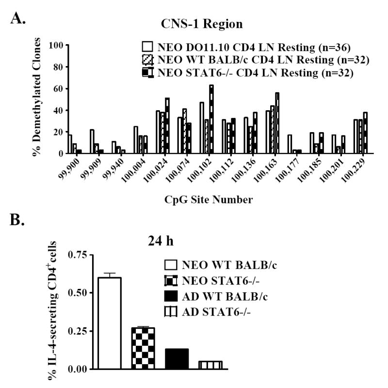
Murine neonates typically mount Th2-biased immune responses. This entails a cell-intrinsic component whose molecular basis is unknown. We found that neonatal CD4(+) T cells are uniquely poised for rapid Th2 function. Within 24 h of activation, neonatal CD4(+) cells made high levels of IL-4 and IL-13 mRNA and protein. The rapid high-level IL-4 production arose from a small subpopulation of cells, did not require cell cycle entry, and was unaffected by pharmacologic DNA demethylation. CpG methylation analyses in resting neonatal cells revealed pre-existing hypomethylation at a key Th2 cytokine regulatory region, termed conserved noncoding sequence 1 (CNS-1). Robust Th2 function and increased CNS-1 demethylation was a stable property that persisted in neonatal Th2 effectors. The transcription factor STAT6 was not required for CNS-1 demethylation and this state was already established in neonatal CD4 single-positive thymocytes. CNS-1 demethylation levels were much greater in IL-4-expressing CD4 single-positive thymocytes compared with unactivated cells. Together, these results indicate that neonatal CD4+ T cells possess distinct qualities that could predispose them toward rapid, effector-like Th2 function.


Figures

Figure 1
Figure 1. Murine neonatal CD4+ cells produce high levels of IL-4 and IL-13 early after activation
CD4+ cells were isolated from day 7 neonatal or adult TCR-transgenic DO11.10 mice. The cells were activated with plate-bound anti-CD3ε plus soluble anti-CD28 mAb (polyclonally). (a) After 24 h, the IL-4 (left graph) or IL-13 (right graph) content in culture supernatants was measured by ELISA. The fold difference in IL-4 or IL-13 secretion between neonatal and adult cells is indicated for each experiment. (b) After 24 h, total RNA was isolated from activated neonatal or adult CD4+ cells. The RNA was reverse transcribed and cDNA products were quantified via real-time PCR using SYBR Green I detection reagents and primers specific for Il4, Il13, or Actb. The levels of Il4 or Il13 mRNA were normalized to those of Actb before making comparisons between samples. The sample with the lowest expression of Il4 (left graph) or Il13 (right graph) was assigned an expression value of 1. The expression values of the other samples are relative to those of the lowest sample. Each reaction was performed in triplicate for both of the depicted experiments. (c) Total RNA was prepared from freshly-isolated neonatal DO11.10 CD4+ cells or from neonatal DO11.10 CD4+ cells that were activated with plate-bound anti-CD3ε plus soluble anti-CD28 mAb for 24 h. Quantitative RT-PCR for Il4 (left graph) and Il13 (right graph) was performed as described above. (d) CD4+ lymph node cells from day 7 neonatal or adult TCR-transgenic DO11.10 mice were activated with cognate ova peptide. After 48 hr, the IL-4 (left graph) or IL-2 (right graph) content in culture supernatants was measured by ELISA. The fold difference in IL-4 or IL-2 secretion between neonatal and adult cells is indicated for each experiment. All results in (a), (b), (c), and (d) are graphed as the mean ± the standard deviation of the triplicate values.
Figure 2
Figure 2. Greater frequencies of neonatal than adult CD4+ cells produce IL-4 early after activation
CD4+ cells were isolated from day 7 neonatal or adult DO11.10 mice. (a) Intracellular staining for IL-4 and IFN-γ was performed on freshly-isolated resting cells (left column) or on cells that were polyclonally activated for 20 h (right column), followed by 4 h of culture with PMA, ionomycin, and monensin. To ensure the specificity of the staining, identically treated cells were stained with control antibodies (smaller inlaid graphs). The depicted experiment is representative of 3 separate experiments. (b) Neonatal or adult CD4+ cells were polyclonally activated for 20 h, washed, and transferred to ELISPOT wells containing PMA and ionomycin for 4 additional h. The frequencies of IL-4-secreting cells were enumerated by ELISPOT. Each sample was plated in quadruplicate and the averages ± the standard deviations are shown. The fold difference in the frequencies of IL-4-producing neonatal and adult CD4+ cells is indicated for each experiment.
Figure 3
Figure 3. Early IL-4 production by neonatal CD4+cells does not require cell cycle entry
CD4+ cells from day 7 neonatal DO11.10 mice were activated polyclonally for 24 h. Some cultures contained the cell cycle inhibitors aphidicolin (G1/S arrest), hydroxyurea (early S arrest), or paclitaxel (G2/M arrest). (a) The IL-4 content in culture supernatants was determined by ELISA. The results are graphed as the mean ± the standard deviation of the triplicate values from a representative experiment. (b) After ethanol fixation, the cells were stained with propidium iodide to measure the percentages of apoptotic cells (left marker) and actively cycling cells (S + G2/M, right marker). (c) Prior to propidium iodide staining, the cells were stained for surface expression of the early activation marker CD69 (solid line). Unactivated cells were stained as controls (dotted line). The experiment in (c) was electronically gated on non-apoptotic cells. All results are representative of at least 3 independent experiments.
Figure 4
Figure 4. Pharmacologic demethylation does not affect IL-4 production by neonatal CD4+ cells
CD4+ cells from day 7 neonatal or adult DO11.10 mice were activated polyclonally in the presence or absence of the demethylating agent 5-aza-deoxycytidine. After 72 h, the (a) IL-4 or (b) IFN-γ content in culture supernatants was determined by ELISA. The results are graphed as the mean ± the standard deviation of the triplicate values from a representative experiment. The fold difference in (a) IL-4 or (b) IFN-γ secretion between treated and untreated neonatal or adult cells is indicated. (c) After 48 h, 5-aza-deoxycytidine (5-aza-dc) treated cells (filled histogram) or untreated cells (open histogram) were stained for surface expression of the activation marker CD25. Unactivated cells were also stained as controls (dotted line). (d) After 48 h, treated and untreated cells were fixed and stained with propidium iodide. The percentages of apoptotic cells (left marker) and actively cycling cells (S + G2/M, right marker) are indicated. The experiment in (c) was electronically gated on non-apoptotic cells. All results are representative of 4 independent experiments.
Figure 5
Figure 5. CNS-1 is highly demethylated in neonatal Th2 effectors
(a) A schematic of the Th2 cytokine genomic locus in adult Th2 effector cells. (b) CD4+ cells were enriched from day 7 neonatal or adult DO11.10 mice. Genomic DNA was isolated from neonatal or adult 5-day Th2 effectors or adult 5-day Th1 effectors. DNA methylation at 14 CpG sites within the CNS-1 region was assessed via bisulfite sequencing. CpG sites are numbered based on sequence AC005742. (c) The percentages of demethylated clones at each CpG site and the numbers of clones sequenced in total (indicated in parentheses) are represented as pooled data from two independent cell preparations. (d) A subset of neonatal and adult effector cells was washed and cultured with PMA, ionomycin, and monensin for 4 h. The cells were stained for intracellular IL-4 and IFN-γ as in Figure 2. The depicted experiment is representative of 5 independent experiments.
Figure 6
Figure 6. CpG demethylation at CNS-1 pre-exists in resting neonatal CD4+cells
(a) As described in Figure 5, CNS-1 CpG methylation levels were assessed in naïve resting adult (sorted CD4+CD44lo) or day 7 neonatal DO11.10 CD4+ cells. (b) The percentages of demethylated clones at each CpG site and the numbers of clones sequenced in total (indicated in parentheses) are represented as pooled data from two independent cell preparations. (c) As described above, CpG methylation at CNS-1 was assessed in sorted (CD44lo) or unsorted resting CD4+ cells from day 7 DO11.10 neonates.
Figure 7
Figure 7. Other regulatory regions of the Th2 cytokine locus are not differentially methylated in neonatal and adult cells
As described in Figure 5, bisulfite methylation analysis was conducted on genomic DNA from (a), (b), (c) naïve resting cells or (a) 5-day Th2 effectors at several CpG sites within the Th2 locus including (a) 6 CpG sites within intron I and exon 2 of the Il4 gene, (b) 9 CpG sites within or adjacent to rad50 RHS7 of the Th2 cytokine LCR, or (c) 13 CpG sites within the CS-1 region of the Il13 promoter. Each CpG site is numbered based on sequence AC005742. The percentages of demethylated clones at each CpG site and the numbers of clones sequenced in total (indicated in parentheses) are represented as pooled data from two independent cell preparations.
Figure 8
Figure 8. The transcription factor STAT6 is not required for CNS-1 demethylation and early IL-4 expression in neonatal CD4+ cells
(a) CNS-1 methylation levels were assessed in resting CD4+ cells from day 7 neonatal DO11.10 TCR-transgenic, BALB/c wild-type (WT), or STAT6-deficient mice. (b) CD4+ cells from day 7 neonatal BALB/c wild-type (WT) or STAT6-deficient mice were polyclonally activated for 24 h. The frequencies of IL-4-secreting cells were enumerated by ELISPOT as described in Figure 2. The depicted experiment is representative of 4 independent experiments.
Figure 9
Figure 9. CNS-1 demethylation is already present in unactivated neonatal CD4 SP thymocytes, and this epigenetic state is greatly expanded in IL-4-producing cells
(a) CNS-1 methylation levels were measured in resting naïve CD4+ CD44lo lymph node (LN) cells from day 7 DO11.10 neonates or from CD4 single positive thymocytes (SP THY) sorted from day 2 DO11.10 neonates. (b) CD4 SP thymocytes from day 3 neonatal DO11.10 mice were enriched and activated for 2 days with plate-bound anti-CDε plus soluble anti-CD28 mAb. After 2 h of restimulation with PMA and ionomycin, the cells were stained for IL-4 expression using Miltenyi IL-4 Cell Enrichment and Detection Kits, followed by staining with anti-CD4 mAb. CD4+IL-4+ cells were sorted and cell preparations from 3 independent experiments were pooled for CNS-1 methylation analysis. CpG methylation levels in resting CD4 SP thymocytes are presented for comparison.

Similar articles

Cited by

References

    1. Adkins B, Leclerc C, Marshall-Clarke S. Neonatal adaptive immunity comes of age. Nat Rev Immunol. 2004;4:553–564. - PubMed
    1. Adkins B, Hamilton K. Freshly isolated, murine neonatal T cells produce IL-4 in response to anti-CD3 stimulation. J Immunol. 1992;149:3448–3455. - PubMed
    1. Adkins B, Bu Y, Guevara P. Murine neonatal CD4+ lymph node cells are highly deficient in the development of antigen-specific Th1 function in adoptive adult hosts. J Immunol. 2002;169:4998–5004. - PubMed
    1. Ansel KM, Lee DU, Rao A. An epigenetic view of helper T cell differentiation. Nat Immunol. 2003;4:616–623. - PubMed
    1. Richter A, Lohning M, Radbruch A. Instruction for cytokine expression in T helper lymphocytes in relation to proliferation and cell cycle progression. J Exp Med. 1999;190:1439–1450. - PMC - PubMed

Publication types

MeSH terms