. 2007 Mar 1;178(5):2987-95.
doi: 10.4049/jimmunol.178.5.2987.

Requirement for a complex of Wiskott-Aldrich syndrome protein (WASP) with WASP interacting protein in podosome formation in macrophages

Affiliations

Requirement for a complex of Wiskott-Aldrich syndrome protein (WASP) with WASP interacting protein in podosome formation in macrophages

Shigeru Tsuboi. J Immunol. .

Abstract

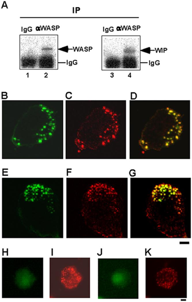
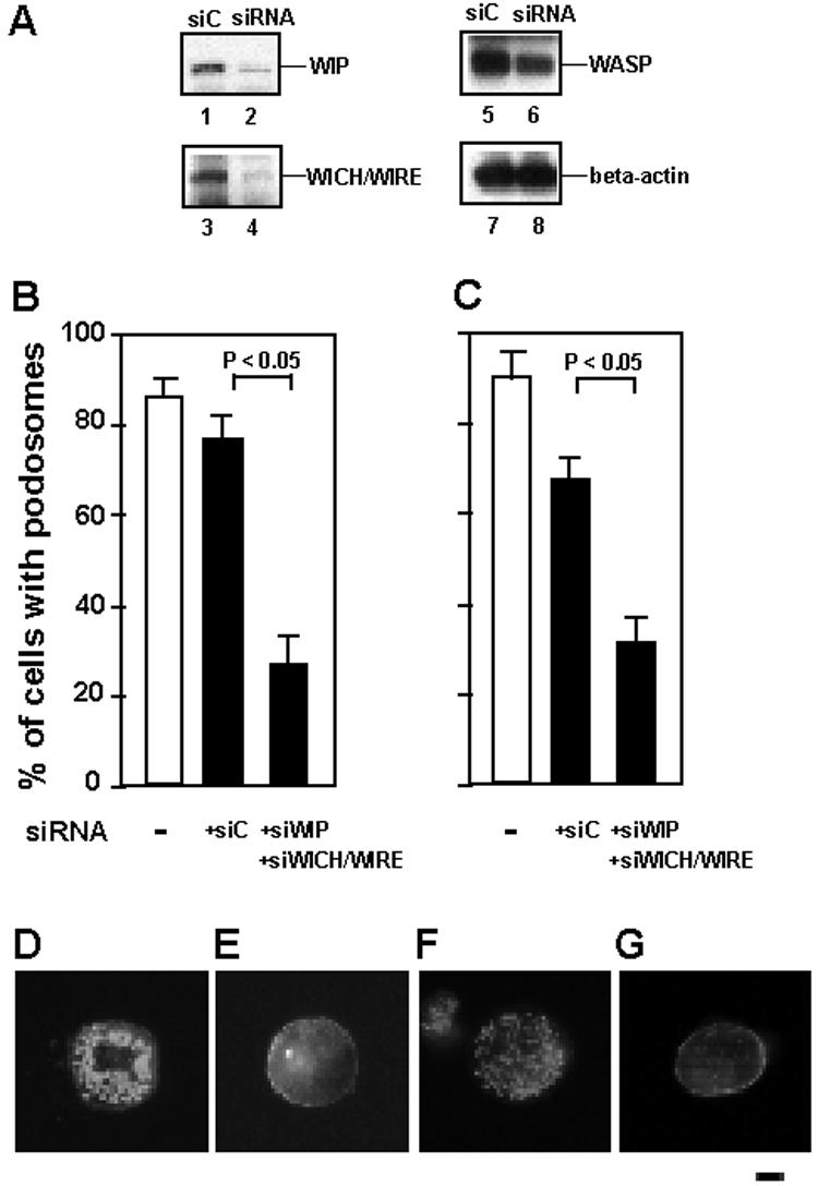
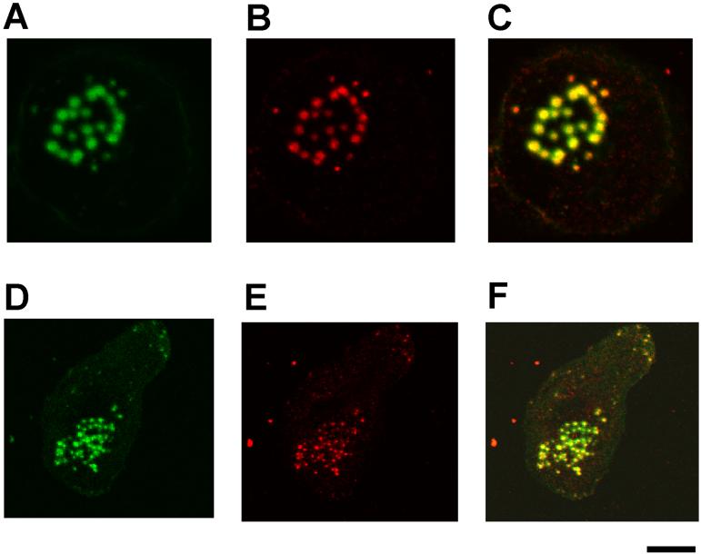
Chemotactic migration of macrophages is critical for the recruitment of leukocytes to inflamed tissues. Macrophages use a specialized adhesive structure called a podosome to migrate. Podosome formation requires the Wiskott-Aldrich syndrome protein (WASP), which is a product of the gene defective in an X-linked inherited immunodeficiency disorder, the Wiskott-Aldrich syndrome. Macrophages from WASP-deficient Wiskott-Aldrich syndrome patients lack podosomes, resulting in defective chemotactic migration. However, the molecular basis for podosome formation is not fully understood. I have shown that the WASP interacting protein (WIP), a binding partner of WASP, plays an important role in podosome formation in macrophages. I showed that WASP bound WIP to form a complex at podosomes and that the knockdown of WIP impairs podosome formation. When WASP binding to WIP was blocked, podosome formation was also impaired. When WASP expression was reduced by small interfering RNA transfection, the amount of the complex of WASP with WIP decreased, resulting in reduced podosome formation. Podosomes were restored by reconstitution of the WASP-WIP complex in WASP knockdown cells. These results indicate that the WASP-WIP complex is required for podosome formation in macrophages. When podosome formation was reduced by blocking WASP binding to WIP, transendothelial migration of macrophages, the most crucial process in macrophage trafficking, was impaired. These results suggest that a complex of WASP with WIP plays a critical role in podosome formation, thereby mediating efficient transendothelial migration of macrophages.


Figures

FIGURE 1
FIGURE 1
WASP binds WIP to form a complex at podosomes. A, Coimmunoprecipitation of WIP with WASP. WASP was immunoprecipitated with anti-WASP mAb or a control IgG from the lysates of human primary macrophages followed by immunoblotting for WASP (lanes 1 and 2) and WIP (lanes 3 and 4). B-G, Confocal laser scanning micrographs of PMA-differentiated THP-1 cells (B-D) and human primary macrophages (E-G). WIP staining (B and E), actin staining (C and F), and overlay of WIP and actin staining (D and G). Yellow color indicates colocalization of green (WIP) and red (actin). THP-1 cells (H and I) and macrophages (J and K) were stained with a control antibody (rabbit IgG) and phalloidin. Bars, 10 μm.
FIGURE 2
FIGURE 2
The role of WIP in podosome formation. A, Expression of WIP and WICH/WIRE was reduced by transfection of siRNA. THP-1 cells were transfected with siRNAs for WIP (siWIP) and WICH/WIRE (siWICH/WIRE) (lanes 2, 4, 6, and 8), or their scrambled control siRNAs (siC) (lanes 1, 3, 5, and 7). Cells were cotranfected with FITC-conjugated control siRNA (Invitrogen). FITC-positive cells were sorted by FACS. Total lysates of sorted cells were analyzed by immunoblotting for WIP (lanes 1 and 2), WICH/WIRE (lanes 3 and 4), WASP (lanes 5 and 6), and β-actin (lanes 7 and 8). B and C, Quantification of podosome formation. The percentage of cells with podosomes per siRNA-transfected cells (FITC-conjugated control siRNA-positive cells) was scored. □, podosome formation of untransfected cells. ■, podosome formation of siRNA-transfected cells. siWIP and WICH/WIRE indicate siRNA for WIP and WICH/WIRE, respectively. siC indicates the mixture of scrambled control siRNAs of siWIP and siWICH/WIRE. Data represent the mean ± SD of triplicate measurements. D-G, Immunofluorescence micrographs of a representative cell of each experiment. Cells were stained for F-actin with Alexa 568-phalloidin. PMA-differentiated THP-1 cells transfected with the scrambled controls (D), or siRNAs for WIP and WICH/WIRE (E); Human primary macrophages transfected with the scrambled controls (F), or siRNAs for WIP and WICH/WIRE (G). Bar is 10 μm.
FIGURE 3
FIGURE 3
The role of WASP-WIP complex in podosome formation. A, Binding of WASP to WIP and WICH/WIRE was blocked by the FLAG-tagged WB fragment. THP-1 cells were transfected with the FLAG-tagged PDZ-GEF (F-C) as a negative control (lanes 1, 3, 5, 7, 9, 11, and 13), or the FLAG-tagged WASP binding site of WIP (residues 321-503) (F-WB) (lanes 2, 4, 6, 8, 10, 12, and 14). Cells were cotransfected with GFP expressing plasmid (pmaxGFP). GFP-positive cells were sorted by FACS. Total lysates of sorted cells were analyzed by immunoblotting for WASP (lanes 1 and 2), WIP (lanes 3 and 4), WICH/WIRE (lanes 5 and 6), and FLAG-tagged proteins (lanes 7 and 8). WASP was immunoprecipitated from the lysates of sorted cells followed by immunoblotting for WASP (lanes 9 and 10), WIP (lanes 11 and 12), and WICH/WIRE (lanes 13 and 14). B and C, Quantification of podosome formation. The percentage of cells with podosomes per transfected cells (GFP-positive cells) was scored. □, podosome formation of cells transfected with FLAG-tagged PDZ-GEF (F-C) or Myc-tagged PDZ-GEF (M-C) as a negative control. ■, podosome formation of cells transfected with FLAG-tagged WASP binding site of WIP (F-WB) or Myc-tagged WIP binding site of the WASP N-terminus (M-WN) to block WASP binding to WIP. Podosome formation of PMA-differentiated THP-1 cells (B) or human primary macrophages (C) was shown. Data represent the mean ± SD of triplicate measurements. D-K, Immunofluorescence micrographs of a representative cell of each experiment. Cells were stained for F-actin with Alexa 568-phalloidin. PMA-differentiated THP-1 cells transfected with F-C (D), F-WB (E), M-C (F), or M-WN (G). Human primary macrophages transfected with F-C (H), F-WB (I), M-C (J), or M-WN (K). Bar is 10 μm. L, THP-1 cells were cotransfected with FLAG-tagged constructs, Myc-tagged constructs and GFP expressing plasmid. M-42 is the Myc-tagged constitutively active form of Cdc42 (V12Cdc42). The total lysates were immunoblotted by anti-FLAG (lanes 1-3) or anti-Myc monoclonal antibodies (lanes 4-6) to detect expression of each protein. M and N, Cells were stained with phalloidin, and podosome and filopodia formation of transfected cells (GFP-positive cells) were examined. The percentages of cells with podosomes (M) and filopodia (N) per transfected cells were scored, respectively. Podosome and filopodia formation of cells transfected with F-C as a negative control (□). Podosome and filopodia formation of cells transfected with F-WB to block WASP binding to WIP (■) (M and N). O-Q, Immunofluorescence micrographs of a representative cell of each experiment. Cells were cotransfected with F-C and M-C (O), F-WB and M-C (P) and F-WB and M-42 (Q), and then stained with Alexa 568-phalloidin. Bar is 10 μm. R, THP-1 cells were cotransfected with FLAG-tagged constructs, Myc-tagged WASP murant constructs and GFP expressing plasmid. dW is a WASP deletion mutant lacking the WIP binding site (residues 171-502). dW Y291E is the deletion mutant with Y291E mutation. dW Y291F is the deletion mutant with Y291F mutation. The total lysates were immunoblotted by anti-FLAG (lanes 1-4) or anti-Myc monoclonal antibodies (lanes 5-8) to detect expression of each protein. S and T, Cells were stained with phalloidin, and podosome and filopodia formation of transfected cells (GFP-positive cells) were examined. The percentages of cells with podosomes (S) and filopodia (T) were scored, respectively. Podosome and filopodia formation of cells transfected with F-C as a negative control (□). Podosome and filopodia formation of cells transfected with F-WB to block WASP binding to WIP (■) (S and T). U-X, Immunofluorescence micrographs of a representative cell of each experiment. Cells were cotransfected with F-C and dW (U), F-WB and dW (V), F-WB and dW Y291E (W) and F-WB and dW Y291F (X). Bar is 10 μm. Data represent the mean ± SD of triplicate measurements.
FIGURE 4
FIGURE 4
Complex formation of WASP with WIP causes podosome formation. A, Knockdown of human WASP and expression of mouse WASP. THP-1 cells were transfected with siRNA for human WASP (siW) or its scrambled control (siC) and FLAG-tagged mouse WASP (F-mW). Cells were cotranfected with FITC-conjugated control siRNA. FITC-positive cells were sorted by FACS. Total lysates of sorted cells were analyzed by immunoblotting with anti-WASP (lanes 1-3), anti-FLAG (lanes 4-6), anti-WIP (lanes 7-9), and anti-β-actin (lanes 10-12). B, Decrease in the amount of the human WASP-WIP complex and reconstitution of the mouse WASP-WIP complex. WIP was immunoprecipitated from the lysates of sorted cells followed by immunoblotting with anti-WIP (lanes 1-3), anti-WASP (lanes 4-6), and anti-FLAG (lanes 7-9). C and D, Quanitfication of podosome formation. The percentage of cells with podosomes per transfected cells (FITC-conjugated control siRNA-positive cells) was scored. □, podosome formation of cells transfected with the scrambled control siRNA for WASP (siC). ■, podosome formation of cells transfected with siRNA for WASP (siW). F-C and F-mW indicate FLAG-tagged PDZ-GEF (a negative control) and FLAG-tagged mouse WASP, respectively. Podosome formation of PMA-differentiated THP-1 cells (C) or human primary macrophages (D) was shown. Data represent the mean ± SD of triplicate measurements. E and F, Immunofluorescence micrographs of a representative cell of each experiment. Cells were stained for F-actin with Alexa 568-phalloidin. PMA-differentiated THP-1 cells transfected with human WASP siRNA (siW) and FLAG-tagged mouse WASP cDNA (F-mW) (E). Human primary macrophages transfected with human WASP siRNA (siW) and FLAG-tagged mouse WASP cDNA (F-mW) (F). Bar is 10 μm.
FIGURE 5
FIGURE 5
Exogenous mouse WASP localizes at restored podosomes. Confocal laser scanning micrographs of cells transfected with human WASP siRNA and FLAG-tagged mouse WASP cDNA. PMA-differentiated THP-1 cells (A-C) and human primary macrophages (D-F). Cells were stained with anti-FLAG mAb for FLAG-tagged mouse WASP (A and D), actin staining (B and E), and overlay of mouse WASP and actin staining. Yellow color indicates colocalization of green (mouse WASP) and red (actin), Bar is 10 μm.
FIGURE 6
FIGURE 6
The role of the WASP-WIP complex in transendothelial migration of macrophages. PMA-differentiated THP-1 cells (A) or human primary macrophages (B) were transfected with FLAG-tagged PDZ-GEF as a negative control (F-C, □), or FLAG-tagged WB (F-WB, ■) to block WASP binding to WIP. Cells were cotransfected with GFP expressing plasmid (pmaxGFP). Cells were cocultured on a monolayer of HUVEC cells for 2 hours. The percentage of transfected cells (GFP-positive cells) transmigrated (Basal), transmigrating (Transmigrating), or retained on the apical surface of the monolayer (Apical) was scored. Data represent the mean ± SD of triplicate measurements.

Similar articles

Cited by

References

    1. Wiskott A. Familiarer, angeborener Morbus Welhofii? Monatsschr Kinderheilkd. 1937;68:212–216.
    1. Aldrich RA, Steinberg AG, Campbell DC. Pedigree demonstrating a sex-linked recessive condition characterized by draining ears, eczematoid dermatitis and bloody diarrhea. Pediatrics. 1954;13:133–139. - PubMed
    1. Ochs HD, Thrasher AJ. The Wiskott-Aldrich syndrome.[see comment] Journal of Allergy & Clinical Immunology. 2006;117:725–738. quiz 739. - PubMed
    1. Derry JM, Ochs HD, Francke U. Isolation of a novel gene mutated in Wiskott-Aldrich syndrome. [Published erratum appears in 1994 Cell 79: following 922.] Cell. 1994;78:635–644. - PubMed
    1. Burns S, Cory GO, Vainchenker W, Thrasher AJ. Mechanisms of WASp-mediated hematologic and immunologic disease. Blood. 2004;104:3454–3462. - PubMed

Publication types

MeSH terms