Clinical Trial
. 2007 Mar 1;178(5):3272-80.
doi: 10.4049/jimmunol.178.5.3272.

Expression profile of FcgammaRIIb on leukocytes and its dysregulation in systemic lupus erythematosus

Affiliations
Clinical Trial

Expression profile of FcgammaRIIb on leukocytes and its dysregulation in systemic lupus erythematosus

Kaihong Su et al. J Immunol. .

Abstract

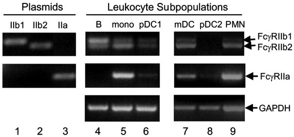
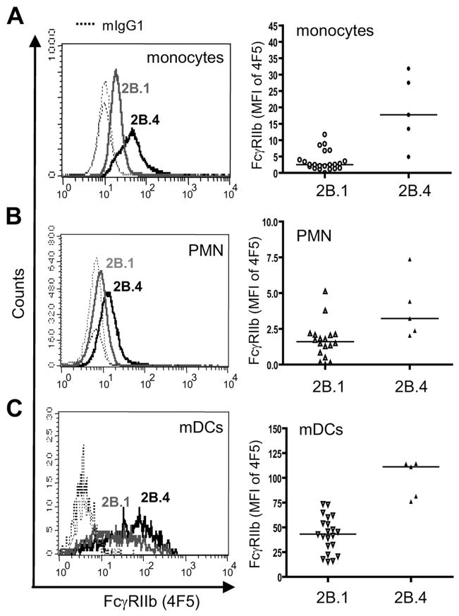
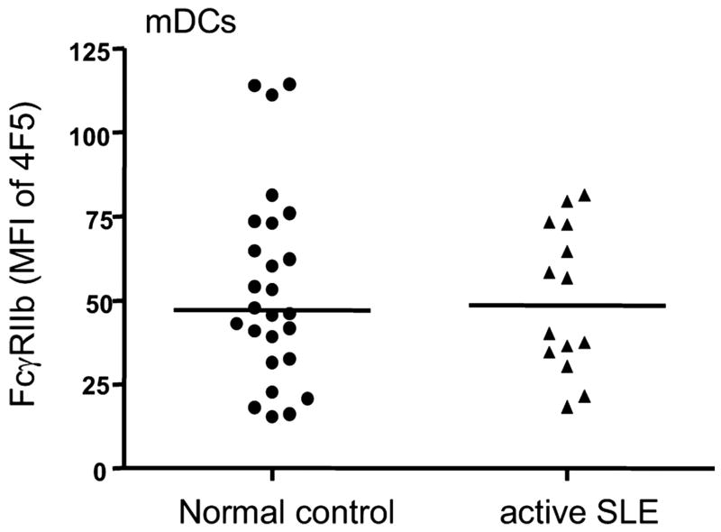
FcgammaRIIb (CD32B, Online Mendelian Inheritance in Man 604590), an IgG FcR with a tyrosine-based inhibitory motif, plays a critical role in the balance of tolerance and autoimmunity in murine models. However, the high degree of homology between FcgammaRIIb and FcgammaRIIa in humans and the lack of specific Abs to differentiate them have hampered study of the normal expression profile of FcgammaRIIb and its potential dysregulation in autoimmune diseases such as systemic lupus erythematosus (SLE). Using our newly developed anti-FcgammaRIIb mAb 4F5 which does not react with FcgammaRIIa, we found that FcgammaRIIb is expressed on the cell surface of circulating B lymphocytes, monocytes, neutrophils, myeloid dendritic cells (DCs), and at very low levels on plasmacytoid DCs from some donors. Normal donors with the less frequent 2B.4 promoter haplotype have higher FcgammaRIIb expression on monocytes, neutrophils, and myeloid DCs similar to that reported for B lymphocytes, indicating that FcgammaRIIb expression on both myeloid and lymphoid cells is regulated by the naturally occurring regulatory single nucleotide polymorphisms in the FCGR2B promoter. FcgammaRIIb expression in normal controls is up-regulated on memory B lymphocytes compared with naive B lymphocytes. In contrast, in active SLE, FcgammaRIIb is significantly down-regulated on both memory and plasma B lymphocytes compared with naive and memory/plasma B lymphocytes from normals. Similar down-regulation of FcgammaRIIb on myeloid-lineage cells in SLE was not seen. Our studies demonstrate the constitutive regulation of FcgammaRIIb by natural gene polymorphisms and the acquired dysregulation in SLE autoimmunity, which may identify opportunities for using this receptor as a therapeutic target.


Conflict of interest statement

Disclosures

The authors have no financial conflict of interest.

Figures

FIGURE 1
FIGURE 1
mAb 4F5 reacts with FcγRIIb not FcγRIIa. A, mAb 4F5 reacts with cell surface FcγRIIb but not FcγRIIa in flow cytometry analysis. A20IIA1.6 cells stably transfected with FcγRIIb or FcγRIIa were incubated with Alexa 488-conjugated mAbs mIgG1 (dashed lines), 4F5 (gray solid line), or AT10 (black solid line). FcγRIIb is monomorphic at amino acid 131 with an arg residue and FcγRIIa is polymorphic at that site with an arg or his residue. Thus, both IIa-R and IIa-H stable transfectants were established and analyzed. B, mAb 4F5 immunoprecipitates FcγRIIb but not FcγRIIa. Whole cell lysate from FcγRIIb, FcγRIIa-R, FcγRIIa-H stable transfectants were immunoprecipitated by mAb mIgG1, 4F5, or IV.3 and subjected to Western blot analysis using rabbit polyclonal Abs specific for the cytoplasmic domain of FcγRIIb (upper) or goat Abs specific for the cytoplasmic domain of FcγRIIa/c (lower) as blotting Abs. C, mAb 4F5 interacts with the recombinant EC of FcγRIIb but not of FcγRIIa. Purified fusion proteins GST-IIb EC or GST-IIa EC (R or H) were incubated with mAb 4F5 or AT10 and the bound proteins were subjected to Western blot analysis by HRP-linked goat anti-mouse IgG Abs. D, mAb 4F5 specifically reacts to denatured FcγRIIb in Western blot analysis. The immunoprecipitates with mAb AT10 or mIgG1 from the whole cell lysate of FcγRIIb, FcγRIIa-R, FcγRIIa-H stable transfectants were subjected to Western blot analysis using mAb 4F5 (upper) or goat polyclonal Abs specific for the cytoplasmic domain of FcγRIIa/c (lower) as blotting Abs.
FIGURE 2
FIGURE 2
Cross-linking of FcγRIIb by mAb 4F5 induces an inhibitory signal for Ca2+ influxes mediated by BCR and FcγRIa (CD64). A, Cross-linking of FcγRIIb by mAb 4F5 inhibited the BCR-mediated Ca2+ influxes. A20IIA1.6-FcγRIIb stable transfectants were preincubated with 4F5 F(ab′)2 (gray line) or its isotype control mIgG1 F(ab′)2 (black line). The endogenous mIgG-BCR and transfected human FcγRIIb were co-cross-linked by F(ab′)2 goat anti-mouse IgG. The Ca2+ responses were recorded using an SLM 8000 spectrofluorometer and presented as the changes in intracellular Ca2+ concentration. B, Cross-linking of FcγRIIb by mAb 4F5 inhibited the FcγRI-mediated Ca2+ influxes. U937 cells were preincubated with primary 32.2 F(ab′)2, IV.3 Fab, and/or 4F5 F(ab′)2 and cross-linked with secondary F(ab′)2 goat anti-mouse IgG for the engagement of FcγRIa alone (black solid line), coengagement of FcγRIa and FcγRIIa (gray dashed line), or coengagement of FcγRIa and FcγRIIb (gray solid line).
FIGURE 3
FIGURE 3
mAb 4F5 partially competes with ahIgG but not with mAb AT10 for receptor binding. A, mAb 4F5 partially competes with ahIgG for receptor binding. Upper panel, IIA1.6-FcγRIIb stable transfectants were preincubated with 30 μg/ml 4F5 F(ab′)2 (thick gray line), IV.3 Fab (thin gray line), or control mIgG1 F(ab′)2 (thick black line). The cells were then washed, stained with 10 μg/ml Alexa 488-conjugated ahIgG, and subjected to flow cytometry analysis. Direct staining of the cells with Alexa 488-conjugated mIgG1 was used as a control (dashed black line). Lower panel, The same experiment was performed on FcγRIIa-R IIA1.6 stable transfectants. B, ahIgG partially competes with mAb 4F5 for receptor binding. The IIA1.6-FcγRIIb stable transfectants were preincubated with 30 μg/ml ahIgG (thick gray line), ahIgA (thin gray line), or no Ab (thick black line). The cells were then washed, stained with 10 μg/ml Alexa 488-conjugated 4F5, and subjected to flow cytometry analysis. Direct staining of the cells with mIgG1-Alexa 488 was used as a control (dashed black line). C, mAb 4F5 does not compete with mAb AT10 for receptor binding. The FcγRIIb-IIA1.6 stable transfectants were preincubated with 30 μg/ml unlabeled mAb 4F5 (thick gray line), AT10 (thin gray line), or no Ab (thick black line). The cells were then washed, stained with 10 μg/ml Alexa 488-conjugated AT10, and subjected to flow cytometry analysis. Direct staining of the cells with mIgG1-Alexa 488 was used as a control (dashed black line).
FIGURE 4
FIGURE 4
The cell surface expression profile of FcγRIIb on circulating leukocytes. PBMCs (A, B, D, and E) or whole blood (C) was incubated with lineage-specific markers and Alexa 488-conjugated mAbs 4F5 (black line), AT10 (dark gray line), IV.3 (light gray line), or mIgG1 isotype control (dashed line) and subjected to multicolor flow cytometry analysis. Left panels, The gating of the cell subpopulations analyzed in the right panel histograms. B lymphocytes are defined as CD19+, mDCs as CD19 BDCA1+, monocytes as CD14+, and pDCs as BDCA2+; neutrophils are defined by side- and forward-scatter.
FIGURE 5
FIGURE 5
The expression of FcγRIIb1 and IIb2 messages in leukocyte subpopulations. Semiquantitative RT-PCR for FcγRIIb and FcγRIIa messages was performed using RNAs isolated from 20,000 purified CD19+ B cells, CD14+ monocytes, CD19BDCA1+ mDCs, or BDCA2+ pDCs of normal donors (lanes 4–9). The PCR using FcγRIIb1, FcγRIIb2, or FcγRIIa cDNA-containing pcDNA3 plasmids as the templates were served as specificity control for primers and PCR conditions (lanes 1–3). The RT-PCR for housekeeping GAPDH gene was used as a loading control.
FIGURE 6
FIGURE 6
The expression of FcγRIIb on pDCs is induced by cytokines. A, PBMCs purified from a representative normal donor were stained with BDCA2-allophycocyanin and 4F5-Alexa 488. Dot plot shows the percentage of pDCs positive for mAb 4F5. B, PBMCs from the same person were cultured in RPMI 1640 plus 10% human AB serum medium with or without indicated cytokines for 22 h and then stained with BDCA2-allophycocyanin and 4F5-Alexa 488.
FIGURE 7
FIGURE 7
The 2B.4 haplotype leads to increased FcγRIIb expression on monocytes, neutrophils, and mDCs. PBMCs from normal donors with the 2B.4 haplotype and with homozygous 2B.1 haplotype were stained with lineage-specific markers and mAb 4F5 for the expression levels of FcγRIIb. A, Normal donors with the 2B.4 haplotype have increased 4F5 staining on CD14+ monocytes than 2B.1 donors. Left panel, A histogram overlay showing 4F5 staining on CD14+ monocytes from a representative 2B.1 donor (gray line) and a representative 2B.4 donor (black line). The dashed lines are mIgG1 isotype controls. Right panel, A summary of the MFI of 4F5 staining on monocytes from 22 2B.1 donors and 5 2B.4 donors (p < 0.002, two-tailed, Mann-Whitney U test). B, Normal donors with the 2B.4 haplotype have increased 4F5 staining on neutrophils compared with 2B.1 donors (p < 0.013, two-tailed, Mann-Whitney U test). Similar analyses were performed as shown in A. C, Donors with the 2B.4 haplotype have increased 4F5 staining on CD19 BDCA1+ mDCs compared with 2B.1 donors (p < 0.001, two-tailed, Mann-Whitney U test). Similar analyses were performed as in A.
FIGURE 8
FIGURE 8
The expression of FcγRIIb is dysregulated on B lymphocytes from active SLE patients. A, Active SLE patients have increased percentage of plasma B lymphocytes compared with normal controls. The percentages of naive B (CD19+CD27), memory B (CD19+CD27+), and plasma B cells (CD19lowCD27high) from a representative normal donor (left) and an active SLE patient (right) are shown in the dot plots. B and C, The expression of FcγRIIb on memory and plasma B cells from active SLE patients is significantly decreased compared both to their own naive B lymphocytes and to memory/plasma B lymphocyte counterparts from normal controls. Histograms in B display the staining of mAb 4F5 on naive and memory B lymphocytes from a representative normal donor (left) and an active SLE patient (right). C, A summary of the MFI of mAb 4F5 staining on naive (N), memory (M), and plasma (P) B lymphocytes from 30 normal controls, 19 active SLE patients (SLEDAI ≥2), and 5 SLE patients in remission (SLEDAI = 0). Two-tailed Student’s t tests were performed to compare the expression levels of FcγRIIb on B cell subpopulations (normal memory B compared with their naive B lymphocytes: p < 0.0001; active SLE memory or plasma B compared with their own naive B lymphocytes: p < 0.05 and p < 0.04, respectively; active SLE memory or plasma B lymphocytes compared with memory or plasma B lymphocyte counterparts from normals: p < 0.0001 and p < 0.03, respectively). There was an increase of 2B.4 haplotype in SLE patients compared with normals (15.8 and 9%, respectively) and the analysis was comparable when the 2B.4 donors were removed from both groups.
FIGURE 9
FIGURE 9
The expression of FcγRIIb is not decreased on mDCs from active SLE patients. The same normal controls and active SLE patients as analyzed in Fig. 8 were also compared for FcγRIIb expression on mDCs by mAb 4F5 staining (p > 0.05, two-tailed, Mann-Whitney U test).

Similar articles

Cited by

References

    1. Daeron M. Fc receptor biology. Annu Rev Immunol. 1997;15:203–234. - PubMed
    1. Ravetch JV, Bolland S. IgG Fc receptors. Annu Rev Immunol. 2001;19:275–290. - PubMed
    1. Amigorena S, Bonnerot C, Drake JR, Choquet D, Hunziker W, Guillet JG, Webster P, Sautes C, Mellman I, Fridman WH. Cytoplasmic domain heterogeneity and functions of IgG Fc receptors in B lymphocytes. Science. 1992;256:1808–1812. - PubMed
    1. Ono M, Bolland S, Tempst P, Ravetch JV. Role of the inositol phosphatase SHIP in negative regulation of the immune system by the receptor FcγRIIB. Nature. 1996;383:263–266. - PubMed
    1. Hunter S, Indik ZK, Kim MK, Cauley MD, Park JG, Schreiber AD. Inhibition of Fcγ receptor-mediated phagocytosis by a nonphagocytic Fcγ receptor. Blood. 1998;91:1762–1768. - PubMed

Publication types

MeSH terms