Comparative Study
. 1992 Jan 16;355(6357):275-8.
doi: 10.1038/355275a0.

Identification of a Fab interaction footprint site on an icosahedral virus by cryoelectron microscopy and X-ray crystallography

Affiliations
Comparative Study

Identification of a Fab interaction footprint site on an icosahedral virus by cryoelectron microscopy and X-ray crystallography

G J Wang et al. Nature. .

Abstract

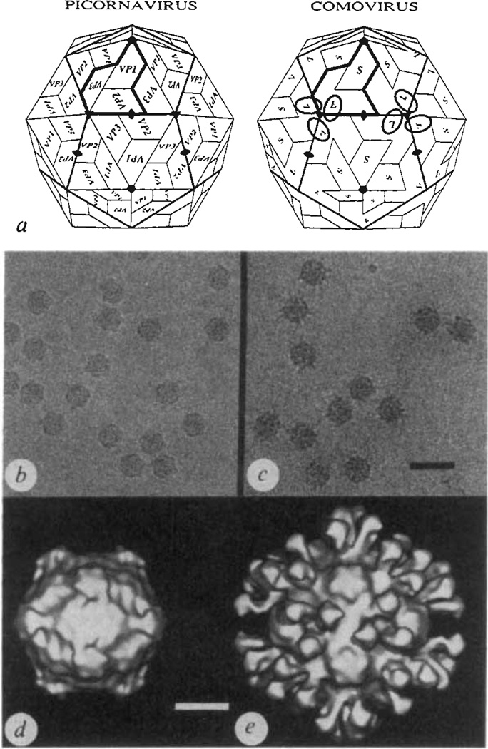
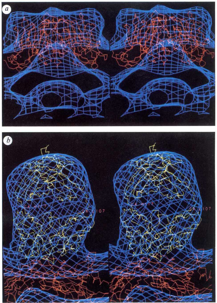
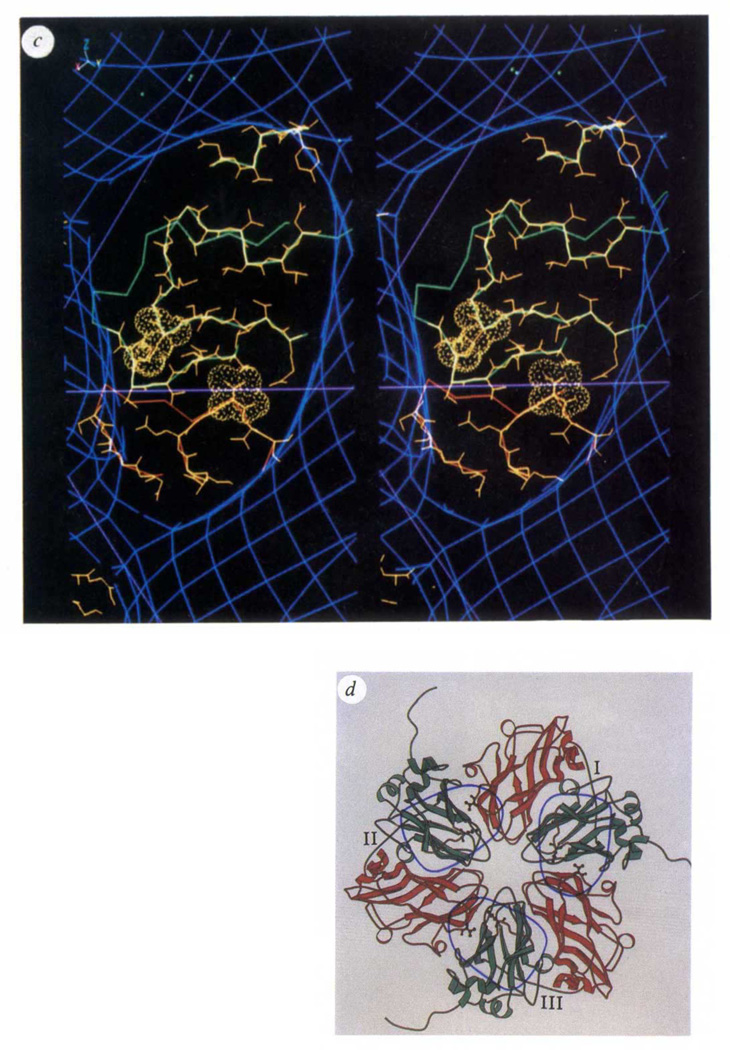
Biological processes frequently require the formation of multi-protein or nucleoprotein complexes. Some of these complexes have been produced in homogeneous form, crystallized, and analysed at high resolution by X-ray crystallography (for example, see refs 1-3). Most, however, are too large or too unstable to crystallize. Individual components of such complexes can often be purified and analysed by crystallography. Here we report how the coordinated application of cryoelectron microscopy, three-dimensional image reconstruction, and X-ray crystallography provides a powerful approach to study large, unstable macromolecular complexes. Three-dimensional reconstructions of native cowpea mosaic virus (CMPV) and a complex of CPMV saturated with a Fab fragment of a monoclonal antibody against the virus have been determined at 23 A resolution from low-irradiation images of unstained, frozen-hydrated samples. Despite the nominal resolution of the complex, the physical footprint of the Fab on the capsid surface and the orientation and position of the Fab have been determined to within a few ångstroms by fitting atomic models of CPMV4 and Fab (Kol)5 to reconstructed density maps.


Figures

FIG. 1
FIG. 1
The capsid architectures of picorna and comoviruses displayed with electron micrographs and three-dimensional image reconstructions of frozen-hydrated native CPMV and CPMV–Fab complexes, a, Schematic comparison of picornavirus and comovirus capsids. In each case one trapezoid represents an eight-stranded antiparallel β-barrel (see Fig. 2d). The icosahedral asymmetric unit of the picornavirus capsid (shaded in the heavy outline) contains three β-barrels, labelled VP1, VP2 and VP3, each with a characteristic amino-acid sequence. The comovirus capsid is similar to the picornavirus capsid except that two of the β-barrels, corresponding to VP2 (N terminus) and VP3 (C terminus), are covalently linked to form a single polypeptide, the large protein subunit (L), and the small protein subunit (S) corresponds to VP1. The ellipsoids on the comovirus capsid show the rough location of 6 of the 60 equivalent Fab binding sites (compare with e). b and c, Electron micrographs of frozen-hydrated samples of CPMV (b) and CPMV complexed with monoclonal antibody (5B2) Fab fragments (c). Scale bar, 500 Å. Both samples were prepared and photographed with established cryomicroscopy procedures. Roughly 4 ml each of a 2.0 mg ml−1 CPMV (b) or 0.5 mg ml−1 CPMV–Fab (c; stoichiometry of Fabs to virus in solution was 600:1) sample at pH 7.4 in PBS was applied to holey-carbon films, blotted with filter paper and rapidly plunged into liquid ethane at about −170 °C. Images were recorded at minimal electron dose (~16 e Å−2) at a nominal magnification of ×49,000 and at 80 kV in a Philips EM420 electron microscope. The images in b and c were recorded at about 0.8 and 0.9 µm underfocus, respectively, d, e, Surface-shaded representations of three-dimensional reconstructions of native virus (d) and virus–Fab complex (e), both viewed in the standard crystallographic 2-fold orientation, were computed with established methods. A total of 17 (d) or 22 (e) particle images were combined to reconstruct both structures to a resolution limit of 23 Å. Scale bar, 100 Å.
FIG 2
FIG 2
Comparisons of CPMV and Fab atomic models with reconstructed electron density derived from electron micrographs of frozen-hydrated specimens, a, b, Stereoviews comparing Cα backbone models of CPMV (red) and Fab (Kol) (yellow) with electron density (blue) derived from the electron-microscopy reconstruction described in Fig. 1. a, Native virions viewed in a direction perpendicular to a vertical fivefold icosahedral symmetry axis. The surface bulge in the density corresponds to amino acids in a β-turn between the outermost strands of the S subunit. b, A close-up view showing the Fab (Kol) and CPMV Cα backbones fitted to the CPMV–Fab reconstruction. The small portions of Kol that protrude outside the Fab electron microscope envelope at the top and into the surface of the CPMV L subunit are regions of the polypeptide that are not conserved in sequences of other IgG molecules, and are therefore likely to be different in the 5B2 Fab structure. When contoured at a higher density level the Fab envelope divides into well-defined globular regions which correspond closely with the Fab two-domain structure. Note that the electron microscope envelope, contoured at a single density level, fits the CPMV and Fab X-ray models equally well. This suggests that the frozen-hydrated CPMV samples imaged were nearly fully saturated with the Fabs and most of the Fab molecules must be fairly rigidly attached in equivalent orientations with respect to the viral surface. c, Stereoview of the Fab footprint on the virion surface showing regions of the CPMV polypeptide chain that lie under the Fab. The region in red corresponds to the ‘VP3’ domain of one L subunit; green denotes the ‘VP2’ domain of a 3-fold-related L subunit. Main chain and side chains of residues that may interact with the Fab are shown in yellow. The amino acids displayed with surface rendering (Lys 34 in the ‘VP2’ domain and Thr 24 in the 3-fold-related ‘VP3’ domain) are spatially equivalent to residues 72 and 76 in the poliovirus VP2 and VP3 domains. Mutations of either of these residues in poliovirus prevents neutralization by poliovirus monoclonal antibodies 10 and 13 (ref. 10), thus defining the 3B antigenic site on serotype I poliovirus. Lines in magenta correspond to the boundaries in Fig. 1a (picornavirus) that separate VP2 from VP3 near the 3-fold axis on the left side of the figure, d, A schematic representation of 3-fold-related L subunits, each numbered near the peptide that connects ‘VP2’ (green) and ‘VP3’ (red) domains. Ellipsoids identify the viral surface in contact with each Fab. The side chains shown correspond to the residues with surface rendering in Fig. 2c. The ellipsoid on the right (covering portions of L subunits I and III) is in the same orientation as the ellipsoid in c.
FIG 2
FIG 2
Comparisons of CPMV and Fab atomic models with reconstructed electron density derived from electron micrographs of frozen-hydrated specimens, a, b, Stereoviews comparing Cα backbone models of CPMV (red) and Fab (Kol) (yellow) with electron density (blue) derived from the electron-microscopy reconstruction described in Fig. 1. a, Native virions viewed in a direction perpendicular to a vertical fivefold icosahedral symmetry axis. The surface bulge in the density corresponds to amino acids in a β-turn between the outermost strands of the S subunit. b, A close-up view showing the Fab (Kol) and CPMV Cα backbones fitted to the CPMV–Fab reconstruction. The small portions of Kol that protrude outside the Fab electron microscope envelope at the top and into the surface of the CPMV L subunit are regions of the polypeptide that are not conserved in sequences of other IgG molecules, and are therefore likely to be different in the 5B2 Fab structure. When contoured at a higher density level the Fab envelope divides into well-defined globular regions which correspond closely with the Fab two-domain structure. Note that the electron microscope envelope, contoured at a single density level, fits the CPMV and Fab X-ray models equally well. This suggests that the frozen-hydrated CPMV samples imaged were nearly fully saturated with the Fabs and most of the Fab molecules must be fairly rigidly attached in equivalent orientations with respect to the viral surface. c, Stereoview of the Fab footprint on the virion surface showing regions of the CPMV polypeptide chain that lie under the Fab. The region in red corresponds to the ‘VP3’ domain of one L subunit; green denotes the ‘VP2’ domain of a 3-fold-related L subunit. Main chain and side chains of residues that may interact with the Fab are shown in yellow. The amino acids displayed with surface rendering (Lys 34 in the ‘VP2’ domain and Thr 24 in the 3-fold-related ‘VP3’ domain) are spatially equivalent to residues 72 and 76 in the poliovirus VP2 and VP3 domains. Mutations of either of these residues in poliovirus prevents neutralization by poliovirus monoclonal antibodies 10 and 13 (ref. 10), thus defining the 3B antigenic site on serotype I poliovirus. Lines in magenta correspond to the boundaries in Fig. 1a (picornavirus) that separate VP2 from VP3 near the 3-fold axis on the left side of the figure, d, A schematic representation of 3-fold-related L subunits, each numbered near the peptide that connects ‘VP2’ (green) and ‘VP3’ (red) domains. Ellipsoids identify the viral surface in contact with each Fab. The side chains shown correspond to the residues with surface rendering in Fig. 2c. The ellipsoid on the right (covering portions of L subunits I and III) is in the same orientation as the ellipsoid in c.

References

    1. Davies DR, Padlan EA, Sheriff S. A. Rev. Biochem. 1990;59:439–473. - PubMed
    1. Ruff M, et al. Science. 1991;252:1682–1689. - PubMed
    1. Rossmann MG, Johnson JE. A. Rev. Biochem. 1989;58:533–573. - PubMed
    1. Chen Z, Stauffacher CV, Johnson JE. Semin. Virol. 1990;1:453–466.
    1. Marquart M, Deisenhofer J, Huber R, Palm W. J. molec. Biol. 1980;141:369–391. - PubMed

Publication types

MeSH terms

Substances