. 2007 Feb 21;99(4):309-21.
doi: 10.1093/jnci/djk053.

Requirement of KISS1 secretion for multiple organ metastasis suppression and maintenance of tumor dormancy

Affiliations

Requirement of KISS1 secretion for multiple organ metastasis suppression and maintenance of tumor dormancy

Kevin T Nash et al. J Natl Cancer Inst. .

Erratum in

  • J Natl Cancer Inst. 2007 Jul 20;99(12):977

Abstract

Background: The KISS1 protein suppresses metastasis of several tumor models without blocking orthotopic tumor growth, but the mechanism remains elusive. For its role in human sexual maturation, KISS1 protein is secreted and processed to kisspeptins, which bind to the G protein-coupled receptor GPR54. We tested the hypothesis that KISS1 secretion is required for metastasis suppression via GPR54.

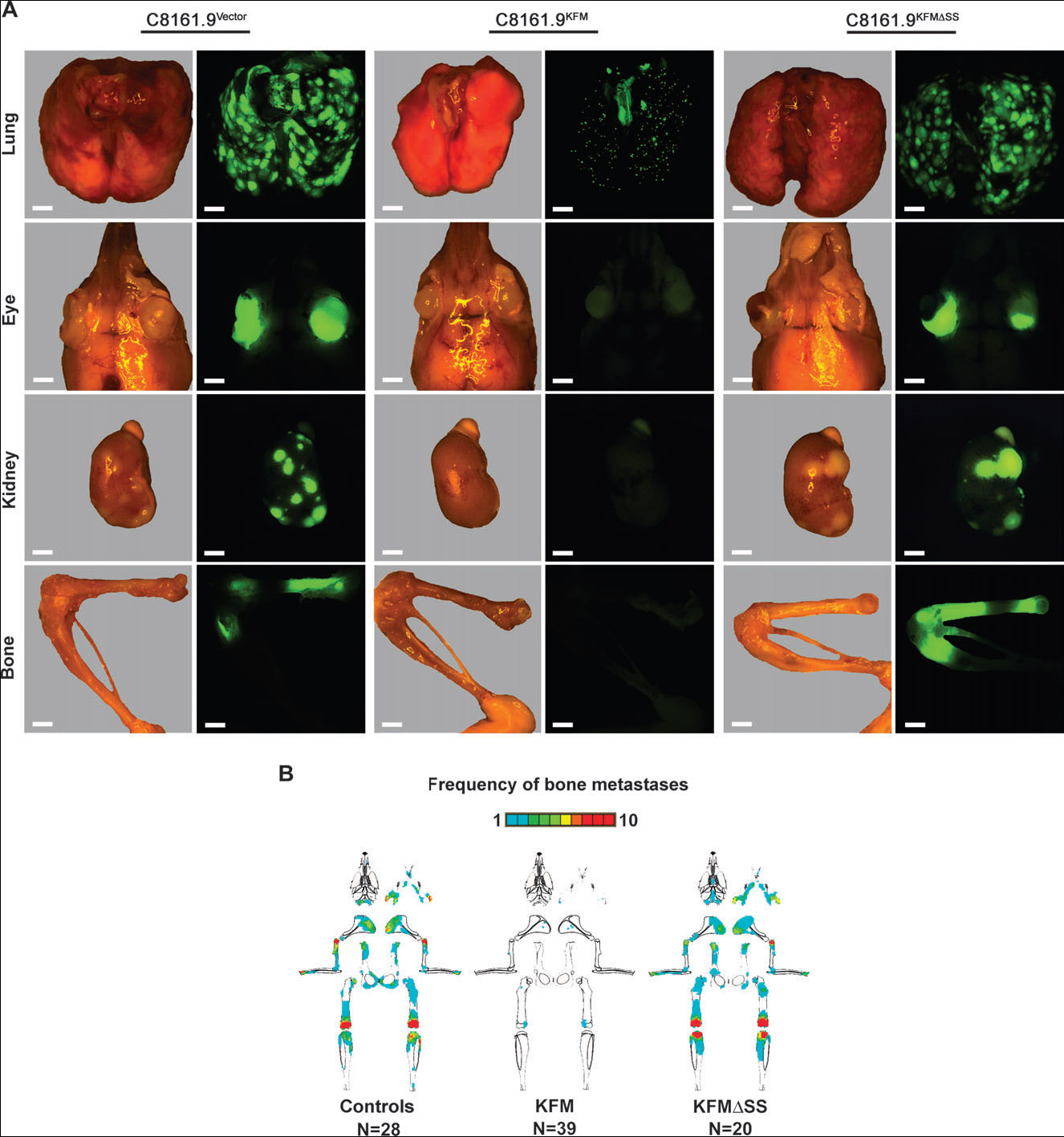
Methods: KISS1 containing an internal FLAG epitope with (KFM) or without (KFMdeltaSS) a signal sequence was transfected into C8161.9 human melanoma cells, which do not express endogenous KISS1. Whole-cell lysates and conditioned medium from C8161.9(KFM) and C8161.9(KFMdeltaSS) cells were collected and analyzed for kisspeptins by immunoprecipitation and enzyme-linked immunosorbent assay. GPR54 levels were measured using real-time reverse transcription-polymerase chain reaction. The ability of conditioned medium from C8161.9(KFM) and C8161.9(KFMdeltaSS) cells to stimulate calcium mobilization in GPR54-expressing Chinese hamster ovary cells (CHO-G) and in C8161.9 cells was evaluated. Metastasis was monitored in athymic mice (groups of 10 per experiment) that were injected with C8161.9(KFM) or C8161.9(KFMdeltaSS) cells labeled with enhanced green fluorescent protein. Survival of mice injected with C8161.9 or C8161.9(KFM) cells was analyzed by Kaplan-Meier methods.

Results: Full-length KFM and KFMdeltaSS were detected in whole-cell lysates of C8161.9(KFM) and C8161.9(KFMdeltaSS) cells, respectively, but kisspeptins were detected only in conditioned medium of C8161.9(KFM) cells. In vivo, C8161.9(KFM), but not C8161.9(KFMdeltaSS), cells were suppressed for metastasis to lung, eye, kidney, and bone, with corresponding differences in mouse survival (median > 120 versus 42 days). C8161.9(KFM) cells seeded mouse lungs but did not form macroscopic metastases. Conditioned medium from C8161.9(KFM), but not C8161.9(KFMdeltaSS), cells stimulated calcium mobilization in CHO-G cells. GPR54 expression was low in C8161.9 cells, which were not stimulated by conditioned medium from C8161.9(KFM) cells.

Conclusions: KISS1 secretion was required for multiple organ metastasis suppression and for maintenance of disseminated cells in a dormant state. The absence of GPR54 expression in C8161.9 cells (whose metastatic spread was suppressed by KFM) suggests that metastasis suppression is not mediated through this receptor. The results imply the existence of another KISS1 receptor and/or paracrine signaling. The findings raise the possibility that soluble KISS1, kisspeptins, or mimetics could be used to maintain tumor dormancy, rendering treatment of already disseminated tumor cells (i.e., micrometastases) a legitimate target.


Figures

Fig. 1
Fig. 1
Detection and processing of KFM, in which an internal FLAG epitope has been inserted into KISS1 upstream of the 54–amino acid metastin sequence. A) Schematic diagrams of KFM and KFMΔSS (secretion signal deleted). Consensus prohormone convertase dibasic recognition sequences (RK, RR, and GKR) are shown. B) Detection of KFM and KFMΔSS in whole-cell C8161.9 cell variant lysates by immunoprecipitation with anti-metastin polyclonal antiserum followed by immunoblotting with anti-FLAG antibody. β-actin was used as a loading control. C) Detection of KISS1 and processed kisspeptins from C8161.9 cell variants in conditioned medium by immunoprecipitation with anti-metastin polyclonal antiserum followed by immunoblotting with anti-FLAG antibody. D) Predicted processing of KFM to kisspeptins and the mature metastin product. The size of kisspeptins detected in (C) approximate the predicted sizes shown. Metastin is not detected because polyacylamide gels are generally not suitable for detection of small polypeptides. E) Sequences of secreted kisspeptins identified in conditioned medium by electrospray ionization mass spectroscopy/mass spectroscopy. All the bands isolated from the sodium dodecyl sulfate–polyacrylamide gel electrophoresis gel contained sequences within the metastin polypeptide. The sequence shown shows that 39 of the 54 metastin amino acids were found and properly oriented. Detection of sequences N-terminal to metastin in several of the larger bands suggests that at least some KISS1 processing occurs extracellularly.
Fig. 2
Fig. 2
Intracellular localization of KISS1 to the Golgi complex of transfected C8161.9 melanoma cells. Each column represents a different C8161.9 transfectant. KGM = C8161.9 cells transfected with KISS1 into which enhanced green fluorescent protein (GFP) was inserted N-terminally to metastin. Vector = C8161.9 cells transfected with empty vector. KFM = C8161.9 cells transfected with KISS1 into which the FLAG epitope was inserted N-terminally to metastin. KFMΔSS = C8161.9 cells transfected with KFM lacking the signal sequence. Cells were stained by immunofluorescence with antibodies against FLAG and/or GM130 or giantin, as indicated. For the KGM transfectants, staining for FLAG was omitted; the fluorescence represents GFP. KFM and KGM colocalize with GM130 and giantin, but KFMΔSS shows diffuse cytoplasmic staining. Scale bar = 16 μm.
Fig. 3
Fig. 3
Effect of kisspeptins on Ca2+ signaling. A and B) Chinese hamster ovary cells expressing the G protein–coupled receptor GPR54 (CHO-G cells) (dashed lines) or type 1 muscarinic acetylcholine receptor (solid lines) were exposed to conditioned medium from C8161.9 melanoma cells transfected with empty vector (VM) (A) or with FLAG-tagged KISS1 (KM) (B). Ca2+ signaling was assayed by ratiometric spectrofluorometry. C and D) C8161.9 cells were treated with stromal-derived factor 1 (SDF-1), fetal bovine serum (FBS), and kisspeptin-10 (KP-10) as indicated, and Ca2+ signaling was assessed. E and F) C8161.9 cells were treated with VM, SDF-1, KM, and FBS as indicated, and Ca2+ signaling was assessed. G and H) AKT activation (i.e., phosphorylation on Ser473) was assayed in C8161.9 cells exposed to SDF-1, epidermal growth factor (EGF), KP-10, KM, and PM (conditioned medium from C8161.9 cells) alone or in combination. I) Relative GPR54 mRNA levels in C8161.9 (81), MelJuSo (Mel), MDA-MB-435 (435), and SKOV3 (V3) cells as determined by real-time reverse transcription–polymerase chain reaction and normalized to S9 ribosomal protein. Placenta (Plac) mRNA is used as a positive control.
Fig. 4
Fig. 4
KISS1 secretion and lung metastasis by C8161.9 human melanoma cells. Cells were not transfected (P) or were transfected with empty vector (V), with FLAG-tagged KISS1 (KFM), or with FLAG-tagged KISS1 lacking the signal sequence (KFMΔSS). Numbers indicate KFM transfectant clones (17, 46, 57, 59, 66) and KFMΔSS transfectant clones (6, 7, 10, 16). Mix indicates uncloned transfectants for each KISS1 construct. A and D) Immunoblot analysis of conditioned medium from P, V, and KFM (A) or P, V, and KFMΔSS (D) cells using immunoprecipitation with anti-metastin polyclonal antiserum followed by immunoblotting with anti–FLAG M2 antibody. B and E) Relative quantification of KISS1 secretion levels by enzyme-linked immunosorbent assay of KFM (B) and KFMΔSS (E) clones. Removal of the putative signal sequence blocks secretion of KISS1 (E). KFM clone 59 is shown on panel E as a positive control for the assay. C and F) Lung colonization in mice injected intravenously with C8161.9 cells transfected with KFM (C) or KFMΔSS (F) was determined by counting surface lung foci. Statistical significance (*** P<.001) was calculated by comparison of experimental values (KFM versus KFMΔSS) with vector control (V) (Holm–Sidak method).
Fig. 5
Fig. 5
KISS1 secretion and maintenance of dormancy of disseminated melanoma cells in the lung. AH) Examination of C8161.9, C8161.9KFM, and C8161KFM.59 cell growth in the lung. Mice were injected into the lateral tail vein with cells transduced with a green fluorescent protein (GFP)–containing lentivirus (AD) or C8161.9 melanoma cells that had been transfected with FLAG-tagged KISS1 (KFM). Images from C8161KFM clone 59 are shown as representative data for the other clones (EH). Representative lung micrographs were taken at 24 hours (A and E), 1 week (B and F), 3 weeks (C and G), and 5 weeks (D and H). Scale bar = 50 μm. I and J) Immunohistochemical visualization of. GFP-labeled C8161.9 cells in lung using an anti-GFP antibody (brown). Arrows identify macroscopic metastatic foci (I) and microscopic metastatic foci (J) at 5 weeks postinjection. Scale bar = 40 μm. K) Growth of fluorescent C8161.9 cells in the lung. Growth of fluorescent lung foci, such as those depicted in panels AH , were plotted for a period of 5 weeks for C8161.9 (closed circle) and C8161.9KFM.59 (open circle) cells. C8161.9KFM.59 foci were statistically significantly smaller than C8161.9 metastases after 3 weeks (*** P<.001, Holm–Sidak Method) and remained smaller until the experiment was terminated after 5 weeks. Data are represented as mean lung focus diameter ± SEM from a minimum of 10 mice. Mean focus diameter for C8161.9 was 1020 μm, 95% confidence interval (CI) = 922 to 1117 μm. Mean focus diameter for C8161.9vector was 930 μm, 95% CI = 833 to 1027 μm. L) Size of tumor (cell) foci at 5 weeks for multiple C8161.9KFM (clone 46; mean = 34 μm, 95% CI = 27 to 41 μm; clone 57; mean = 25, 95% CI = 22 to 28 μm; clone 59, mean = 27 μm, 95% CI = 24 to 31 μm) and C8161.9KFMΔSS (clone 6; mean = 1148 μm, 95% CI = 1053 to 1244 μm; clone 7; mean = 1103 μm, 95% CI = 1015 to 1191 μm; clone 16, mean = 1074 μm, 95% CI = 988 to 1134 μm) clones were determined by fluorescence microscopy (as above) following intravenous inoculation of athymic mice. Lung foci (metastases) from C8161.9KFM were statistically significantly smaller than those formed following injection of C8161.9 or C8161KFMΔSS cells (*** P<.001, Holm–Sidak Method). Each group in the experiment contained 10 mice. Determination of KISS1 mRNA levels by real-time reverse transcription–polymerase chain reaction (RT–PCR) in C8161.9KFM and C8161.9KFMΔSS cells isolated from lung tissue 5 weeks after intravenous inoculation of cells. Fluorescent cells were established as cell cultures by combining explants from 2 to 3 lungs each for C8161.9KFM.59 and C8161.9KFMΔSS clones 6, 7, and 16. KISS1 levels were then determined by real-time RT–PCR; KISS1 mRNA levels were normalized to C8161.9vector (V) cells before injection. N) Orthotopic tumor growth of C8161.9 and C8161.9KFM.59 cells isolated from lungs. C8161.9 and C8161.9KFM.59 cells were injected intravenously into athymic mice and cells harvested from the lungs of mice 5 weeks after inoculation. Following establishment of cell cultures, tumor cells (C8161.9 and C8161.9KFM.59) were injected orthotopically into five athymic mice per group, and mean tumor diameters of C8161.9 (closed square) and C8161.9KFM.59 (open square) were determined weekly. O) Kaplan–Meier survival analysis of mice receiving C8161.9 (P), C8161.9vector (V), and KFM-expressing clones (KFM.17, KFM.46, and KFM.59; 10 mice per clone). The experiment was terminated at 120 days after intravenous inoculation or if mice appeared moribund. Differences in survival between mice receiving C8161.9 (mean survival = 38.8 days, 95% CI = 37.8 to 39.9 days), C8161.9vector (mean survival = 42.6 days, 95% CI = 41.6 to 43.4 days), and C8161.9KFM (mean survival clone 17 = 66.7 days, 95% CI = 51.6 to 81.8 days; mean survival clones 46 and 59 ≥ 120 and 118.6 days) cell clones were determined using Kaplan–Meier survival analysis log-rank test, and statistical significance (* P<.001) was calculated by comparing values for KFM clones to C8161.9vector (Holm–Sidak method). P) Frequency of lung metastases in moribund (i.e., cachexic) mice compared to healthy survivors from panel O (10 mice per group). Mean number of lung metastases for C8161.9 (mean = 63 metastases, 95% CI = 50 to 76 metastases), C8161.9vector (mean = 128 metastases, 95% CI = 117 to 140 metastases), and C8161.9KFM clones (clone 17; mean = 13 metastases, 95% CI = 7 to 19 metastases; clone 46; mean = 0 metastases, 95% CI = 0 to 1 metastases; clone 59, mean = 1 metastasis, 95% CI = 0 to 2 metastases) are shown. Statistical significance (*** P<.001) was calculated by comparison of experimental values for KFM versus C8161.9vector control (V) (Holm–Sidak method).
Fig. 6
Fig. 6
KISS1 suppression of metastasis to multiple organs in mice. Mice were injected in the left cardiac ventricle with C8161.9 cells that had been transfected with empty vector or with FLAG-tagged KISS1 containing (KFM) or lacking (KFMΔSS) the signal sequence and then infected with a green fluorescent protein–containing lentivirus. A) Representative lung, eye, kidney, and bone photomicrographs and fluorescent photomicro-graphs showing metastatic lesions 5 weeks after injection. Most extra-pulmonary sites were devoid of green cells. Scale bar = 2000 μm. B) Graphical mapping of the distribution, relative size, and frequency of fluorescent metastatic foci in bone. Bone maps reflect a data merge of all mice (10 mice per experimental group) from two or three independent experiments. Mice received C8161.9 and C8161.9vector cells (controls); C8161.9KFM clones 46, 57, and 59 (KFM); and C8161.9KFMΔSS clones 6, 7, and 16 (KFMΔSS). The total number of mice examined is shown. A frequency scale is depicted above bone schematic diagrams. If more than 10 mice in a group had metastases at a particular location, the scale reads red .

References

    1. Jiang Y, Berk M, Singh LS, Tan HY, Yin LH, Powell CT, et al. KiSS1 suppresses metastasis in human ovarian cancer via inhibition of protein kinase C alpha. Clin Exp Metastasis. 2005;22:369–76. - PubMed
    1. Lee J-H, Miele ME, Hicks DJ, Phillips KK, Trent JM, Weissman BE, et al. KiSS-1, a novel human malignant melanoma metastasis-suppressor gene. J Natl Cancer Inst. 1996;88:1731–7. - PubMed
    1. Lee J-H, Welch DR. Identification of highly expressed genes in metastasis-suppressed chromosome 6/human malignant melanoma hybrid cells using subtractive hybridization and differential display. Int J Cancer. 1997;71:1035–44. - PubMed
    1. Lee J-H, Welch DR. Suppression of metastasis in human breast carcinoma MDA-MB-435 cells after transfection with the metastasis suppressor gene, KiSS-1. Cancer Res. 1997;57:2384–7. - PubMed
    1. Lee J-H, Miele ME, Hicks DJ, Phillips KK, Trent JM, Weissman BE, et al. KiSS-1, a novel human malignant melanoma metastasis-suppressor gene [erratum] J Natl Cancer Inst. 1997;89:1549. - PubMed

Publication types

MeSH terms