. 2007 Feb;25(3):837-46.
doi: 10.1111/j.1460-9568.2007.05312.x.

Impaired spatial working memory but spared spatial reference memory following functional loss of NMDA receptors in the dentate gyrus

Affiliations

Impaired spatial working memory but spared spatial reference memory following functional loss of NMDA receptors in the dentate gyrus

B Niewoehner et al. Eur J Neurosci. 2007 Feb.

Abstract

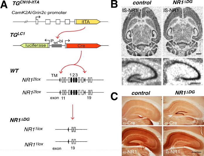
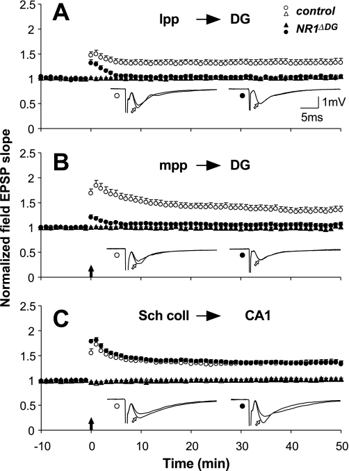
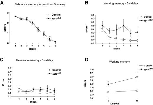
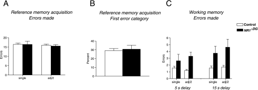
Novel spatially restricted genetic manipulations can be used to assess contributions made by synaptic plasticity to learning and memory, not just selectively within the hippocampus, but even within specific hippocampal subfields. Here we generated genetically modified mice (NR1(deltaDG) mice) exhibiting complete loss of the NR1 subunit of the N-methyl-D-aspartate receptor specifically in the granule cells of the dentate gyrus. There was no evidence of any reduction in NR1 subunit levels in any of the other hippocampal subfields, or elsewhere in the brain. NR1(deltaDG) mice displayed severely impaired long-term potentiation (LTP) in both medial and lateral perforant path inputs to the dentate gyrus, whereas LTP was unchanged in CA3-to-CA1 cell synapses in hippocampal slices. Behavioural assessment of NR1(deltaDG) mice revealed a spatial working memory impairment on a three-from-six radial arm maze task despite normal hippocampus-dependent spatial reference memory acquisition and performance of the same task. This behavioural phenotype resembles that of NR1(deltaCA3) mice but differs from that of NR1(deltaCA1) mice which do show a spatial reference memory deficit, consistent with the idea of subfield-specific contributions to hippocampal information processing. Furthermore, this pattern of selective functional loss and sparing is the same as previously observed with the global GluR-A L-alpha-amino-3-hydroxy-5-methyl-4-isoxazelopropionate receptor subunit knockout, a mutation which blocks the expression of hippocampal LTP. The present results show that dissociations between spatial working memory and spatial reference memory can be induced by disrupting synaptic plasticity specifically and exclusively within the dentate gyrus subfield of the hippocampal formation.


Figures

F<sc>ig</sc>. 1
Fig. 1
Depletion of functional NMDAR in NR1ΔDG mice. (A) Genetic elements used for cell-specific NR1 gene deletion. Expression of itTA from the CamK2A/Grin2c hybrid promoter (transgene from line TGCN10–itTA) drives Cre-recombinase expression by activation of the bidirectional Ptetbi promoter of the luciferase/Cre tet-responder (transgene from line TGLC1). Cre-recombinase excises the loxP (black triangle) flanked exons 11–18 (boxes) of the gene-targeted modified NR12lox alleles. Exons encoding membrane regions 1–3 (TM1–3) are given in black. In cells with active Cre the NR12lox alleles are converted to NR11lox alleles. (B) In situ hybridization of littermate control animals (left) and DG-specific NR1 deletion mice (NR1ΔDG) (right) with NR1-specific probe. The lower panels give a zoom of the hippocampus from the respective upper panels (scale bars: 1 mm). (C) Cre immunohistochemistry (top panels) of littermate NR12lox (control, left) and NR1ΔDG mice (right) and with anti-NMDAR1 antibody (bottom panels) (scale bar: 1 mm).
F<sc>ig</sc>. 2
Fig. 2
Specific loss of LTP in DG granule cell synapses in NR1ΔDG mice. Summary graphs of normalized field EPSP slopes evoked in the control (open circles) and the NR1ΔDG mice (filled circles) in the lateral perforant path (A), medial perforant path (B), and in the CA1 region (C) For the sake of clarity, only the non-tetanized pathways of NR1ΔDG mice are shown in A and B (filled triangles), whereas the non-tetanized pathways (filled and open triangles) for both groups of animals are shown in C. The insets show superimposed means of six consecutive synaptic responses in the tetanized pathway before and 40 min after (open arrows) tetanization from control (left) and NR1ΔDG mice (right). Filled arrows indicate the time of tetanic stimulation. Vertical bars indicate SEM.
F<sc>ig</sc>. 3
Fig. 3
NR1ΔDG mice display normal spatial reference memory but impaired spatial working memory on a 3/6 radial arm maze task. (A) Mean (± SEM) number of reference memory errors per trial (maximum of three) for control (white circles; n = 16) and NR1ΔDG mice (black squares; n = 8) during reference memory acquisition in the 3/6 radial arm maze task (doors prevented working memory errors in this phase of the experiment). Each block consisted of four trials. (B) Mean (± SEM) number of working memory errors per trial during simultaneous assessment of working and reference memory performance on the task (doors no longer prevented working memory errors). The inter-choice interval was 5 s. Each block consisted of four trials. (C) Mean (± SEM) number of reference memory errors per trial during simultaneous assessment of working and reference memory performance. (D) Mean (± SEM) number of working memory errors per trial (averaged over 24 trials) during testing with an inter-choice interval of either 5 or 15 s.
F<sc>ig</sc>. 4
Fig. 4
NR1ΔDG mice display normal spatial pattern separation on the radial arm maze task. (A) Mean (± SEM) total number of reference memory errors during acquisition (32 trials) to the single non-baited arm and adjacent non-baited arms (divided by 2) for control (white bars; n = 16) and NR1ΔDG mice (black bars; n = 8). (B) Mean (± SEM) percentage of trials during acquisition (32 trials) on which the first reference memory error is to the single non-baited arm. (C) Mean (± SEM) total number of working memory errors to the single-baited arm and adjacent baited arms (divided by 2) during testing with either a 5- or 15-s inter-choice interval (24 trials for both).

References

    1. Aggleton JP, Neave N, Nagle S, Sahgal A. A comparison of the effects of medial prefrontal, cingulate cortex, and cingulum bundle lesions on tests of spatial memory: evidence of a double dissociation between frontal and cingulum bundle contributions. J.Neurosci. 1995;15:7270–7281. - PMC - PubMed
    1. Bannerman DM, Good MA, Butcher SP, Ramsay M, Morris RGM. Distinct components of spatial learning revealed by prior training and NMDA receptor blockade. Nature. 1995;378:182–186. - PubMed
    1. Bannerman DM, Rawlins JNP, Good MA. The drugs don't work – or do they? a review of pharmacological studies assessing the contribution of NMDA receptors to hippocampal-dependent forms of memory. Psychopharmacology. 2006;188:552–566. - PubMed
    1. Bannerman DM, Rawlins JNP, McHugh SB, Deacon RM, Yee BK, Bast T, Zhang WN, Pothuizen HH, Feldon J. Regional dissociations within the hippocampus-memory and anxiety. Neurosci.Biobehav.Rev. 2004;28:273–283. - PubMed
    1. Bast T, da Silva BM, Morris RGM. Distinct contributions of hippocampal NMDA and AMPA receptors to encoding and retrieval of one-trial place memory. J.Neurosci. 2005;25:5845–5856. - PMC - PubMed

Publication types

Substances