. 2007 Feb 21:7:11.
doi: 10.1186/1471-213X-7-11.

Term Amniotic membrane is a high throughput source for multipotent Mesenchymal Stem Cells with the ability to differentiate into endothelial cells in vitro

Affiliations

Term Amniotic membrane is a high throughput source for multipotent Mesenchymal Stem Cells with the ability to differentiate into endothelial cells in vitro

Francesco Alviano et al. BMC Dev Biol. .

Abstract

Background: Term Amniotic membrane (AM) is a very attractive source of Mesenchymal Stem Cells (MSCs) due to the fact that this fetal tissue is usually discarded without ethical conflicts, leading to high efficiency in MSC recovery with no intrusive procedures. Here we confirmed that term AM, as previously reported in the literature, is an abundant source of hMSCs; in particular we further investigated the AM differentiation potential by assessing whether these cells may also be committed to the angiogenic fate. In agreement with the recommendation of the International Society for Cellular Therapy, the mesenchymal cells herein investigated were named Amniotic Membrane-human Mesenchymal Stromal Cells (AM-hMSC).

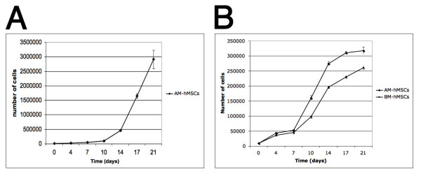
Results: The recovery of hMSCs and their in vitro expansion potential were greater in amniotic membrane than in bone marrow stroma. At flow cytometry analysis AM-hMSCs showed an immunophenotypical profile, i.e., positive for CD105, CD73, CD29, CD44, CD166 and negative for CD14, CD34, CD45, consistent with that reported for bone marrow-derived MSCs. In addition, amniotic membrane-isolated cells underwent in vitro osteogenic (von Kossa stain), adipogenic (Oil Red-O stain), chondrogenic (collagen type II immunohistochemichal detection) and myogenic (RT-PCR MyoD and Myogenin expression as well as desmin immunohistochemical detection) differentiation. In angiogenic experiments, a spontaneous differentiation into endothelial cells was detected by in vitro matrigel assay and this behaviour has been enhanced through Vascular Endothelial Growth Factor (VEGF) induction. According to these findings, VEGF receptor 1 and 2 (FLT-1 and KDR) were basally expressed in AM-hMSCs and the expression of endothelial-specific markers like FLT-1 KDR, ICAM-1 increased after exposure to VEGF together with the occurrence of CD34 and von Willebrand Factor positive cells.

Conclusion: The current study suggests that AM-hMSCs may emerge as a remarkable tool for the cell therapy of multiple diseased tissues. AM-hMSCs may potentially assist both bone and cartilage repair, nevertheless, due to their angiogenic potential, they may also pave the way for novel approaches in the development of tissue-engineered vascular grafts which are useful when vascularization of ischemic tissues is required.


Figures

Figure 1
Figure 1
Human amniotic membrane. Amniotic membrane sheet as seen by light microscopy. The sample has been stained with Mallory's stain to highlight the connective tissue elements (stained red) as indicated by the arrows (A). Morphology of AM-hMSCs subconfluent at third passage. Magnification ×40. Arrows indicate mitotic figures (B).
Figure 2
Figure 2
Oct-4 expression. RT-PCR analysis of Oct-4 expression in 5th passage AM-hMSCs and BM-hMSCs. Samples are as follows: M: Marker; P: Positive control (HeLa cells); BM: BM-hMSCs; AM: AM-hMSCs; N: Negative control (reagent control). Beta-actin was used as a house-keeping gene. Amplicon lengths: Beta actin 236 bp; Oct-4 249 bp. The data are representative of a set of at least three experiments.
Figure 3
Figure 3
hMSCs Proliferation assay. In vitro expansion of AM-hMSC. Passage three AM-hMSCs were seeded at an initial concentration of 1000 cells/cm2 (time 0). At day 4, cells were harvested, counted with a hemocytometer and then re-plated at sub-confluent density. The same procedure was repeated at days 7, 10, 14, 17 e 21 (A). Comparison between growth kinetics of amniotic membrane derived cells and bone marrow derived cells. The figure shows the mean numbers of hMSCs obtained by hemocytometer counting on days 0, 4, 7, 10, 14, 17, 21. At t = 0 1000 cells per cm2 were seeded in 6 well plates. Triplicate cultures were harvested for each point. The values represent the mean and SD of three separate experiments (B).
Figure 4
Figure 4
Immunophenotypical characterization of amniotic membrane derived cells. Cells at the 5th culture passage were trypsinized, labelled with antibodies against the antigens indicated and analysed by flow cytometry. AM-hMSCs expressed SH2, SH3, SH4, CD29, CD44, CD166, while CD45, CD34, CD14 were negative. A representative example of 5 amniotic membrane samples is shown.
Figure 5
Figure 5
AM-hMSCs multi-lineage differentiations in vitro: chondrogenic (A), osteogenic (B), adipogenic (C-H) commitments. Chondrogenic differentiation revealed by immunohistochemical stain for collagen II in induced AM-hMSCs. Original magnification ×40 (A). Osteogenic differentiation evidenced by the formation of mineralized matrix as shown by von Kossa staining. Original magnification ×10 (B).Small colonies with lipid secretion during the first week of adipogenic induction as highlighted by Red Oil staining for neutral lipids. Magnification ×4 (C); at higher magnification, multivacuolar adipogenic cells. Magnification ×10 and ×25 respectively (D, E). Big aggregates with intensive and massive lipid secretion at the third week of adipogenic induction. Magnification ×10, ×10 and ×40 respectively (F, G, H).
Figure 6
Figure 6
Skeletal muscle differentiation of AM-hMSCs. RT-PCR for skeletal muscle transcription factors MyoD and Myogenin. MyoD appears after 1 week of induction while Myogenin is expressed in the second week of induction. Samples are as follows: lane 1: AM-hMSCs cultured in control medium; lane 2: induced AM-hMSCs after 7 days; lane 3: induced AM-hMSCs after 14 days; lane 4: positive control (RD18 cell line); lane 5: reagent control. Beta-actin was used as a house-keeping gene (A). Immunocytochemical staining for Desmin after 3 weeks' induction: uninduced AM-hMSCs (B) and Desmin positive induced cells (C).
Figure 7
Figure 7
Angiogenic commitment: light microscopic analysis of AM-hMSCs after incubation on Matrigel. (A) Spontaneous organization in capillary-like structures on semisolid medium after 2 (A1), 4 (A2) and 20 (A3) hours of incubation. (B) Increased capillary-like structure formation in AM-hMSCs cultured in angiogenic medium supplemented by VEGF 50 ng/mL after 2 (B1), 4 (B2) and 20 (B3) hours' incubation. Within 4 hours of incubation on semisolid medium, cells preserve a round shape and homogeneous distribution (A1 and B1).
Figure 8
Figure 8
Endothelial specific markers after AM-hMSCs angiogenic differentiation. Flow cytometry analysis for FLT-1, KDR, CD34, ICAM-1, vWF expression in AM-hMSCs cultured in absence and in presence of VEGF (50 ng/ml for 7 days). Uninduced cells in black.
Figure 9
Figure 9
Immunofluorescence detection of vWF, FLT-1 and KDR expression. AM-hMSCs cultured in standard medium for 7 days did not show vWF expression (A, Magnification ×40). AM-hMSCs stimulated with medium containing 50 ng/ml of VEGF for 7 days showed vWF expression (B, Magnification ×40); the immunostaining revealed a cytoplasmatic granular positivity at higher magnification (C, Magnification ×100). FLT-1 and KDR expression in induced AM-hMSCs (D, E, Magnification ×40). Nuclei are stained with DAPI (blue).

References

    1. Prockop DJ. Marrow Stromal cells as stem cells for non hematopoietic tissues. Science. 1997;276:71–74. doi: 10.1126/science.276.5309.71. - DOI - PubMed
    1. Pittenger MF, Mackay AM, Beck SC, Jaiswal RK, Douglas R, Mosca JD, Moorman MA, Simonetti DW, Craig S, Marshak DR. Multilineage potential of adult human mesenchymal stem cells. Science. 1999;284:143–147. doi: 10.1126/science.284.5411.143. - DOI - PubMed
    1. Baksh D, Song L, Tuan RS. Adult mesenchymal stem cells: characterization, differentiation, and application in cell and gene therapy. J Cell Mol Med. 2004;8:301–316. - PMC - PubMed
    1. Oswald J, Boxberger S, Jorgensen B, Feldmann S, Ehninger G, Bornhauser M, Werner C. Mesenchymal stem cells can be differentiated into endothelial cells in vitro. Stem Cells. 2004;22:377–384. doi: 10.1634/stemcells.22-3-377. - DOI - PubMed
    1. Heil M, Eitenmuller I, Schmitz-Rixen T, Schaper W. Arteriogenesis versus angiogenesis: similarities and differences. J Cell Mol Med. 2006;10:45–55. - PMC - PubMed

Publication types