Comparative Study
. 2007 May;81(9):4732-43.
doi: 10.1128/JVI.02476-06. Epub 2007 Feb 21.

Live covisualization of competing adeno-associated virus and herpes simplex virus type 1 DNA replication: molecular mechanisms of interaction

Affiliations
Comparative Study

Live covisualization of competing adeno-associated virus and herpes simplex virus type 1 DNA replication: molecular mechanisms of interaction

Daniel L Glauser et al. J Virol. 2007 May.

Abstract

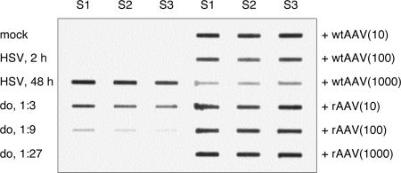
We performed live cell visualization assays to directly assess the interaction between competing adeno-associated virus (AAV) and herpes simplex virus type 1 (HSV-1) DNA replication. Our studies reveal the formation of separate AAV and HSV-1 replication compartments and the inhibition of HSV-1 replication compartment formation in the presence of AAV. AAV Rep is recruited into AAV replication compartments but not into those of HSV-1, while the single-stranded DNA-binding protein HSV-1 ICP8 is recruited into both AAV and HSV-1 replication compartments, although with differential staining patterns. Slot blot analysis of coinfected cells revealed a dose-dependent inhibition of HSV-1 DNA replication by wild-type AAV but not by rep-negative recombinant AAV. Consistent with this, Western blot analysis indicated that wild-type AAV affects the levels of the HSV-1 immediate-early protein ICP4 and the early protein ICP8 only modestly but strongly inhibits the accumulation of the late proteins VP16 and gC. Furthermore, we demonstrate that the presence of Rep in the absence of AAV DNA replication is sufficient for the inhibition of HSV-1. In particular, Rep68/78 proteins severely inhibit the formation of mature HSV-1 replication compartments and lead to the accumulation of ICP8 at sites of cellular DNA synthesis, a phenomenon previously observed in the presence of viral polymerase inhibitors. Taken together, our results suggest that AAV and HSV-1 replicate in separate compartments and that AAV Rep inhibits HSV-1 at the level of DNA replication.


Figures

FIG. 1.
FIG. 1.
Schematic representation of live covisualization of competing viral replication origins. Replication of an HSV-1 amplicon vector is visualized with plasmid pHSV-tetO, which contains the HSV-1 oriS, the HSV-1 DNA packaging/cleavage signal (pac), and five reiterations of the seven-copy teto sequence, comprising a total of 35 TetR binding sites. In the presence of HSV-1 replication factors, the accumulation of concatemeric HSV-1 amplicon replication products (HSV Cc.) is visualized by binding of an EYFP-TetR or ECFP-TetR fusion protein. Visualization of rAAV replication, which employs interactions of an mRFP-LacI or EYFP-LacI fusion protein with laco repeats present in an rAAV genome (rAAVlacO), has been described previously (13). Double-stranded monomeric (AAV ITRm) and dimeric (AAV ITRd) rAAV replication intermediates are indicated.
FIG. 2.
FIG. 2.
Live covisualization of HSV-1 amplicon replication and HSV-1 ICP4. (A) Nuclear distribution of EYFP-TetR and ECFP-ICP4. Vero cells were cotransfected with pHSV-tetO (a to f), pBstetO (g to i), or pHSVPrPUC (k to m) and pEYFPTetR (a to m). On the following day, the cells were infected with recombinant HSV-1 expressing ECFP fused to ICP4 (vECFP-ICP4) at an MOI of 5 PFU (a to c and g to m) or were mock infected (d to f). Live cells were observed by CLSM at 12 to 16 h p.i., with settings specific for EYFP (EYFP-TetR fusion protein) and ECFP (ECFP-ICP4 fusion protein). The images represent projections through three-dimensional reconstructions of the nuclei. (B) Nuclear distribution of EYFP-TetR and the nucleolus marker ECFP-NHPX. Vero cells were cotransfected with pEYFPTetR and pALZ14ECFP-NHPX. On the following day, live cells were observed as described for panel A, using settings specific for EYFP (EYFP-TetR fusion protein) and ECFP (ECFP-NHPX fusion protein).
FIG. 3.
FIG. 3.
Live covisualization of HSV-1 amplicon and rAAV DNA replication. (A) Independent formation of HSV-1 amplicon and rAAV RCs. HeLa cells were cotransfected with pHSV-tetO, pAAVlacO, pEYFPTetR, pCMVmRFPLacI, and pRep. On the following day, the cells were infected with HSV-1 at an MOI of 5 PFU. Live cells treated with 1 μg/ml Hoechst 33342 were observed by CLSM at 12 to 16 h p.i., with settings specific for EYFP (pHSV-tetO RCs), mRFP (rAAVlacO RCs), and Hoechst. The images represent single z stacks of the nuclei. (B) Formation of HSV-1 amplicon and rAAV RCs and nuclear distribution of AAV Rep. HeLa cells were cotransfected with pHSV-tetO, pAAVlacO, pECFP-TetR, pSV2-EYFP/lacI, and p5CR. On the following day, the cells were infected with HSV-1 at an MOI of 1 PFU. Live cells were observed by CLSM at 48 to 72 h p.i., with settings specific for ECFP (pHSV-tetO RCs), EYFP (rAAVlacO RCs), and mCherry (mCherry-Rep68/78 fusion proteins). The images represent projections through three-dimensional reconstructions of the nuclei. (C) Three-dimensional views of the nucleus shown in panel B. pHSV-tetO RCs are stained blue, rAAVlacO RCs are stained green, and mCherry-Rep68/78 proteins are stained red. Deconvolved three-dimensional reconstructions of the nucleus were processed in Imaris software, using the surpass view mode.
FIG. 4.
FIG. 4.
rAAV DNA replication and nuclear distribution of HSV-1 ICP8 and ICP4. (A) Covisualization of rAAV DNA and ICP8 protein. Vero cells were cotransfected with pAAVlacO, pSV2-EYFP/lacI, and pRep. On the following day, the cells were infected with HSV-1 at an MOI of 5 PFU. At 14 h p.i., the cells were fixed and stained with anti-ICP8 MAb 7381 and an AF594-conjugated secondary antibody, as well as DAPI. The cells were then observed by CLSM, with settings specific for EYFP (rAAVlacO RCs), AF594 (ICP8), and DAPI. Images represent single z stacks of the nuclei. (B) Live covisualization of rAAV DNA and ICP8. Vero cells were cotransfected with pAAVlacO, pSV2-EYFP/lacI, pCMVUL29-mRFP, and pRep. On the following day, the cells were infected with HSV-1 at an MOI of 5 PFU. Live cells treated with 1 μg/ml Hoechst 33342 were observed by CLSM at 12 to 16 h p.i., with settings specific for EYFP (rAAVlacO RCs), mRFP (ICP8-mRFP fusion protein), and Hoechst. Images represent a single z stack of a nucleus. (C) Covisualization of rAAV DNA and ICP4. Vero cells were transfected, infected, stained, and observed as described for panel A, except that a MAb specific for ICP4 was used. (D) Live covisualization of rAAV DNA and ICP4. Vero cells were cotransfected with pAAVlacO, pCMVmRFPLacI, and pRep. On the following day, the cells were infected with recombinant HSV-1 expressing EYFP fused to ICP4 (vEYFP-ICP4) at an MOI of 5 PFU. Live cells were observed as described for panel B, with settings specific for mRFP (rAAVlacO RCs), EYFP (EYFP-ICP4 fusion protein), and Hoechst.
FIG. 5.
FIG. 5.
Influence of AAV2 coinfection on HSV-1 DNA replication. Triplicate wells of HeLa cells were mock infected, infected with HSV-1 (MOI, 1 PFU), coinfected with HSV-1 (MOI, 1 PFU) and wt AAV2 (MOI, 10, 100, or 1,000 IU), or coinfected with HSV-1 (MOI, 1 PFU) and rAAV2-GFP vector (MOI, 10, 100, or 1,000 TU). At 48 h p.i., the cells were harvested, and HSV-1 DNA was detected by slot blotting and hybridization with a probe specific for HSV-1 UL35. Threefold serial dilutions (do) of HSV-1 replication products in the absence of AAV infection were used for quantification of HSV-1 replication products in the presence of AAV coinfection. S1 to S3, sample numbers for triplicate samples; HSV, HSV-1 infected; +wtAAV, coinfected with HSV-1 and wt AAV2; +rAAV, coinfected with HSV-1 and rAAV2-GFP. The numbers in parentheses indicate the MOIs of AAV2 and rAAV2-GFP in IU and TU, respectively.
FIG. 6.
FIG. 6.
Influence of AAV2 coinfection on HSV-1 protein levels. HeLa cells were mock infected, infected with HSV-1 (MOI, 1 PFU), coinfected with HSV-1 (MOI, 1 PFU) and increasing amounts of wt AAV2 (MOI, 10, 100, and 1,000 IU), or coinfected with HSV-1 (MOI, 1 PFU) and increasing amounts of rAAV2-GFP vector (MOI, 10, 100, and 1,000 TU). At 48 h p.i., the cells were analyzed by SDS-polyacrylamide gel electrophoresis and Western blotting with antibodies specific for (A) HSV-1 ICP4, HSV-1 ICP8, HSV-1 VP16, and HSV-1 gC or (B) AAV Rep and GFP. Detection of actin served as a loading control. A sample of HSV-1-infected cells were lysed after 2 h of absorption, representing the input HSV-1 virus. In panel A, the relative signal intensities (normalized to that for actin) are indicated as percentages. The signal intensity of mock-infected cells was set to 0%, while that of cells infected with HSV-1 alone was set to 100%. M, mock infected; 2 h, HSV-1 infected and harvested after 2 h of absorption; wtAAV, wt AAV2 infected; rAAV, rAAV2-GFP infected.
FIG. 7.
FIG. 7.
Influence of AAV Rep68/78 proteins on HSV-1 RC formation. (A) Vero cells were transfected with plasmid pCMVrep68/78_AL, encoding Rep68/78 under the control of the CMV promoter, or plasmid pEGFP-N3, containing the same vector backbone and encoding EGFP instead of Rep68/78. On the following day, the cells were infected with HSV-1 at an MOI of 10 PFU. Cells were fixed at 0, 4, 8, and 12 h p.i. and stained with anti-ICP8 MAb 7381 and an AF594-conjugated secondary antibody (a to k; red). The cells in panels f to k were also stained with a rabbit serum specific for Rep and a FITC-conjugated secondary antibody (f to k; green). pEGFP-N3-tranfected cells were identified by EGFP fluorescence (a to e; green). Cells were observed by epifluorescence microscopy, and stages of HSV-1 replication were assessed according to the ICP8 staining pattern, as previously described (5, 27). The numbers indicate the proportions of cells in the respective stages at 12 h p.i. and are means ± standard deviations for triplicate experiments. (B) Time course of HSV-1 RC formation in transfected cells expressing EGFP or Rep68/78. The bars show the proportions of cells in the respective stages and represent the mean values for triplicate experiments.
FIG. 8.
FIG. 8.
Rep-induced numerous ICP8 foci are sites of active DNA synthesis. Vero cells were transfected with plasmid pCMVrep-ECFP-N3, encoding a Rep-ECFP fusion protein (a to d), or left untransfected (e to h). On the following day, the cells were infected with HSV-1 at an MOI of 10 PFU in the absence (a to d) or presence (e to h) of 400 μg/ml PAA. The cells were pulse labeled with 1 mM BrdU for 30 min before fixation at 12 h p.i. The cells were then stained with the rabbit anti-HSV-1 ICP8 serum 4-83 and a FITC-conjugated secondary antibody as well as with an anti-BrdU MAb and an AF594-conjugated secondary antibody. Cells were observed by CLSM, with settings specific for ECFP (Rep-ECFP fusion), FITC (ICP8), and AF594 (BrdU). The images represent projections through three-dimensional reconstructions of the nuclei.

Similar articles

Cited by

References

    1. Bantel-Schaal, U., and H. zur Hausen. 1988. Adeno-associated viruses inhibit SV40 DNA amplification and replication of herpes simplex virus in SV40-transformed hamster cells. Virology 164:64-74. - PubMed
    1. Beard, P., S. Faber, K. W. Wilcox, and L. I. Pizer. 1986. Herpes simplex virus immediate early infected-cell polypeptide 4 binds to DNA and promotes transcription. Proc. Natl. Acad. Sci. USA 83:4016-4020. - PMC - PubMed
    1. Berthet, C., K. Raj, P. Saudan, and P. Beard. 2005. How adeno-associated virus Rep78 protein arrests cells completely in S phase. Proc. Natl. Acad. Sci. USA 102:13634-13639. - PMC - PubMed
    1. Buller, R. M., J. E. Janik, E. D. Sebring, and J. A. Rose. 1981. Herpes simplex virus types 1 and 2 completely help adenovirus-associated virus replication. J. Virol. 40:241-247. - PMC - PubMed
    1. Burkham, J., D. M. Coen, and S. K. Weller. 1998. ND10 protein PML is recruited to herpes simplex virus type 1 prereplicative sites and replication compartments in the presence of viral DNA polymerase. J. Virol. 72:10100-10107. - PMC - PubMed

Publication types

LinkOut - more resources