Comparative Study
. 2007 May;81(9):4766-75.
doi: 10.1128/JVI.02608-06. Epub 2007 Feb 21.

Primary CD4+ T-cell responses provide both helper and cytotoxic functions during Epstein-Barr virus infection and transformation of fetal cord blood B cells

Affiliations
Comparative Study

Primary CD4+ T-cell responses provide both helper and cytotoxic functions during Epstein-Barr virus infection and transformation of fetal cord blood B cells

Georgina J MacArthur et al. J Virol. 2007 May.

Abstract

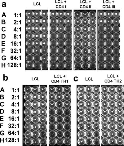
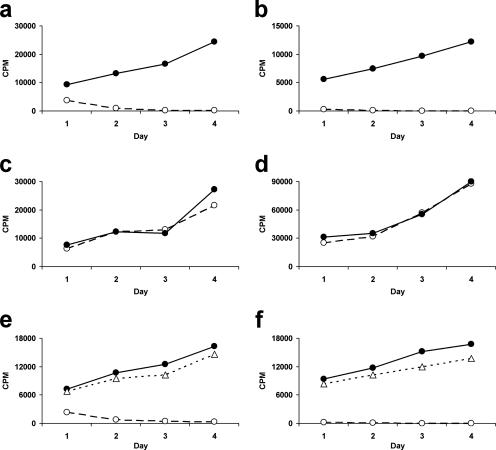
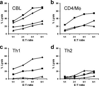
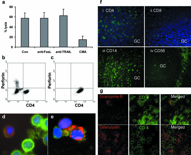
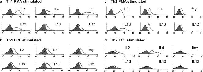
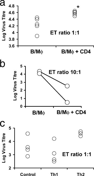
Most humans carry Epstein-Barr virus (EBV) in circulating memory B cells as a latent infection that is controlled by an immune response. When infected by EBV, B lymphocytes in fetal cord blood are readily transformed to lymphoblastoid cell lines (LCL). It is frequently assumed that this high efficiency of transformation is due to the absence of a primary immune response. However, cord blood lymphocytes stimulated with autologous LCL yield CD4+ T cells that can completely inhibit the growth of LCL by a major histocompatibility complex-restricted cytotoxic mechanism mediated by granulysin and granzyme B. Because EBV-transformed B cells maintain the phenotype of antigen-activated B-cell blasts, they can potentially receive inhibitory or helper functions from CD4+ T cells. To assess these functions, the effect of EBV-specific CD4+ T cells on the efficiency of virus transformation of autologous B cells was assayed. Paradoxically, although the cytotoxic CD4+ T-cell lines reduced EBV B-cell transformation at a high effector/target ratio of 10:1, they caused a twofold increase in B-cell transformation at the lower effector/target ratio of 1:1. Th1-polarized CD4+ T cells were more effective at inhibiting B-cell transformation, but Th2-polarized cell lines had reduced cytotoxic activity, were unable to inhibit LCL growth, and caused a 10-fold increase in transformation efficiency. Tonsil lymphoid follicles lacked NK cells and CD8+ T cells but contained CD4+ T cells. We propose that CD4+ T cells provide helper or cytotoxic functions to EBV-transformed B cells and that the balance of these functions within tonsil compartments is critical in establishing asymptomatic primary EBV infection and maintaining a stable lifelong latent infection.


Figures

FIG. 1.
FIG. 1.
Inhibition of LCL outgrowth by coculture with anti-EBV CD4+ T-cell lines. (a) (LCL) Doubling dilutions of LCL seeded in triplicate U-bottomed wells beginning at 104 cells/well in row A and decreasing to 78 cells per well in row H. The photograph on day 21 of culture shows distinct pellets of growing cells in rows A to F. LCL in rows E to H seeded with fewer cells (<624/well) grew poorly, with pellets of viable cells present in only some of the wells. (LCL + CD4 i) Triplicate wells seeded with identical dilutions of LCL and 104 CD4+ T cells/well of a line derived from unfractionated CBL stimulated with LCL as described in Materials and Methods, giving an E:T ratio from 1:1 in row A to 128:1 in row H, as indicated on the left-hand side. After 21 days, visible pellets of LCL were seen only in rows A and B; the remaining rows had no live cells, indicating complete inhibition of LCL growth. (LCL + CD4 ii) Wells seeded with LCL and a T-cell line derived from purified CD4+ CBL stimulated with LCL. No inhibition of LCL growth is seen. (LCL + CD4 iii) Wells seeded with LCL and T cells derived from purified CD4+ CBL stimulated with LCL and CD14+ monocytes. Inhibition of LCL growth is seen at E:T ratios of 2:1 or greater. (b) (LCL) Triplicate control LCL and LCL CD4+ T-cell cocultures set up as for panel a. (LCL + CD4 TH1) CD4+ T-cell lines derived from unfractionated CBL polarized toward a Th1 phenotype by the addition of IFN-γ and neutralizing antibody to IL-4. LCL growth is inhibited at E:T ratios greater that 4:1. (c) (LCL) Triplicate control LCL and LCL CD4+ T-cell cocultures set up as for panel a. (LCL + CD4 TH2) Growth of LCL cultured in the presence of CD4+ T cells polarized to a Th2 phenotype by the addition of IL-4 and neutralizing antibody to IFN-γ was not inhibited. All live cells at the conclusion of the experiments were confirmed to be LCL by flow cytometry.
FIG. 2.
FIG. 2.
Inhibition of LCL proliferation by CBL-derived CD4+ T-cell lines over the short term. LCL were cultured either alone (•) or in the presence of CD4+ T cells (○) at an E:T ratio of 16:1. LCL were seeded at 5 × 10³/ml in 2-ml wells of 24-well tissue culture plates. Proliferation was measured by tritiated thymidine incorporation over a 24-h period in triplicate 100-μl aliquots of the cultures taken daily from days 1 to 4. Data represent mean values of triplicates for representative CD4+ T-cell lines. (a) Inhibition of LCL growth by CBL-derived CD4+ T-cell lines. (b) Inhibition of LCL growth by Th1-polarized CD4+ cell lines. (c) No inhibition of LCL growth by Th2-polarized CD4+ cell lines. (d) No inhibition of nonautologous LCL by CBL-derived CD4+ T-cell lines. (e) Inhibition of LCL growth is contact dependent. LCL were cultured alone (•), in contact with CBL-derived CD4+ T-cell lines (○), or separately from the CD4+ T cells by a Transwell (▵). (f) T-cell-derived soluble factors do not contribute to inhibition. Cultures were set up as for panel e, but additional LCL were placed in the Transwell to stimulate cytokine production by the CD4+ T cells.
FIG. 3.
FIG. 3.
Cytotoxicity of CD4+ T-cell lines toward autologous LCL. CD4+ T-cell-mediated cytotoxicity was assessed at E:T ratios between 1:1 and 16:1 by 51Cr release assays. LCL were labeled with 100 μCi of chromium for 1 h, seeded at 104/well in U-bottomed 96-well plates, and cultured with CD4+ T-cell lines over a 16-h period. Data represent specific lysis of autologous LCL by (a) CD4+ T-cell lines derived from unfractionated CBL by stimulation with autologous LCL, (b) T-cell lines derived from purified CBL CD4+ T cells and CD14+ monocytes stimulated with autologous LCL, (c) CD4+ T-cell lines derived from unfractionated CBL stimulated with autologous LCL and polarized toward a Th1 phenotype by the addition of IFN-γ and neutralizing antibody to IL-4, and (d) CD4+ T-cell lines derived from unfractionated CBL and polarized toward a Th2 phenotype by the addition of IL-4 and neutralizing antibody to IFN-γ. Different graph symbols are used simply to discriminate between different lines in the same panel. Mφ, macrophage.
FIG. 4.
FIG. 4.
Mechanism of CD4+ T-cell cytotoxicity. (a) CD4+ T-cell-mediated cytotoxicity measured by a 16-h chromium release assay at an E:T ratio of 16:1. Killing was not inhibited by neutralizing antibodies to Fas ligand or TRAIL but was markedly reduced by the addition of the granule-disrupting antibiotic CMA. Con, control. Means ± standard errors of the means for six CD4+ T-cell lines. (b and c) Flow cytometry of formalin-fixed cells, dual stained for CD4+ and intracellular perforin, showed the absence of perforin in both (b) freshly isolated resting cord blood CD4+ T cells and (c) cytotoxic CD4+ T-cell lines. (d) Acetone-fixed cytospin of cytotoxic CD4+ T-cell lines stained with CD4 (FITC) and granzyme B (TRITC). In flow cytometry using a log scale, the intensity of CD4 on the cell lines varies by nearly an order of magnitude (c). By comparison, in the linear scaling of fluorescence microscopy, these cells show the same range from very bright CD4 to dim CD4. Dual staining with granzyme B TRITC showed that a minority of CD4+ T cells contained granzyme B, and this was mainly in CD4 dim cells (d). (e) Dual staining of a cytotoxic CD4+ T-cell line with CD4 FITC and granulysin Texas Red, showing that a proportion of CD4+ T cells also contain granulysin. (f) Acetone-fixed frozen sections of adult tonsil stained with (i) anti-CD4, (ii) anti-CD8, (iii) anti-CD14, and (iv) anti-CD56. Sections i and ii are counterstained with DAPI (4′,6′-diamidino-2-phenylindole) to highlight the nuclei. The germinal center is marked GC. (g) Dual staining of interfollicular T-cell areas of the tonsil sections with anti-CD4 FITC and anti-granzyme B TRITC or anti-granulysin Texas Red. Scattered CD4+ T cells that stained for granzyme B were detected; a greater number of granulysin-positive cells were seen, but the majority of CD4+ T cells were negative.
FIG. 5.
FIG. 5.
Flow cytometric determination of intracellular cytokine profiles of CD4+ T-cell lines following stimulation with PMA and ionomycin or autologous LCL. CD4+ T cells were cultured in the presence of PMA and ionomycin or autologous LCL for 6 h and in the presence of monensin for the last 5 h. CD4+ T cells were subsequently fixed, made permeable, stained, and analyzed by flow cytometry. A gate was set on live cells according to forward and side scatter properties, and a second gate was set around the CD4+ T cells. Histograms represent staining of the gated CD4+ T cells with an isotype control antibody (shaded area) and staining for IL-2, IL-4, IFN-γ, IL-13, IL-10, and IL-12, depicted by the black line, as labeled above the appropriate histogram.
FIG. 6.
FIG. 6.
Effect of autologous cord blood-derived CD4 T-cell lines on the titer of EBV measured by transformation of B cells. (a) (B/Mφ) Titer of EBV assayed in cultures containing equal numbers of purified B cells and CD14 monocytes from five different samples of cord blood. (B/Mφ + CD4) Titration of the same samples of virus and cord blood cells in the presence of cytotoxic CD4 T cells added at a T-cell/resting B-cell ratio of 1:1. *, the median titer of virus was significantly raised after the addition of cytotoxic CD4 T cells (P = 0.009, Mann-Whitney U test). (b) (B/Mφ) Two further titrations of EBV assayed in cultures containing equal numbers of purified B cells and CD14 monocytes. (B/Mφ + CD4) The addition of cytotoxic CD4 T cells at the higher ratio of 10:1 substantially reduced the transformation efficiency in both cases. (c) (Control) Titer of EBV assayed in cultures containing equal numbers of purified B cells and CD14 monocytes from three different samples of cord blood. (Th1) Titration of the same samples of virus and cord blood cells in the presence of cytotoxic CD4 T cells derived from unfractionated CBL stimulated with LCL polarized to a Th1 phenotype by the addition of IL-12 and anti-IL-4. (Th2) Titration of the same samples of virus and cord blood cells in the presence of cytotoxic CD4 T cells derived from unfractionated CBL stimulated with LCL polarized to a Th2 phenotype by the addition of IL-4 and anti-IL-12. In every case, the addition of Th2 cells substantially raised the transformation efficiency. B, B cell; Mφ, macrophage.

Similar articles

Cited by

References

    1. Babcock, G. J., L. L. Decker, R. B. Freeman, and D. A. Thorley-Lawson. 1999. Epstein-Barr virus-infected resting memory B cells, not proliferating lymphoblasts, accumulate in the peripheral blood of immunosuppressed patients. J. Exp. Med. 190:567-576. - PMC - PubMed
    1. Babcock, G. J., D. Hochberg, and A. D. Thorley-Lawson. 2000. The expression pattern of Epstein-Barr virus latent genes in vivo is dependent upon the differentiation stage of the infected B cell. Immunity 13:497-506. - PubMed
    1. Babcock, G. J., and D. A. Thorley-Lawson. 2000. Tonsillar memory B cells, latently infected with Epstein-Barr virus, express the restricted pattern of latent genes previously found only in Epstein-Barr virus-associated tumors. Proc. Natl. Acad. Sci. USA 97:12250-12255. - PMC - PubMed
    1. Banchereau, J., F. Briere, C. Caux, J. Davoust, S. Lebecque, Y. J. Liu, B. Pulendran, and K. Palucka. 2000. Immunobiology of dendritic cells. Annu. Rev. Immunol. 18:767-811. - PubMed
    1. Bickham, K., and C. Munz. 2003. Contrasting roles of dendritic cells and B cells in the immune control of Epstein-Barr virus. Curr. Top. Microbiol. Immunol. 276:55-76. - PubMed

Publication types

MeSH terms