. 2007 Apr;14(4):391-6.
doi: 10.1128/CVI.00403-06. Epub 2007 Feb 21.

Use of a novel chimeric mouse model with a functionally active human immune system to study human immunodeficiency virus type 1 infection

Affiliations

Use of a novel chimeric mouse model with a functionally active human immune system to study human immunodeficiency virus type 1 infection

Dong Sung An et al. Clin Vaccine Immunol. 2007 Apr.

Abstract

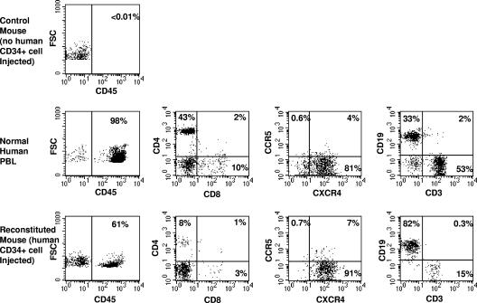
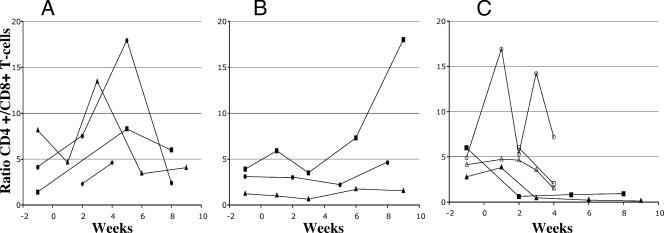
The goal of this study was to develop a small-animal model to study human immunodeficiency virus type 1 (HIV-1) pathogenesis in blood and primary and secondary lymphoid organs. Rag2(-/-)gamma(c)(-/-) mice that are neonatally injected with human CD34(+) cells develop a functional human immune system (HIS), with human hematopoietic cells being found in the thymuses, peripheral blood, spleens, and bone marrow of the animals (hereafter these animals are referred to as HIS-Rag2(-/-)gamma(c)(-/-) mice). HIS-Rag2(-/-)gamma(c)(-/-) mice were infected with small amounts of CCR5-tropic HIV-1. Viral replication and immunophenotypic changes in the human cells in peripheral blood and lymphoid organs were examined. The productive infection of human cells in peripheral blood, thymus and spleen tissue, and bone marrow was detected. Ratios of CD4(+) T cells to CD8(+) T cells in the infected animals declined. Although no specific anti-HIV-1 immune responses were detected, immunoglobulin M (IgM) and IgG antibodies to an unidentified fetal calf serum protein present in the virus preparation were found in the inoculated animals. Thus, we have shown that the HIS-Rag2(-/-)gamma(c)(-/-) mouse model can be used for infection with low doses of CCR5-tropic HIV-1, which is most commonly transmitted during primary infections. HIS-Rag2(-/-)gamma(c)(-/-) mice can serve as a small-animal model for investigating HIV-1 pathogenesis and testing potential HIV-1 therapies, and studies with this model may replace some long and costly studies with nonhuman primates.


Figures

FIG. 1.
FIG. 1.
Reconstitution of human lymphocytes in the Rag2−/−γc−/− mouse. At 22 weeks postinjection with human CD34+ cells, peripheral blood was collected and cells were stained for human CD45, CD4, CD8, CCR5, CXCR4, CD19, and CD3 lymphoid cell surface markers, as indicated, and analyzed by flow cytometry. The control mouse (top panel) was an animal that did not receive human CD34+ cells, and as a result, no human CD45+ cells, including human T and B cells, could be detected in this mouse. Normal human blood was analyzed in parallel for comparison (middle panels). The injection of human CD34+ cells into a neonatal Rag2−/−γc−/− mouse reconstitutes a lymphocyte subset in the peripheral blood with a profile similar to that of human peripheral blood lymphocytes (PBL) when gated on human CD45+ cells (bottom panels). FSC, forward scatter.
FIG. 2.
FIG. 2.
Kinetics of ratios of CD4+ T cells to CD8+ T cells in uninfected and HIV-1-infected mice. Mice were mock injected or injected with heat-inactivated (HI) or 500 or 5,000 TCID50 of HIV-1NFN-SX(SL9). At various time points pre- or postinjection, as indicated, the percentages of CD4+ and CD8+ T cells among human CD45+ lymphocytes were obtained by flow cytometric analysis and the ratio of CD4+ T cells to CD8+ T cells was calculated. Results from four independent experiments are shown. (A) Animals inoculated with heat-inactivated HIV-1 (black circles) and uninfected animals (other symbols). (B) HIV-1-inoculated, nonproductively infected (500 TCID50) animals. (C) HIV-1-inoculated, productively infected animals. Closed symbols, 500 TCID50; open symbols, 5,000 TCID50.
FIG. 3.
FIG. 3.
Western blot analysis using serum from an HIV-1-inoculated HIS-Rag2−/−γc−/− mouse (mouse 5 in Tables 1 and 2) for the assessment of reactive proteins present in media from mock-transfected 293T cells (lane 1) and HIV-1NFN-SX-transfected 293T cells (lane 2), Iscove's medium alone (lane 3), and 10% fetal bovine serum (lanes 4 and 5). Molecular size standards are indicated on the right. Goat anti-human IgM was used as a secondary antibody for lanes 1 to 4, and goat anti-human IgG was used as a secondary antibody for lane 5. Western blotting of sera from mice productively infected with HIV-1 or uninfected mice did not show an antibody response to fetal bovine serum proteins.

References

    1. Aldrovandi, G. M., G. Feuer, L. Gao, B. Jamieson, M. Kristeva, I. S. Y. Chen, and J. A. Zack. 1993. The SCID-hu mouse as a model for HIV-1 infection. Nature 363:732-736. - PubMed
    1. Baenziger, S., R. Tussiwand, E. Schlaepfer, L. Mazzucchelli, M. Heikenwalder, M. O. Kurrer, S. Behnke, J. Frey, A. Oxenius, H. Joller, A. Aguzzi, M. G. Manz, and R. F. Speck. 2006. Disseminated and sustained HIV infection in CD34+ cord blood cell-transplanted Rag2−/−γc−/− mice. Proc. Natl. Acad. Sci. USA 103:15951-15956. - PMC - PubMed
    1. Baumann, J. G., D. Unutmaz, M. D. Miller, S. K. Breun, S. M. Grill, J. Mirro, D. R. Littman, A. Rein, and V. N. KewalRamani. 2004. Murine T cells potently restrict human immunodeficiency virus infection. J. Virol. 78:12537-12547. - PMC - PubMed
    1. Berges, B. K., W. H. Wheat, B. E. Palmer, E. Connick, and R. Akkina. 2006. HIV-1 infection and CD4 T cell depletion in the humanized Rag2−/−gamma c−/− (RAG-hu) mouse model. Retrovirology 3:76. - PMC - PubMed
    1. Bonyhadi, M. L., L. Rabin, S. Salimi, D. A. Brown, J. Kosek, J. M. McCune, and H. Kaneshima. 1993. HIV induces thymus depletion in vivo. Nature 363:728-732. - PubMed

Publication types