. 2007 Feb 21;27(8):1933-41.
doi: 10.1523/JNEUROSCI.5471-06.2007.

Apolipoprotein E-containing lipoproteins protect neurons from apoptosis via a signaling pathway involving low-density lipoprotein receptor-related protein-1

Affiliations

Apolipoprotein E-containing lipoproteins protect neurons from apoptosis via a signaling pathway involving low-density lipoprotein receptor-related protein-1

Hideki Hayashi et al. J Neurosci. .

Abstract

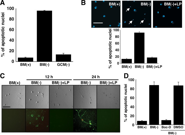
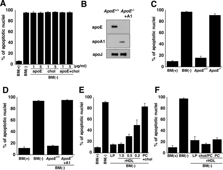
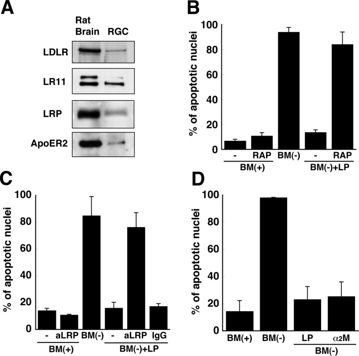
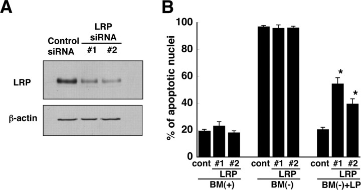
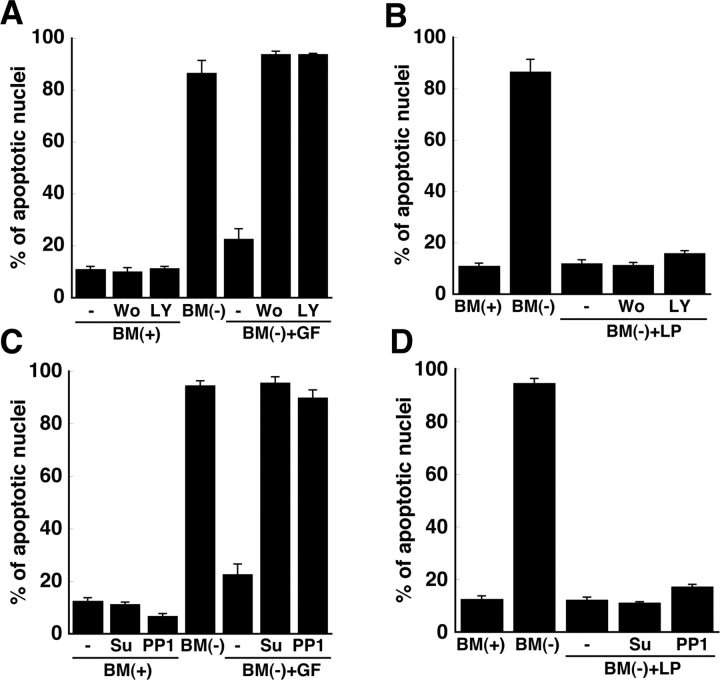
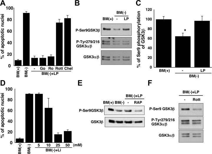
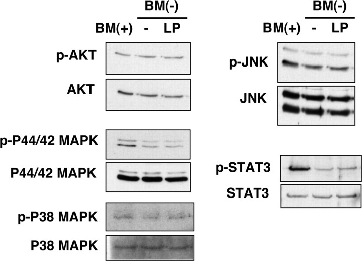
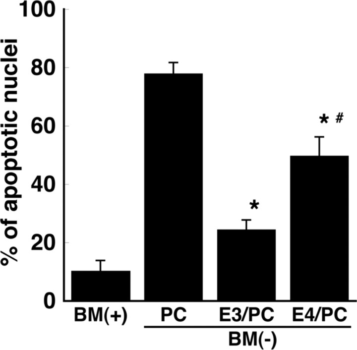
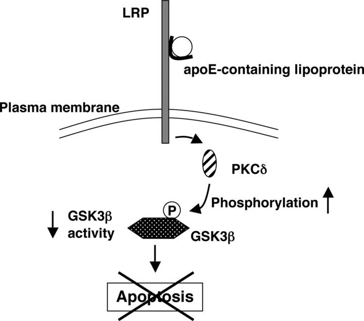
Apolipoprotein E (apoE)-containing lipoproteins (LPs) are secreted by glia and play important roles in lipid homeostasis in the CNS. Glia-derived LPs also promote synaptogenesis and stimulate axon growth of CNS neurons. Here, we provide evidence that glia-derived LPs protect CNS neurons from apoptosis by a receptor-mediated signaling pathway. The protective effect was greater for apolipoprotein E3 than for apolipoprotein E4, the expression of which is a risk factor for Alzheimer's disease. The anti-apoptotic effect of LPs required the association of apolipoprotein E with lipids but did not require cholesterol. Apoptosis was not prevented by lipids alone or by apoA1- or apoJ-containing lipoproteins. The prevention of neuronal apoptosis was initiated after the binding of LPs to the low-density lipoprotein receptor-related protein (LRP), a multifunctional receptor of the low-density lipoprotein receptor family. We showed that inhibition of LRP activation, by treatment of neurons with receptor-associated protein or anti-LRP antibodies, or by LRP gene-silencing experiments, reduced the protective effect of LPs. Furthermore, another LRP ligand, alpha2-macroglobulin, also protected the neurons from apoptosis. After binding to LRP, LPs initiate a signaling pathway that involves activation of protein kinase Cdelta and inactivation of glycogen synthase kinase-3beta. These findings indicate the potential for using glial lipoproteins or an activator of the LRP signaling pathway for treatment for neurodegenerative disorders such as Alzheimer's disease.


Figures

Figure 1.
Figure 1.
ApoE-containing glial lipoproteins prevent neuronal apoptosis induced by withdrawal of trophic additives. A, B, Fragmented/shrunken nuclei were detected by Hoechst staining of RGCs after incubation for 24 h in BM(+)] or BM(−). Glia-conditioned medium without trophic additives [GCM(−)] or basal medium without trophic additives but with apoE-containing LPs was added to RGCs. The final cholesterol concentration of LPs in the media was 1 μg/ml. The number of apoptotic neurons was quantified as a percentage of the total number of neurons. Data are means ± SE from five independent experiments. B, Typical images of Hoechst-stained RGCs are shown. Typical apoptotic nuclei are indicated by arrows. C, Shown are phase-contrast (top) and fluorescence (bottom) images of RGCs stained with annexin V–fluorescein (green) and propidium iodide (red) after incubation for 12 and 24 h under the same conditions as for B. Representative images are shown from one of three independent experiments with similar results. Scale bars: B, C, 50 μm. D, Apoptosis induced by withdrawal of trophic additives is inhibited by a broad-spectrum caspase inhibitor. RGCs were incubated for 24 h in BM(+) or BM(−). The medium added to some cultures contained the broad-spectrum caspase inhibitor Boc-D fmk (Boc-D) (100 μm) dissolved in dimethylsulfoxide. Medium provided to control cells (DMSO) contained an equivalent amount (2 μl/ml) of dimethylsulfoxide without inhibitor. Apoptotic neurons were detected by Hoechst staining. Data are means ± SE from four independent experiments.
Figure 2.
Figure 2.
ApoE-containing lipoproteins, but not apoE alone, cholesterol alone, or apoA1/apoJ-containing lipoproteins, prevent apoptosis of RGCs. A, Recombinant human apoE3 and/or cholesterol (5 and 1 μg/ml, respectively) were added to BM(−) and incubated with RGCs for 24 h. Control cultures were given BM(−) or BM(+). The number of apoptotic neurons as a percentage of all neurons was determined by Hoechst staining. B, Immunoblot of apoE, apoA1, and apoJ in lipoproteins isolated by ultracentrifugation from cultures of ApoE +/+ and ApoE −/− glia. ApoA1-containing lipoproteins (+A1) were generated by addition of human apoA1 to glia isolated from ApoE −/− mice. C, Apoptosis was assessed in RGCs incubated in BM(−) supplemented with lipoproteins (120 μg of protein/ml) isolated from glial cultures from ApoE +/+ and ApoE −/− mice. D, BM(−) was supplemented with apoE-containing lipoproteins (ApoE +/+) or apoA1-containing lipoproteins (ApoE −/− + A1) so that the concentration of cholesterol in the medium was 1 μg/ml. E, rHDLs that contained apoE, phosphatidylcholine (PC), and cholesterol (molar ratio, 1:100:10) were prepared. BM(−) was supplemented with rHDLs so that the amount of apoE was the same as (1.0), 50% (0.5), or 20% (0.2) of that in glial LPs. Liposomes lacking apoE, but containing the same amount of phosphatidylcholine and cholesterol as in rHDLs (PC + chol), did not prevent apoptosis. F, RGCs were incubated in BM(−) for 24 h with rHDLs that contained the same amount of apoE as in LPs and either contained (chol/PC) or lacked (PC) cholesterol. For all experiments, fragmented/shrunken nuclei were detected by Hoechst staining and designated as apoptotic cells. Data are means ± SE from three or four independent experiments. chol, Cholesterol.
Figure 3.
Figure 3.
LRP mediates the protective effect of apoE-containing lipoproteins. A, Receptors of the low-density lipoprotein receptor family were detected by immunoblotting of 20 μg of protein from lysates of RGCs. Rat brain proteins (20 μg) were used as a positive control. Data are from one experiment representative of three experiments with similar results. BD, RGCs were incubated for 24 h with BM(+) or BM(−) in the presence of apoE-containing LPs and/or 300 nm RAP (B), 10 μg/ml anti-LRP antibody (aLRP) (C), or 10 μg/ml nonspecific IgG (C), after a 2 h pretreatment with RAP, antibodies, or 100 nm α2-macroglobulin (α2M) (D). The percentage of cells containing apoptotic nuclei was determined by Hoechst staining. Data are means ± SE of at least three independent experiments.
Figure 4.
Figure 4.
Reduction of LRP-1 in RGCs by siRNA. A, After 3 d in culture, RGCs were transfected with 20 pmol of control siRNA or two different LRP-1 siRNAs (LRP #1 and LRP #2). RGCs were harvested 3 d after transfection, and proteins were separated by 0.1% SDS-PAGE. LRP was detected by immunoblotting. Immunoblotting of β-actin was used as a loading control. Data are from one experiment that is representative of three experiments with similar results. B, RGCs were transfected with control siRNA (cont) or two different siRNAs (LRP #1 and #2) that were specific for LRP-1. After 3 d, the neurons were incubated for 24 h with BM(+), BM(−), or BM(−) in the presence of apoE-containing lipoproteins [BM(−) + LP]. The percentage of cells containing apoptotic nuclei was determined by Hoechst staining. Data are means ± SE of four independent experiments. *p < 0.0001 for control versus LRP #1; *p = 0.0003 for control versus LRP #2. Statistical analysis was performed using one-way ANOVA followed by Bonferroni's multiple comparison.
Figure 5.
Figure 5.
Phosphatidylinositol-3-kinase and Src family kinases do not contribute to the anti-apoptotic effect of apoE-containing lipoproteins. Retinal ganglion cells were incubated in BM(+), BM(−), or BM(−) plus apoE-containing lipoproteins [BM(−) + LP] for 24 h. Apoptosis was assessed by Hoechst staining. The medium added to some neuronal cultures contained brain-derived neurotrophic factor (50 ng/ml), ciliary neurotrophic factor (50 ng/ml), and forskolin (10 μm; +GF; A, C), and/or the phosphatidylinositol-3-kinase inhibitors wortmannin (Wo; 100 nm) or LY294002 (LY; 50 μm; A, B) or Src kinase inhibitors PP1 (10 μm) or Su6656 (Su; 10 μm; C, D). Data are means ± SE of three independent experiments.
Figure 6.
Figure 6.
PKCδ and GSK3β contribute to the anti-apoptotic effect of apoE-containing lipoproteins. A, RGCs were incubated with inhibitors of PKC: Gö6976 (Go; 20 nm), Ro32-0432 (Ro; 50 nm), rottlerin (Rott; 2 μm), and chelerythrine (Chel; 1 μm). BM(−) was supplemented with apoE-containing LPs and PKC inhibitors and then supplied to RGCs for 24 h after a 2 h pretreatment of the cells with PKC inhibitors. Apoptosis was assessed by Hoechst staining. Data are means ± SE from three independent experiments. B, Immunoblotting of GSK3α/β with antibodies directed against GSK3β phosphorylated at Ser-9 (P-Ser9GSK3β), GSK3α/β phosphorylated at Tyr-279/216 (P-Tyr279/216 GSK3α/β), or total GSK3α/β protein, from the same experiment. RGCs were incubated in BM(+) or BM(−) with or without apoE-containing lipoproteins. Data are from one experiment representative of six independent experiments with similar results. C, Quantitation of phosphorylation at Ser-9 of GSK3β from immunoblotting experiments shown in B as a percentage of the phosphorylation in the presence of trophic additives. Data are means ± SE from six independent experiments. *p = 0.016 for BM(+) versus BM(−); p = 0.029 for BM(−) versus BM(−) plus LP. Statistical analysis was performed using one-way ANOVA followed by Bonferroni's multiple comparison. D, Apoptosis was assessed by Hoechst staining of neurons incubated for 24 h in BM(+) or BM(−) and the indicated concentrations of lithium chloride (Li), an inhibitor of GSK3. Data are means ± SE from three independent experiments. E, RGCs were incubated as in B, but in the presence or absence of RAP, and then immunoblotted for total GSK3β protein and P-Ser9GSK3β. Data are representative of four independent experiments with similar results. F, Immunoblotting of GSK3α/β, GSK3β phosphorylated at Ser-9, and GSK3α/β phosphorylated at Tyr-279/216 in neurons incubated as in B in BM(−) plus LP in the absence or presence of rottlerin. Data are representative of three independent experiments with similar results.
Figure 7.
Figure 7.
Phosphorylation of signaling molecules in RGCs in response to apoE-containing lipoproteins. Apoptosis was initiated in RGCs by withdrawal of trophic additives [BM(−)] from basal medium [BM(+)]. RGCs were incubated for 9 h in the presence (LP) or absence (−) of apoE-containing lipoproteins and then harvested and analyzed by immunoblotting using antibodies directed against phosphorylated (p) Akt (phosphorylated on Ser-473), P44/42 MAPK (phosphorylated on Thr-202/Tyr-204), P38 MAP kinase (phosphorylated on Thr-180/Tyr-182), STAT3 (phosphorylated on Tyr-705), and JNK (phosphorylated on Thr-183/Tyr-185). Total amounts of the proteins were detected by immunoblotting. Data are from one experiment representative of three experiments with similar results.
Figure 8.
Figure 8.
ApoE4-containing rHDLs are less protective against apoptosis than are apoE3-containing rHDLs. rHDLs containing recombinant human apoE3 or apoE4 were prepared with phosphatidylcholine (PC) (molar ratio, 1:100). The apoE3- (E3/PC) or apoE4 (E4/PC)-containing rHDLs were added to BM(−) and incubated with retinal ganglion cells for 24 h. Control cultures were given BM(+). Liposomes that lacked apoE but contained the same amount of PC as in rHDLs (+PC) were used as a negative control. The number of apoptotic neurons was determined by Hoechst staining as a percentage of the total number of neurons. Data are means ± SE from five independent experiments. *p < 0.005 for BM(−) plus PC versus BM(−) plus E3/PC and BM(−) plus E4/PC. # p = 0.0057 for E3/PC versus E4/PC. Statistical analysis was performed using one-way ANOVA followed by Bonferroni's multiple comparison.
Figure 9.
Figure 9.
Proposed anti-apoptotic signaling pathway induced in RGCs by apoE-containing lipoproteins. When apoptosis is induced in RGCs by removal of trophic additives, apoE-containing lipoproteins secreted by glia protect the RGCs from apoptosis. After binding of apoE-containing lipoproteins to LRP on the cell surface, activation of PKCδ, either directly or indirectly, increases the phosphorylation (P) of GSK3β, thereby inactivating GSK3β and protecting the RGCs from apoptosis.

References

    1. Bacskai BJ, Xia MQ, Strickland DK, Hyman BT. The endocytic receptor protein LRP also mediates neuronal calcium signaling via N-methyl-d-aspartate receptors. Proc Natl Acad Sci USA. 2000;97:11551–11556. - PMC - PubMed
    1. Barres BA, Silverstein BE, Corey DP, Chun LLY. Immunological, morphological, and electrophysiological variation among retinal ganglion cells purified by panning. Neuron. 1988;1:791–803. - PubMed
    1. Bondy CA, Cheng CM. Signaling by insulin-like growth factor 1 in brain. Eur J Pharmacol. 2004;490:25–31. - PubMed
    1. Bright R, Mochly-Rosen D. The role of protein kinase C in cerebral ischemic and reperfusion injury. Stroke. 2005;36:2781–2790. - PubMed
    1. Brown MS, Goldstein JL. Receptor-mediated control of cholesterol metabolism. Science. 1976;191:150–154. - PubMed

Publication types

MeSH terms

LinkOut - more resources