. 2007 Feb 21;27(8):1942-53.
doi: 10.1523/JNEUROSCI.3208-06.2007.

Kv4.3-mediated A-type K+ currents underlie rhythmic activity in hippocampal interneurons

Affiliations

Kv4.3-mediated A-type K+ currents underlie rhythmic activity in hippocampal interneurons

Mathieu L Bourdeau et al. J Neurosci. .

Abstract

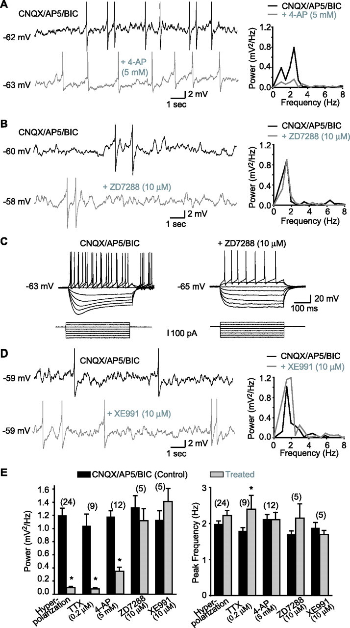
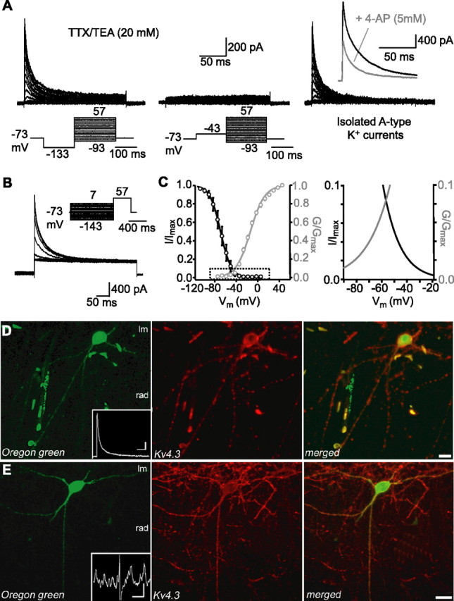
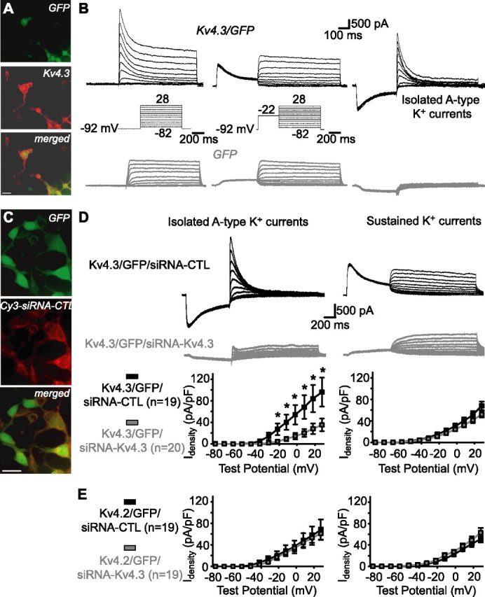
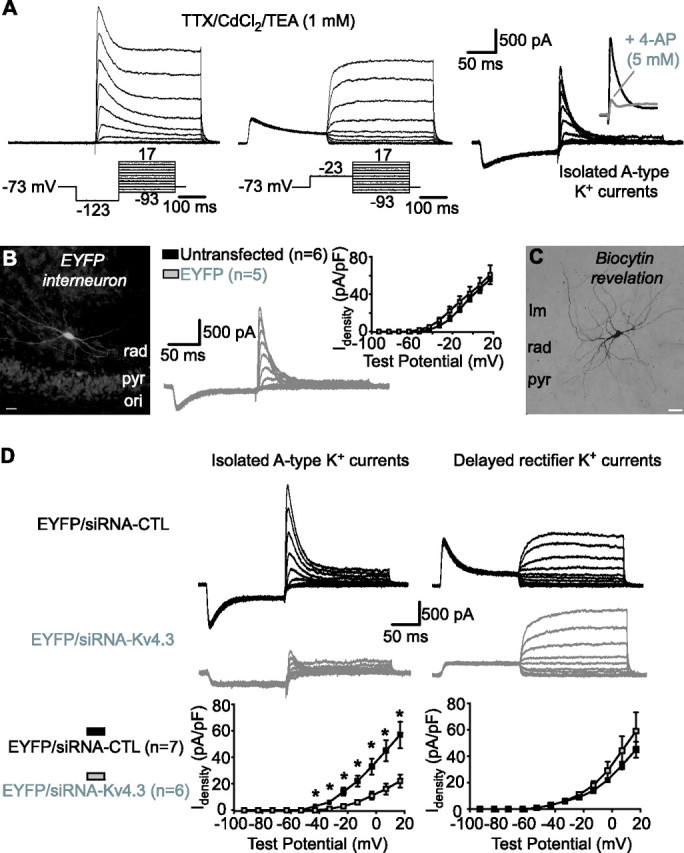
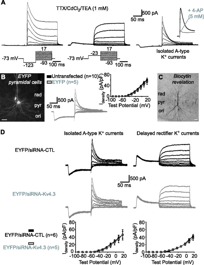
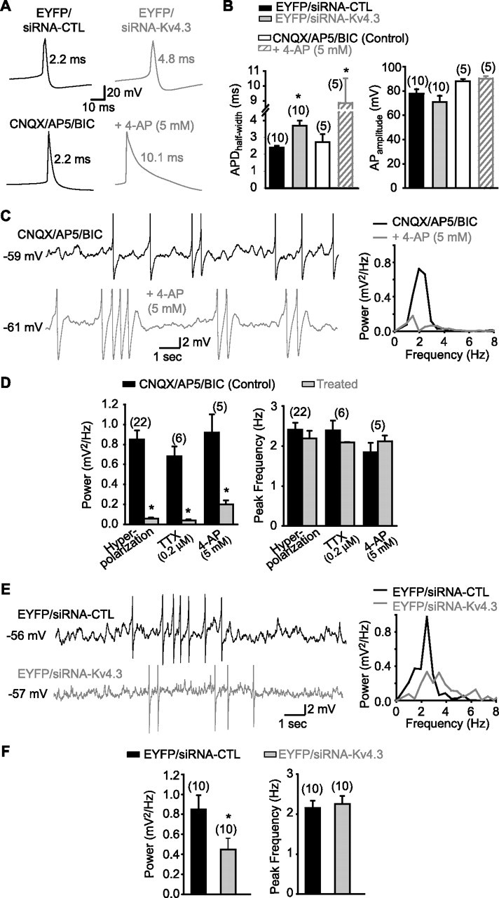
Hippocampal-dependent learning and memory processes are associated with theta frequency rhythmic activity. Interneuron and pyramidal cell network interactions underlie this activity, but contributions of interneuron voltage-gated membrane conductances remain unclear. We show that interneurons at the CA1 lacunosum-moleculare (LM) and radiatum (RAD) junction (LM/RAD) display voltage-dependent subthreshold membrane potential oscillations (MPOs) generated by voltage-gated tetrodotoxin-sensitive Na+ and 4-aminopyridine (4-AP)-sensitive K+ currents. They also exhibit prominent 4-AP-sensitive A-type K+ currents, with gating properties showing activation at subthreshold membrane potentials. We found that LM/RAD cells are part of specific interneuron subpopulations expressing the K+ channel subunit Kv4.3 and their transfection with Kv4.3 small interfering RNA selectively impaired A-type K+ currents and MPOs. Thus, our findings reveal a novel function of Kv4.3-mediated A-type K+ currents in the generation of intrinsic MPOs in specific subpopulations of interneurons that may participate in hippocampal theta-related rhythmic activity.


Figures

Figure 1.
Figure 1.
MPOs in LM/RAD interneurons in acute slices are dependent on 4-AP-sensitive K+ currents but not on Ih or IM. A, MPOs from a representative interneuron (left) recorded at membrane potential near spike threshold in the presence of non-NMDA, NMDA, and GABAA receptor antagonists [CNQX, 20 μm; AP5, 50 μm; bicuculline (BIC), 25 μm; top trace]. In the same cell, MPOs are significantly reduced by application of 4-AP (5 mm; bottom trace). In this and other figures with traces showing MPOs, action potentials are truncated. Power spectra (right) of records from the same cell show the reduction of the power of MPOs by 4-AP. B, Similar data from another cell showing that MPOs recorded near spike threshold are not diminished by ZD7288, a blocker of the h current (Ih). C, Positive control experiments from the same cell as in B showing that the sag in the membrane response elicited by hyperpolarizing current injections and produced by Ih (control in CNQX/AP5/BIC; left) was blocked by ZD7288 (10 μm; right). D, Representative traces from another interneuron showing that MPOs and corresponding power spectra are not reduced by XE991 (10 μm), a selective blocker of muscarine-sensitive K+ current (IM). E, Summary bar graphs showing that the power of MPOs was significantly reduced by membrane hyperpolarization (Vm threshold −10 mV), TTX (0.2 μm), and 4-AP (5 mm) but not by ZD7288 (10 μm) or XE991 (10 μm), whereas the peak frequency of MPOs was generally unchanged. *p < 0.05.
Figure 2.
Figure 2.
Properties of A-type K+ currents and expression of Kv4.3 in LM/RAD interneurons in acute slices. A, K+ currents were recorded from outside-out patches pulled from somata of interneurons in the presence of TTX (0.5 μm) and TEA (20 mm). K+ currents composed of rapidly inactivating and noninactivating components were evoked by test pulses to potentials between −93 and 57 mV (200 ms) from a potential of −133 mV (150 ms) (left), whereas currents composed of only a noninactivating component were evoked from a potential of −43 mV (middle). Digital subtraction resulted in the isolation of A-type K+ currents (right) and inset shows reduction of A-type K+ currents from another interneuron (elicited by a test pulses to 57 mV) by 5 mm 4-AP. B, Inactivation of A-type K+ currents was studied by applying 1 s prepulses between −143 and 7 mV followed by a pulse to 57 mV (400 ms). C, Mean activation (n = 14) and inactivation (n = 10) curves of A-type K+ currents were fitted using a Boltzmann function (left). Boxed area is shown enlarged at the right, illustrating the intersection of activation and inactivation curves and window current near threshold. D, Confocal image of an Oregon green-filled interneuron (left) from which A-type K+ currents were recorded in outside-out patch (inset; calibration: 200 pA, 25 ms). Confocal image shows immunolabeling for Kv4.3 in the same section (middle) and indicates that the protein is found in the soma and dendritic comportments. Merged images (right) show colocalization. E, Example of a different Oregon green-labeled interneuron (left), which displayed MPOs during whole-cell recordings (inset; calibration: 2 mV, 1 s). This interneuron was also immunopositive for Kv4.3 (middle and right images). Scale bars: D, 20 μm; E, 10 μm.
Figure 3.
Figure 3.
Kv4.3-mediated A-type K+ currents and functional knockdown by siRNA in HEK293 cells. A, Fluorescence images showing that GFP-expressing cells cotransfected with Kv4.3 (top) were immunopositive for Kv4.3 (middle and bottom). Scale bar, 25 μm. B, In cells cotransfected with Kv4.3 and GFP (top traces), prominent A-type K+ currents were isolated (right) by subtracting the sustained K+ currents elicited by voltage jumps from a depolarized potential (middle) from the total K+ currents evoked by voltage steps from a hyperpolarized potential (left). A-type K+ currents were absent in cells transfected with GFP only (bottom traces). C, Fluorescence images showing that GFP-expressing cells (top) cotransfected with Kv4.3 and a cyanine3-tagged nontargeting control siRNA (Cy3-siRNA-CTL) colocalized the fluorescently tagged siRNA (middle and bottom). Scale bar, 25 μm. D, Traces from representative cells (top and middle) and summary graphs for all cells (bottom; *p < 0.05) illustrating that cotransfection of Kv4.3, GFP, and a nontargeting control (siRNA-CTL) resulted in large A-type K+ currents, whereas cotransfection of Kv4.3, GFP, and siRNA targeting Kv4.3 (siRNA-Kv4.3) prevented expression of A-type K+ currents (left). In contrast, endogenous sustained K+ currents were not different in the same cells (right), indicating a selective functional knockdown of A-type currents. E, Summary graphs for all cells from control experiments with transfection of Kv4.2, showing that Kv4.2-mediated A-type K+ currents were similar in cells cotransfected with siRNA-CTL or siRNA-Kv4.3 (left). Sustained K+ currents were also similar in both groups (right), indicating a selective functional knockdown of Kv4.3-mediated A-type K+ currents.
Figure 4.
Figure 4.
Kv4.3 siRNA reduces A-type K+ currents in interneurons in slice cultures. A, Whole-cell K+ currents recorded from interneurons in TTX (1 μm), CdCl2 (150 μm), and a low concentration of TEA (1 mm). Total K+ currents activated by test pulses from a hyperpolarized potential consisted of rapidly inactivating and delayed components (left). Delayed rectifier K+ currents evoked by test pulses from a depolarized potential (middle) were subtracted to isolate A-type K+ currents (right). Inset shows sensitivity of A-type K+ currents to 5 mm 4-AP. B, Fluorescence image of an EYFP-expressing LM/RAD interneuron (left). Traces from a representative EYFP-expressing interneuron showing isolated A-type K+ currents are shown in the middle. A summary graph illustrating similar A-type K+ current density in untransfected and EYFP-expressing interneurons is shown at the right. C, Example of biocytin labeling of an EYFP-expressing interneuron showing typical nonpyramidal morphology of LM/RAD interneurons. D, Traces from representative interneurons (top) and summary graphs for all cells (bottom), indicating that transfection of Kv4.3 siRNA selectively reduced A-type K+ current density (left) and did not affect delayed rectifier K+ currents (right), compared with transfection with control siRNA. Scale bars: B, C, 25 μm. *p < 0.05.
Figure 5.
Figure 5.
Kv4.3 siRNA did not affect A-type K+ currents in CA1 pyramidal cells in slice cultures. A, Total whole-cell K+ currents (left) were composed of rapidly inactivating A-type (right) and delayed rectifier (middle) K+ currents in pyramidal cells. A-type K+ currents were inhibited by 4-AP (inset). B, Fluorescence image of EYFP-expressing pyramidal cells (left). Traces from a representative EYFP-expressing pyramidal cell (middle) and summary graph for all cells (right) showing similar A-type K+ current density in untransfected and EYFP-expressing pyramidal cells are shown. C, Example of biocytin labeling of an EYFP-expressing pyramidal cell. D, Traces from representative pyramidal cells (top) and summary graphs (bottom) illustrating that A-type (left) and delayed rectifier (right) K+ current density were unchanged by Kv4.3 compared with control siRNA transfection. Scale bars: B, C, 25 μm.
Figure 6.
Figure 6.
Kv4.3 siRNA broadens action potentials and inhibits MPOs in interneurons in slice cultures. A, Representative traces of spontaneous action potentials in interneurons illustrating that Kv4.3 siRNA (top) and 4-AP (bottom) increase action potential duration. B, Summary bar graphs indicating that APDhalf-width was significantly increased by either Kv4.3 siRNA compared with control siRNA or by 5 mm 4-AP (left), whereas action potential amplitude was unaffected by Kv4.3 siRNA or by 4-AP (right). C, Representative traces (in presence of CNQX, AP5, and bicuculline) and corresponding power spectra for the same cell illustrating MPOs and the inhibitory effect of the K+ channel blocker 4-AP (5 mm). D, Summary bar graphs indicating that the power (left) but not the peak frequency (right) of MPOs is reduced by hyperpolarization, TTX, and 4-AP. E, Representative traces and corresponding power spectra for the same cells showing that MPOs are decreased in interneurons transfected with Kv4.3 siRNA (bottom) compared with interneurons transfected with control siRNA (top). F, Summary bar graphs demonstrating the significant reduction in MPO power (left) and unchanged peak frequency (right) by Kv4.3 siRNA compared with the control siRNA. *p < 0.05.
Figure 7.
Figure 7.
Specific subpopulations of CA1 interneurons coexpressing Kv4.3 and Ca2+-binding proteins or peptides. A, Confocal image of parvalbumin-immunopositive interneurons located in stratum pyramidale (left). Confocal image of immunolabeling for Kv4.3 in the same section (middle) and the merged images (right) indicate that parvalbumin-positive interneurons mostly did not colocalize Kv4.3. B, Calbindin-positive neurons (left) in stratum radiatum and lacunosum-moleculare often colocalized Kv4.3 (middle, right). Arrows point to double-labeled interneurons. C, Somatostatin-positive interneurons (left) in stratum oriens (ori) and alveus (alv) mostly did not colocalize Kv4.3 (middle, right). D, Cholecystokinin-immunopositive interneurons in stratum radiatum and lacunosum-moleculare (left) highly colocalized Kv4.3 (middle, right). Scale bars, 15 μm.

References

    1. Alonso A, Llinas RR. Subthreshold Na+-dependent theta-like rhythmicity in stellate cells of entorhinal cortex layer II. Nature. 1989;342:175–177. - PubMed
    1. Alonso A, Llinas RR. Electrophysiology of the mammillary complex in vitro. II. Medial mammillary neurons. J Neurophysiol. 1992;68:1321–1331. - PubMed
    1. Barry DM, Xu H, Schuessler RB, Nerbonne JM. Functional knockout of the transient outward current, long-QT syndrome, and cardiac remodeling in mice expressing a dominant-negative Kv4 alpha subunit. Circ Res. 1998;83:560–567. - PubMed
    1. Birnbaum SG, Varga AW, Yuan LL, Anderson AE, Sweatt JD, Schrader LA. Structure and function of Kv4-family transient potassium channels. Physiol Rev. 2004;84:803–833. - PubMed
    1. Bland BH. The physiology and pharmacology of hippocampal formation theta rhythms. Prog Neurobiol. 1986;26:1–54. - PubMed

Publication types

MeSH terms

LinkOut - more resources