. 2007 Feb 21;27(8):2074-80.
doi: 10.1523/JNEUROSCI.3251-06.2007.

Long-term depression of a dopamine IPSC

Affiliations

Long-term depression of a dopamine IPSC

Michael J Beckstead et al. J Neurosci. .

Abstract

Two determinants of dopamine release from terminals in striatal and limbic structures are the pattern and rate of dopamine neuron firing in the ventral midbrain. This activity is regulated in part by somatodendritic release of dopamine and subsequent feedback inhibition through activation of D2 receptors on dopamine neuron cell bodies and dendrites. This study describes stimulus-dependent long-term depression (LTD) of IPSCs mediated by dopamine. This LTD was blocked by chelation of postsynaptic intracellular calcium, was dependent on the activation of D2 receptors and was independent of glutamate-mediated transmission. Application of a high concentration of dopamine mimicked depression of the IPSC and prevented additional attempts to induce LTD, suggesting that the mechanism of the depression is agonist-dependent receptor activation. Using extracellular recording, there is an inhibition of firing that follows electrical stimulation, and after the induction of LTD the duration of that inhibition was decreased. Reduced inhibition could increase burst firing and action potential-dependent release of dopamine in terminal regions in vivo.


Figures
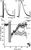
Figure 1.
Figure 1.
Low-frequency stimulation induces LTD of dopamine but not GABAB IPSCs. a, Low-frequency stimulation (LFS, 0.5 ms, 2 Hz for 300 s) induced depression of dopamine IPSCs (1, pre-LFS; 2, post-LFS; n = 13). b, In contrast, low-frequency stimulation produced temporary depression but failed to induce LTD of GABAB receptor-mediated IPSCs (1, pre-LFS; 2, post-LFS; n = 5; p = 0.0005). c, Depression of the dopamine IPSC persisted for the length of the experiment (c), and administration of the antagonist CGP 56999a (100 nm) or sulpiride (50–200 nm) confirmed that the IPSCs were mediated by GABAB or D2 receptors, respectively.
Figure 2.
Figure 2.
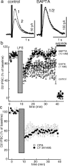
LTD of the dopamine IPSC is blocked by chelating intracellular calcium. a, b, Strongly chelating intracellular calcium with BAPTA (10 mm) prevented induction of LTD (a; 1, pre-LFS; 2, post-LFS), and mildly chelating intracellular calcium with EGTA (1 mm) reduced the amplitude of the synaptic depression (b). Sulpiride (50–200 nm) was used to confirm that the IPSCs were mediated by D2 receptors. Dunnett's test indicated a nonsignificant difference from control conditions for EGTA (1 mm; p = 0.068) and a significant difference from control conditions for BAPTA (10 mm; p = 0.00002). n = 8–13 in each group. c, LTD induction was maintained in the presence of the mGluR antagonist LY 341495 (100 μm, n = 6) or after depletion of intracellular calcium stores with cyclopiazonic acid (CPA) (10 μm; n = 6).
Figure 3.
Figure 3.
Low-frequency stimulation does not affect currents elicited by exogenous dopamine. a, One of the tests used to confirm that neurons being recorded were dopaminergic (see Materials and Methods) was a brief application of exogenous dopamine delivered via an iontophoretic pipette at the beginning of each recording (DA ionto arrowhead). In some neurons, a second iontophoretic pulse was applied at the end of the experiment (second arrowhead) to assess changes in maximum dopamine current. b, This sample trace is from a neuron that expressed LTD (1, before LFS; 2, after LFS). c, There was no consistent change in the iontophoretic current throughout the course of these experiments (average + 4.7 ± 4.9%; n = 17).
Figure 4.
Figure 4.
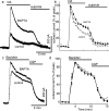
Chelating calcium increases the amplitude of GIRK currents and decreases the desensitization rate of D2 receptor-mediated currents. a, b, When intracellular calcium was chelated with BAPTA (10 mm), bath perfusion of dopamine (DA; 100 μm) elicited larger D2 receptor-mediated outward currents (a) that desensitized less (a, b; p = 0.00093; F(9,144) = 3.35 for time-chelation interaction; n = 8–10). c, d, GABAB receptor-mediated outward currents elicited by baclofen (30 μm) were also larger in the presence of calcium chelation (t(40) = 2.52; p = 0.016; n = 18–24), but there was no difference in desensitization rate (c, d; p = 0.507 for time-chelator interaction; F(6,168) = 0.884; n = 13–17). Perfusion of CGP 56999a (100 nm) or sulpiride (50–200 nm) confirmed that the currents were mediated by GABAB or D2 receptors, respectively.
Figure 5.
Figure 5.
LTD is induced by D2 receptor activation. a, A 4 min bath application of a high concentration of dopamine (DA, 100 μm) produced an outward current that occluded the dopamine IPSC for the first 10–20 min of washout. a–d, The amplitude of the IPSC remained depressed because of receptor desensitization, even after dopamine was washed out of the slice (1, before dopamine; 2, after 20 min washout). c, d, The amplitude of the dopamine IPSC recovered completely only when intracellular calcium was chelated with BAPTA (10 mm; t(11) = 2.58; p = 0.026; n = 6–7) (Supplemental Fig. 4, available at www.jneurosci.org as supplemental material).
Figure 6.
Figure 6.
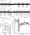
LTD of the dopamine IPSC increases neuron firing. Recordings were obtained in the loose cell-attached patch configuration, and the pacemaker firing pattern that dopamine neurons exhibit in vitro was analyzed. a, As reported previously (Beckstead et al., 2004), electrical stimulation (5 stims) produced a pause in firing that lasted ∼1 s, a result of D2 receptor activation (top trace, 13 sweeps overlaid). b, Low-frequency stimulation (0.5 ms, 2 Hz, 300 s) shortened the duration of the pause (a, bottom trace, 13 sweeps overlaid) in eight of eight cells tested (b; t(8) = 5.92; p = 0.0004). c, This change in firing produced by synaptic depression did not recover for the duration of the experiment.

References

    1. Adell A, Artigas F. The somatodendritic release of dopamine in the ventral tegmental area and its regulation by afferent transmitter systems. Neurosci Biobehav Rev. 2004;28:415–431. - PubMed
    1. Beckstead MJ, Grandy DK, Wickman K, Williams JT. Vesicular dopamine release elicits an inhibitory postsynaptic current in midbrain dopamine neurons. Neuron. 2004;42:939–946. - PubMed
    1. Bellone C, Luscher C. mGluRs induce a long-term depression in the ventral tegmental area that involves a switch of the subunit composition of AMPA receptors. Eur J Neurosci. 2005;21:1280–1288. - PubMed
    1. Berridge KC, Robinson TE. What is the role of dopamine in reward: hedonic impact, reward learning, or incentive salience? Brain Res Brain Res Rev. 1998;28:309–369. - PubMed
    1. Bjorklund A, Lindvall O. Dopamine in dendrites of substantia nigra neurons: suggestions for a role in dendritic terminals. Brain Res. 1975;83:531–537. - PubMed

Publication types

MeSH terms

LinkOut - more resources