. 2007 Apr;18(4):1530-42.
doi: 10.1091/mbc.e06-10-0902. Epub 2007 Feb 21.

Impaired fertility and spermiogenetic disorders with loss of cell adhesion in male mice expressing an interfering Rap1 mutant

Affiliations

Impaired fertility and spermiogenetic disorders with loss of cell adhesion in male mice expressing an interfering Rap1 mutant

Evanthia Aivatiadou et al. Mol Biol Cell. 2007 Apr.

Abstract

The guanosine trisphosphatase Rap1 serves as a critical player in signal transduction, somatic cell proliferation and differentiation, and cell-cell adhesion by acting through distinct mechanisms. During mouse spermiogenesis, Rap1 is activated and forms a signaling complex with its effector, the serine-threonine kinase B-Raf. To investigate the functional role of Rap1 in male germ cell differentiation, we generated transgenic mice expressing an inactive Rap1 mutant selectively in differentiating spermatids. This expression resulted in a derailment of spermiogenesis due to an anomalous release of immature round spermatids from the seminiferous epithelium within the tubule lumen and in low sperm counts. These spermiogenetic disorders correlated with impaired fertility, with the transgenic males being severely subfertile. Because mutant testis exhibited perturbations in ectoplasmic specializations (ESs), a Sertoli-germ cell-specific adherens junction, we searched for expression of vascular endothelial cadherin (VE-cadherin), an adhesion molecule regulated by Rap1, in spermatogenic cells of wild-type and mutant mice. We found that germ cells express VE-cadherin with a timing strictly related to apical ES formation and function; immature, VE-cadherin-positive spermatids were, however, prematurely released in the transgenic testis. In conclusion, interfering with Rap1 function during spermiogenesis leads to reduced fertility by impairment of germ-Sertoli cell contacts; our transgenic mouse provides an in vivo model to study the regulation of ES dynamics.


Figures

Figure 1.
Figure 1.
Characterization of transgenic iRap1 mice. (A) PCR genotyping with selected primers to detect the 350-base pair transgenic fragment in the founder lines (TGRap1-TGRap11); as a positive control, the bPGV-mPI-Rap1S17N-HA plasmid (first lane); as a negative control, DNA extracted from a wild-type mouse (second lane). (B) The 400-base pair transgenic transcript was detected by RT-PCR analysis in germ cells of F1TGRap3, F1TGRap9, and F1TGRap11 male mice. A wild-type (WT) male gave no transcript. (C) Western blots of COS cells, transfected (+) or not (−) with pCDNA3-Rap1-HA plasmid, probed with anti-Rap1 antibodies (left), monoclonal (mHA), anti-HA antibodies (middle), and polyclonal (pHA) anti-HA antibodies (right). (D) Western blots of germ cells from 16-, 28-, 35-, and 60-d-old transgenic testes probed with anti-Rap1 antibodies (left) and anti-HA antibodies (right). The mutant protein is detectable starting from 28-d-old testis.
Figure 2.
Figure 2.
Analysis of testis histology and epididymal spermatozoa of the TGRap1 male founder. (A) Hematoxylin and eosin-stained section showing a heavily altered histology of the seminiferous epithelium and numerous immature cells released into the tubule lumen. Bar, 16 μm. (B) Cauda epididymal spermatozoa display severe head abnormalities, as a hammer-shaped or ovoid head. Bar, 10 μm. (C–E) Scanning electron micrographs of spermatozoa from TGRap1 founder (C and D), showing an ovoid and hammer-shaped head, respectively, and a wild-type head (E).
Figure 3.
Figure 3.
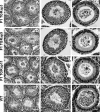
Histological examination of adult testis and epididymis. Sections of transgenic testes from F1 TGRap3 (A and B), F1 TGRap9 (D and E), and F1 TGRap11 (G and H) mice are characterized by common abnormalities as the unusual presence of immature spermatids and occasional enlarged round cells inside the tubule lumen and few released spermatozoa. Such abnormalities are found also in the epididymal lumen (C; F1 TGRap3; F, F1 TGRap9; and I, F1 TGRap11). For comparison, wild-type testis (K and L) and epididymis (M). Bar, 50 μm (A, D, G, and K), 27 μm (B, E, H, and L); and 18 μm (C, F, I, and M).
Figure 4.
Figure 4.
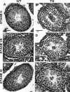
Histological comparison of stages of the seminiferous epithelial cycle between wild-type (WT column) and transgenic (TG column) testes. (A–F) Representative cross sections of stage II tubules (A and B), stage VII tubules (C and D), and stage XI tubules (E and F) are shown. Transgenic testes at all spermatogenic stages exhibit abnormal tubules, with a reduction in ordinary classes of expected spermatids, low numbers of spermatozoa, and the constant presence of differentiating spermatids immaturely released into the lumen. Moreover (D, boxed inset provides a higher magnification), Sertoli–germ cell contacts are often disturbed with a more or less serious loss of adhesion. Bar, 20 μm (A, C, and E) and 16 μm (B, D, and F).
Figure 5.
Figure 5.
Histological comparison between wild-type (WT column) and transgenic (TG column) epididymes. (A and C) Low magnifications providing a general view of sectioned epididymes. (B and D) Higher magnification of a representative cross section from cauda epididimys. Notice the low sperm numbers in the transgenic epididymis. Bar, 65 μm (A and C) and 19 μm (B and D).
Figure 6.
Figure 6.
Immunocytochemical detection of mutant Rap1-HA. Transgenic testis sections immunostained with HA-antibody. (A) A 16-d-old testis, no HA-immunoreactive cells are detectable. (B and C) A 28-d-old testis. Haploid cells have made their first appearance and are differentiating; these cells are HA-Rap1 immunostained. (D–F) Adult testis. (D) Control section immunostained with presaturated HA antibody. (E and F) Mutant Rap1 is restricted to haploid germ cells, including spermatids immaturely released within the lumen. (C and F) Arrows indicate detachments in cell–cell contacts. Bar, 16 μm (A), 37 μm (B), 20 μm (C), 50 μm (D and E), and 10 μm (F).
Figure 7.
Figure 7.
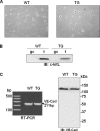
VE-cadherin expression in male germ cells. (A) Assessment of purity of germ cell preparations from WT and transgenic (TG) testis by light microscopy under phase contrast. (B) Western immunoblot analysis for the Sertoli cell marker c-kit Ligand; gc, germ cells; t, total testis homogenate. (C) RT-PCR (left) and Western immunoblot (right; 110 μg/each lane) analysis for VE-cadherin expression in germ cells of wild-type and transgenic mouse.
Figure 8.
Figure 8.
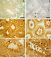
Immunocytochemical detection of VE-cadherin in wild-type seminiferous epithelium. (A–G) Representative cross sections of tubules immunostained with the VE-cadherin antibody. (E–G) Sections further counterstained with hematoxylin. (H) Control, treated with preimmune serum and then counterstained with hematoxylin. Numbers inside the tubule lumen indicate the stages of the epithelial cycle. VE-cadherin immunoreactivity is restricted to the haploid population of germ cells; notice the stage specificity of its expression. Arrow in D points to the basal soma of Sertoli cell, that is clearly VE-cadherin positive at stage VIII when spermiation occurs and VE-cadherin is undetectable in germ cells. Bar, 50 μm (A and E) and 27 μm (B, C, D, F, G, and H).
Figure 9.
Figure 9.
Localization of VE-cadherin by immunofluorescence analysis. VE-cadherin (green), DAPI (blue); superimposition of the two fluorochromes (merged images). (A–C) A testis cross section. VE-cadherin (A and C) stains the adluminal compartment where differentiating spermatids are located; the nucleus staining (B and C) allows assessing of differentiation of round-to-elongated spermatids. VE-cadherin signal delineates neatly the cell periphery where there are the regions of contacts between spermatids and Sertoli cells. (D) A 3× magnification of the boxed area in C. (E–M) Spermatogenic cells isolated by enzymatic treatment of testis. (E–G) Round spermatids are VE-cadherin positive, whereas the spermatocyte at the upper of the image is not labeled. (G) Bright field image. (H–K) A late round spermatid that shows a bright VE-cadherin signal, in particular, some spots orderly distributed along the cap head. (K) Bright field. (L–M) Control sample, where L is the merged image, and M is the bright field. Bar, 28 μm (A–C), 10 μm (E–G), 4 μm (H–K), and 6 μm (L and M).
Figure 10.
Figure 10.
Immunocytochemical detection of VE-cadherin in mutant seminiferous epithelium. (A–G) Representative cross sections of tubules immunostained with the VE-cadherin antibody. (F and G) Sections further counterstained with hematoxylin. (H) Control, treated with preimmune serum and then counterstained with hematoxylin. (B–D) For stage-specific immunoreactivity, VE-cadherin exhibits the same pattern as in the wild type. (E) Higher magnification showing exfoliated immature spermatids that are VE-cadherin positive, within the lumen. Bar, 50 μm (A and F), 16 μm (B, C, D, G, and H), and 13 μm (E).
Figure 11.
Figure 11.
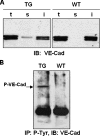
Partition of VE-cadherin between soluble and insoluble fractions and its tyrosine phosphorylation in sexually immature testis. (A) Lysates (120 μg/lane) of TG and WT P30 seminiferous tubules obtained as described in Materials and Methods were subjected to SDS-gel electrophoresis and electroblotted to be probed with antibodies to VE-cadherin. t, total homogenate fraction; s, soluble homogenate fraction; and i, detergent homogenate fraction. The soluble homogenate fraction from the transgenic tubules was VE-cadherin positive, whereas in the wild-type VE-cadherin was extracted only under higher detergent conditions. (B) Total homogenate fractions (600 μg/each) treated with PV (see Materials and Methods) from TG and WT P30 seminiferous tubules were immunoprecipitated with anti-phospho-tyrosine antibody, separated by SDS-gel electrophoresis and Western blotted to be probed with antibodies to VE-cadherin. Tyrosine-phosphorylated VE-cadherin (P-VE-cad) was recovered from the TG tubules only. (A and B) Typical results of one of two independent experiments are shown.
Figure 12.
Figure 12.
Schematic drawing of how Rap1 may regulate spermatid adhesion to the supporting Sertoli cell at the apical ES. (A) A Sertoli cell factor triggers the activation, cAMP/Epac-mediated, of Rap1 in differentiating spermatids. The Rap1-dependent cytoskeletal reorganization promotes and/or enhances VE-cadherin–mediated spermatid–Sertoli cell adhesion. (B) Next to spermiation, Rap1 function is switched off (in terminally differentiated spermatids Rap1 is no more present being eliminated by the way of the cytoplasmic droplet and/or degraded by the proteasome; see Berruti, 2000). Consequently, sperm adhesion at the apical ES is disrupted and spermiation can occur with the release of spermatozoa.

References

    1. Aivatiadou E., Brunetti F., Berruti G. cAMP promotes an Epac2 redistribution where Rap1 is located in differentiating male germ cells. FEBS J. 2005;272(Supplement 1):326.
    1. Andersson A. M., Edvardsen K., Skakkebaek N. E. Expression and localization of N- and E-cadherin in the human testis and epididymis. Int. J. Androl. 1994;17:174–180. - PubMed
    1. Aravindan G. R., Pineau C. P., Bardin C. W., Cheng C. Y. Ability of trypsin in mimicking germ cell factors that affect Sertoli cell secretory function. J. Cell Physiol. 1996;168:123–133. - PubMed
    1. Asha H., de Ruiter N. D., Wang M. G., Hariharan I. K. The Rap1 GTPase functions as a regulator of morphogenesis in vivo. EMBO J. 1999;18:605–615. - PMC - PubMed
    1. Beardsley A., O'Donnell L. Characterization of normal spermiation and spermiation failure induced by hormone suppression in adult rats. Biol. Reprod. 2003;68:1299–1307. - PubMed

Publication types

MeSH terms