. 2007 Apr;18(4):1497-506.
doi: 10.1091/mbc.e06-10-0885. Epub 2007 Feb 21.

Fatty acid remodeling of GPI-anchored proteins is required for their raft association

Affiliations

Fatty acid remodeling of GPI-anchored proteins is required for their raft association

Yusuke Maeda et al. Mol Biol Cell. 2007 Apr.

Abstract

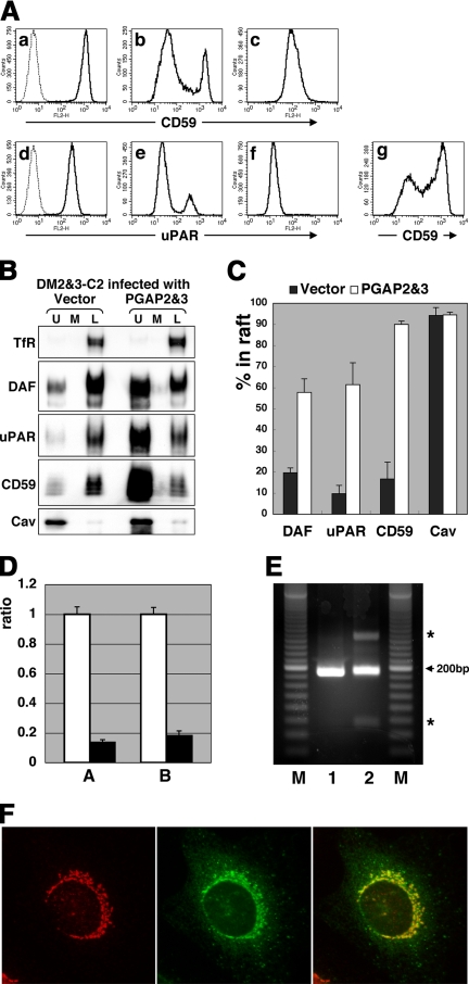
Whereas most of the cellular phosphatidylinositol (PI) contain unsaturated fatty chains and are excluded from rafts, GPI-anchored proteins (APs) unusually contain two saturated fatty chains in their PI moiety, and they are typically found within lipid rafts. However, the origin of the saturated chains and whether they are essential for raft association are unclear. Here, we report that GPI-APs, with two saturated fatty chains, are generated from those bearing an unsaturated chain by fatty acid remodeling that occurs most likely in the Golgi and requires post-GPI-attachment to proteins (PGAP)2 and PGAP3. The surface GPI-APs isolated from the PGAP2 and -3 double-mutant Chinese hamster ovary (CHO) cells had unsaturated chains, such as oleic, arachidonic, and docosatetraenoic acids in the sn-2 position, whereas those from wild-type CHO cells had exclusively stearic acid, a saturated chain, indicating that the sn-2 chain is exchanged to a saturated chain. We then assessed the association of GPI-APs with lipid rafts. Recovery of unremodeled GPI-APs from the double-mutant cells in the detergent-resistant membrane fraction was very low, indicating that GPI-APs become competent to be incorporated into lipid rafts by PGAP3- and PGAP2-mediated fatty acid remodeling. We also show that the remodeling requires the preceding PGAP1-mediated deacylation from inositol of GPI-APs in the endoplasmic reticulum.


Figures

Figure 1.
Figure 1.
PGAP2&3 double-mutant cells are defective in fatty acid remodeling of GPI-APs. (A) Restored surface expression of CD59 on double-mutant DM2&3-C2 cells derived from C84 PGAP2-deficient cells. C84 PGAP2-single-mutant (parent for DM2&3-C2; top), DM2&3-C2 double-mutant (middle), and GD3S-C37 wild-type (parent for C84; bottom) cells were stained with antibody 5H8 against human CD59 and analyzed by FACS. A dotted line in C84 cells represents background staining without anti-CD59 antibody. (B) MS analysis of PI of epitope-tagged CD59 isolated from the plasma membrane. Top (A) and bottom (B), wild-type 3B2A and DM2&3-C2 cells, respectively. Alk and acyl represent alkyl-acyl (or alkenyl-acyl) and diacyl glycerols, respectively, and figures before and after colon represent the number of total carbons and unsaturated bonds, respectively. Neutral loss scanning of 277 Da (phosphorylinositol + NH4) was used for the detection of positive molecular weight-related ions. (C) Summary of the MS/MS analysis (Supplemental Figure S1) of various precursor PI species of epitope-tagged CD59 detected in the first negative ion mode MS (precursor ion scanning of m/z 241). Stearic acid (C18:0) was exclusively used at sn-2 position of PI in wild-type cells (left), whereas oleic (18:1), arachidonic (20:4) and docosatetraenoic (C22:4) acids were used at the same position in DM2&3-C2 cells (right). Alkyl chains are underlined.
Figure 2.
Figure 2.
Fatty acid remodeling is critical for the raft association of GPI-APs. (A) Proteins in wild-type GD3S-C37 and DM2&3-C2 cells were fractionated by sucrose density gradient ultracentrifugation after solubilization in 1% TX-100 at 4°C. Aliquots of top (U), middle (M), and bottom (L) fractions after ultracentrifugation were analyzed by SDS-PAGE/Western blotting with antibodies against TfR, DAF, uPAR, CD59, and caveolin-1 (Cav). U and L correspond to raft (detergent-resistant) and nonraft (detergent-soluble) fractions, respectively. This picture is representative of three independent experiments. (B) Amounts of DAF, uPAR, CD59, and Cav in U and L fractions were measured on Western blots with a CCD camera, and the percentage of proteins in the raft was calculated as the ratio of intensity in L to that of U+L fractions. Results are means and standard deviations from three independent experiments.
Figure 3.
Figure 3.
PGAP3, the gene responsible for the second defect in DM2&3-C2, encodes a protein mainly expressed in the Golgi. (A) FACS analysis of the surface expression of CD59 (a–c and g) and uPAR (d–f) on DM2&3-C2 cells transiently transfected with control (a and d), human PGAP3 (b and e), or yeast PER1 (g) expression vector and on C84 PGAP2-single-mutant cells transfected with control vector (c and f). Dotted lines (a and d) represent background staining without antibodies. (B) Proteins in DM2&3-C2 cells stably transfected with PGAP2 and PGAP3 were fractionated as described in Figure 2. Fractions obtained from two independent experiments showed a quite similar pattern to fractions obtained from wild-type cells, as shown in Figure 2. (C) The percentages of proteins in rafts were quantified as described in Figure 2. Results are means and standard deviations from two independent experiments. (D) The amounts of PGAP3 mRNA expressed in GD3S-C37 (white bars) and DM2&3-C2 (black bars) were evaluated by real-time RT-PCR. Two different primer sets (set A, left and set B, right) were used for PCR of PGAP3, and the amount was normalized by the amount of β-actin. The relative ratio was calculated with the amount in GD3S-C37 being set as 1. Bars represent SD of n = 3. (E) PGAP3 cDNAs obtained from GD3S-C37 (lane 1) and DM2&3-C2 (lane 2) were amplified by PCR with primer set A. Twenty base-pair ladder maker DNA was used as reference (lanes M). Asterisks, two abnormal bands in lane 2. (F) Expression of PGAP3 in the Golgi. DM2&3-C2 cells transiently transfected with HA-tagged PGAP3 were fixed, permeabilized with TX-100, and stained for GPP130, a Golgi protein, (left) and HA (middle). (Right) Merged profile showing colocalization of GPP130 and PGAP3.
Figure 4.
Figure 4.
A defect in PGAP1 inhibits fatty acid remodeling mediated by PGAP3 and PGAP2. (A) Surface expression of CD59 was examined by FACS in AM-B, another PGAP2-single-mutant (a), wild-type 3B2A (b), C10 PGAP1-single mutant (c), and DM1&2-C14 (d–f) cells. DM1&2-C14 cells were either nontransfected (d), transiently transfected with PGAP1 expression vector (e), or cotransfected with PGAP1 and PGAP2 expression vectors (f). A dotted line (a) represents background staining. Thick gray lines (b–d and f) indicate the cells treated with PI-PLC. (B) 3B2A wild-type, C10 PGAP1-single-mutant, and DM1&2-C17 cells were transiently transfected with VSVGts-FF-mEGFP-GPI expression vector and the processed FLAG-mEGFP-GPI was collected from the cell lysates with anti-FLAG beads and eluted with a FLAG-peptide (Tashima et al., 2006). FLAG-mEGFP-GPIs were chromatographed in an octyl-FF column with a 5–40% gradient of 1-propanol. Fractions were subjected to SDS-PAGE and analyzed by Western blotting with an anti-FLAG antibody.
Figure 5.
Figure 5.
Current model of fatty acid remodeling of GPI-APs in mammalian cells. GPI is synthesized from PI with an unsaturated fatty acid at sn-2 position and has an acylated (usually palmitoylated) inositol. Immediately after attachment of GPI to newly synthesized proteins, inositol-linked palmitate is removed by PGAP1, the deacylase, in the ER (step a). The inositol-deacylation is essential for subsequent steps in fatty acid remodeling in the Golgi. An unsaturated fatty chain at sn-2 is removed, resulting in lyso-GPI-APs. PGAP3 is involved in this step (step b). Reacylation of sn-2 with stearic acid follows and PGAP2 is involved in this step (step c). A donor of stearoyl chain is yet to be determined. GPI-APs that have undergone fatty acid remodeling are competent for the integration into lipid rafts (step d). In PGAP1-deficient cells, GPI-APs before inositol deacylation are expressed on the cell surface without any further modification (step e). In PGAP3-deficient cells, GPI-APs carrying an unsaturated fatty chain at sn-2 are expressed on the cell surface (step f). In PGAP2-deficient cells, lyso-form GPI-APs are transported to the cell surface (step g), rapidly cleaved by surface (lyso-) PLD, and secreted (step h). PGAP1, PGAP3, and PGAP2 are epistatic in this order.

References

    1. Bagnat M., Keranen S., Shevchenko A., Shevchenko A., Simons K. Lipid rafts function in biosynthetic delivery of proteins to the cell surface in yeast. Proc. Natl. Acad. Sci. USA. 2000;97:3254–3259. - PMC - PubMed
    1. Benting J., Rietveld A., Ansorge I., Simons K. Acyl and alkyl chain length of GPI-anchors is critical for raft association in vitro. FEBS Lett. 1999;462:47–50. - PubMed
    1. Bosson R., Jaquenoud M., Conzelmann A. GUP1 of Saccharomyces cerevisiae encodes an O-acyltransferase involved in remodeling of the GPI anchor. Mol. Biol. Cell. 2006;17:2636–2645. - PMC - PubMed
    1. Brewis I. A., Ferguson M. A., Mehlert A., Turner A. J., Hooper N. M. Structures of the glycosyl-phosphatidylinositol anchors of porcine and human renal membrane dipeptidase. Comprehensive structural studies on the porcine anchor and interspecies comparison of the glycan core structures. J. Biol. Chem. 1995;270:22946–22956. - PubMed
    1. Brown D. A., Rose J. K. Sorting of GPI-anchored proteins to glycolipid-enriched membrane subdomains during transport to the apical cell surface. Cell. 1992;68:533–544. - PubMed

Publication types

MeSH terms

Substances

Associated data

LinkOut - more resources