. 2007 May;18(5):1609-20.
doi: 10.1091/mbc.e06-06-0518. Epub 2007 Feb 21.

The N-terminus of Dictyostelium Scar interacts with Abi and HSPC300 and is essential for proper regulation and function

Affiliations

The N-terminus of Dictyostelium Scar interacts with Abi and HSPC300 and is essential for proper regulation and function

Diana Caracino et al. Mol Biol Cell. 2007 May.

Abstract

Scar/WAVE proteins, members of the conserved Wiskott-Aldrich syndrome (WAS) family, promote actin polymerization by activating the Arp2/3 complex. A number of proteins, including a complex containing Nap1, PIR121, Abi1/2, and HSPC300, interact with Scar/WAVE, though the role of this complex in regulating Scar function remains unclear. Here we identify a short N-terminal region of Dictyostelium Scar that is necessary and sufficient for interaction with HSPC300 and Abi in vitro. Cells expressing Scar lacking this N-terminal region show abnormalities in F-actin distribution, cell morphology, movement, and cytokinesis. This is true even in the presence of wild-type Scar. The data suggest that the first 96 amino acids of Scar are necessary for participation in a large-molecular-weight protein complex, and that this Scar-containing complex is responsible for the proper localization and regulation of Scar. The presence of mis-regulated or unregulated Scar has significant deleterious effects on cells and may explain the need to keep Scar activity tightly controlled in vivo either by assembly in a complex or by rapid degradation.


Figures

Figure 1.
Figure 1.
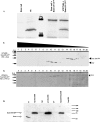
Endogenous Scar is present in a large-molecular-weight complex. Clarified cell lysate from growing HPS400 cells was loaded onto a 7–20% sucrose gradient. Fractions of 0.5 ml were collected and immunoblotted for Scar (A) or PIR121 (B). Numbers above blots indicate fraction number with the bottom of the gradient on the left. The majority of endogenous Scar is present in fractions 9–14 (A), which contain protein complexes of ∼400–600 kDa. The majority of PIR121 protein was detected in the same fractions (B).
Figure 2.
Figure 2.
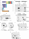
The N-terminus of Scar binds HSPC300 and Abi in vitro. (a) Constructs used in these experiments. Numbers represent position of amino acids relative to the N-terminus. Relative molecular weights are indicated on the right. All Scar constructs contain C-terminal HA tag. FL, full-length Scar; SHD, construct containing the Scar homology domain (SHD) and Basic (B) regions; PWA, construct containing the poly-proline region, WH2, connecting (C), and acidic (A) regions; SHDΔ44, SHD construct lacking the first 44 amino acids; SHDΔ96, SHD construct lacking the first 96 amino acids. SHD1-96, Scar construct containing only amino acids 1–96; HSPC300, HSPC300 protein with C-terminal myc tag; Abi protein, amino acids 1–333 with C-terminal T7 peptide tag. (b) SDS-PAGE of in vitro binding and coimmunoprecipitation assays. Scar constructs with a C-terminal HA tag and HSPC300 with C-terminal myc tag were separately transcribed and translated in vitro, mixed, and then immunoprecipitated with protein G agarose beads conjugated to anti-HA (lanes 1–6) or anti-myc antibodies (lanes 7–12). Bead only controls representing lysates mixed with agarose beads are shown in lanes 1 and 7. Immunoprecipitates of lysate containing no added DNA are shown in lanes 2 and 8. Lysates containing PCR-generated DNAs of untagged Scar (lane 3) or untagged HSPC300 (lane 9) immunoprecipitated with anti-HA or anti-myc–conjugated beads, respectively, are shown. Transcription and translation of untagged proteins was verified (data not shown). Lysates containing both Scar-HA and HSPC300-myc immunoprecipitated with anti-HA (lane 5) or anti-myc (lane 11) are shown. FL Scar-HA migrates at ∼66 kDa; HSPC300 migrates at ∼11 kDa. Controls for anti-HA bead specificity are shown in lanes 4 and 6. Controls for anti-myc bead specificity are shown in lanes 10 and 12. (c) Scar-HA, Abi-T7, and HSPC300-myc were cotranslated and immunoprecipitated in various combinations. Lysates immunoprecipitated with anti-T7 (Abi), anti-HA (Scar), or anti-myc–conjugated agarose beads are shown. Assays were done as described in b. Molecular-weight marker sizes are indicated on the left. (d) Amino acids 1–44 of Scar are necessary to bind HSPC300 in vitro. Assay was done as described in b. Proteins added in binding assay are indicated for each lane. Lysates immunoprecipitated for HSPC300 using anti-myc–conjugated agarose beads (lanes 1–6) or for Scar using anti-HA–conjugated agarose beads (lanes 7–12) are shown. (e) Amino acids 1–96 of Scar are sufficient to bind HSPC300 in vitro. Assays were as in b. Lysates were immunoprecipitated for Scar using anti-HA bound agarose beads. The gels were either detected for 35S-Met (top panels) or immunoblotted with anti-HA antibody (bottom panels). Control lysate containing no added DNA is shown in lane 1. HSCP300 control is shown in lane 2. Lane 3 represents lysate containing both SHD-HA and HSPC300-myc proteins. Lane 4 represents lysate containing 1–96 fragment of SHD domain and HSPC300. (f) Amino acids 1–44 of Scar are necessary to bind to Abi. ScarΔ44-HA (full-length Scar minus the first 44 amino acids), Abi-T7, and HSPC300-myc were cotranslated and immunoprecipitated with anti-T7– conjugated agarose beads (lane 1), anti-HA–conjugated agarose beads (lane 2), or anti-myc–conjugated agarose beads (lane 3). ScarΔ44-HA (full-length Scar minus the first 44 amino acids) and Abi-T7 were cotranslated and immunoprecipitated with anti-HA–conjugated agarose beads (lane 4) or anti-T–conjugated agarose beads (lane 5). (g) Amino acids 1–96 of Scar are sufficient to bind Abi in the presence of HSPC300. Scar SHD1-96-HA and Abi-T7 were cotranslated and immunoprecipitated with anti-HA–conjugated agarose beads (lane 1) or anti-T7–conjugated agarose beads (lane 2). Scar SHD1-96-HA, Abi-T7, and HSPC300-myc were cotranslated and immunoprecipitated with anti-T7–conjugated agarose beads (lane 3), anti-HA–conjugated agarose beads (lane 4), or anti-myc–conjugated agarose beads (lane 5).
Figure 3.
Figure 3.
ScarΔ96-GFP is stably expressed and complexes differently than wild-type Scar. (A) Growing HPS400 cells expressing Scar (∼66 kDa) and/or ScarΔ96-GFP (∼75 kDa) were collected and Western blotted for Scar. ScarΔ96-GFP is stably expressed in both backgrounds. Control cells are HPS400 and Scar null cells expressing empty vector. Clarified cell lysate from parental cells expressing ScarΔ96-GFP was loaded onto a 7–20% sucrose gradient. Fractions of 0.5 ml, were collected and immunoblotted for Scar (B) or PIR121 (C). Numbers above blots indicate fraction number. The majority of endogenous Scar is present in fractions 9–12 as in Figure 1. ScarΔ96-GFP is present in fractions 14–18 (B), which contain protein complexes of ∼150–300 kDa. PIR121 protein is not detected in the fractions containing ScarΔ96-GFP but continues to track with wild-type Scar (C). (D) Cells null for each of the Scar complex members with or without expressed ScarΔ96-GFP were collected and Western blotted as in A. ScarΔ96-GFP is stably expressed in each background.
Figure 4.
Figure 4.
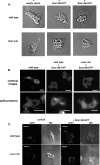
Expression of ScarΔ96-GFP results in an abnormal morphology. (A) Axenically grown parental (HPS400) and scar null cells expressing empty vector or ScarΔ96-GFP were allowed to adhere to glass coverslips and observed with Nomarski differential interference contrast. Movies of cells are available in the Supplementary Material. (B) ScarΔ96-GFP does not localize to leading edges of newly forming pseudopods. Full-length Scar-GFP was expressed in scar null cells developed for 6 h (leftmost panels) and ScarΔ96-GFP was expressed in similarly developed parental (middle panels), and scar null backgrounds (right panels). The GFP in the cells was visualized using confocal microscopy (top panels) and epifluorescence microscopy (bottom panels). Scale bar, 10 μm. (C) Cells expressing ScarΔ96-GFP have aberrant actin staining. Six-hour developed parental and scar− cells expressing ScarΔ96-GFP were stained with TRITC-phalloidin. DIC images of cells are also shown. Bar, 10 μm.
Figure 5.
Figure 5.
Cells expressing ScarΔ96 show no dramatic increases in F-actin levels. Cellular F-actin levels were measured as described in Materials and Methods. Basal F-actin levels in all ScarΔ96-GFP–expressing cells measured were not statistically different from levels in wild-type cells. The difference between scar null and scar null/Scar PWA-GFP was statistically significant. The Y-axis is fold change in F-actin polymerization relative to parental control. Data represent results from three separate experiments.
Figure 6.
Figure 6.
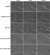
Cells expressing ScarΔ96-GFP have motility and chemotaxis defects. Chemotaxis assays were performed on 8-h developed HPS400, Scar null, and HPS400 and Scar null cells expressing ScarΔ96-GFP and Scar null cells expressing full-length Scar-GFP. Times of exposure to a cAMP gradient are shown at the top of the figure.
Figure 7.
Figure 7.
Induction of cytokinesis defect by extended induction of Δ96 constructs. (A–C, I, and J) Axenically growing strains with the ScarΔ96-GFP construct uninduced; (D–H) each strain after 72 h of ScarΔ96 induction. A–F, DIC images; G–J, epifluorescent images with nuclei stained with DAPI and cells outlined with TRITC-phalloidin.

Similar articles

Cited by

References

    1. Baba Y., et al. Involvement of wiskott-aldrich syndrome protein in B-cell cytoplasmic tyrosine kinase pathway. Blood. 1999;93:2003–2012. - PubMed
    1. Bear J. E. Atlanta, GA: Emory University; 1998. SCAR Defines a New Family of WASP-related Actin Cytoskeleton Regulators.
    1. Bear J. E., Rawls J. F., Saxe C. L., III SCAR, a WASP-related protein, isolated as a suppressor of receptor defects in late Dictyostelium development. J. Cell Biol. 1998;142:1325–1335. - PMC - PubMed
    1. Bishop A. L., Hall A. Rho GTPases and their effector proteins. Biochem. J. 2000;348(Pt 2):241–255. - PMC - PubMed
    1. Blagg S. L., Stewart M., Sambles C., Insall R. H. PIR121 regulates pseudopod dynamics and SCAR activity in Dictyostelium. Curr. Biol. 2003a;13:1480–1487. - PubMed

Publication types

MeSH terms

LinkOut - more resources