Review
. 2007:40:132-57.
doi: 10.1159/000100353.

Mechanics of the orbita

Affiliations
Review

Mechanics of the orbita

Joseph L Demer. Dev Ophthalmol. 2007.

Abstract

The oculomotor periphery was formerly regarded as a simple mechanism executing complex behaviors explicitly specified by innervation. It is now recognized that several fundamental aspects of ocular motility are properties of the extraocular muscles (EOMs) and their associated connective tissue pulleys. The Active Pulley Hypothesis proposes that rectus and inferior oblique EOMs have connective tissue soft pulleys that are actively controlled by the action of the EOMs' orbital layers. Functional imaging and histology have suggested that the rectus pulley array constitutes an inner mechanism, similar to a gimbal, that is rotated torsionally around the orbital axis by an outer mechanism driven by the oblique EOMs. This arrangement may mechanically account for several commutative aspects of ocular motor control, including Listing's law, yet permits implementation of noncommutative motility as during the vestibulo-ocular reflex. Recent human behavioral studies, as well neurophysiology in monkeys, are consistent with mechanical rather than central neural implementation of Listing's law. Pathology of the pulley system is associated with predictable patterns of strabismus that are surgically treatable when the pathologic anatomy is characterized by imaging. This mechanical determination may imply limited possibilities for neural adaptation to some ocular motor pathologies, but indicates greater potential for surgical treatments.


Figures
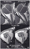
Fig. 1
Fig. 1
Axial MRI images of a right orbit taken at the level of the lens, fovea, and optic nerve (top row), and simultaneously in an inferior plane along the IR muscle path (bottom row), in abduction (left) and adduction (right). Note the bisegmental IR path, with an inflection corresponding to the IR pulley. For this 73° horizontal gaze shift, there was a corresponding 36° shift in IR muscle path anterior to the inflection at its pulley. By permission from Demer [19].
Fig. 2
Fig. 2
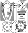
Diagram of the orbita. Coronal views are depicted at levels indicated on axial view. Functional pulleys are at level depicted at lower right. LG = Lacrimal gland; LPS = levator palpebrae superioris muscle; SOT = superior oblique tendon. By permission from Demer [31].
Fig. 3
Fig. 3
Diagram of EOM and pulley behavior for half angle kinematics conforming to LL. a Lateral view. Rotational velocity axis of the EOM is perpendicular to the segment from pulley to scleral insertion. The velocity axis for the LR is vertical in primary position. b Lateral view. In supraduction to angle alpha, the LR velocity axis tilts posteriorly by angle alpha/2 if distance D1 from pulley to globe center is equal to distance D2 from globe center to insertion. c Lateral view. In primary position, terminal segment of the IO muscle lies in the plane containing the LR and IR pulleys into which the IO’s orbital layer inserts. The IO velocity axis parallels primary gaze. d Superior view of rectus EOMs and pulleys in primary position, corresponding to a. e Superior view. In order for adduction to maintain D1 = D2 in an oculocentric reference, the MR pulley must shift posteriorly in the orbit, and the LR pulley anteriorly. This is proposed to be implemented by the orbital layers of these EOMs, working against elastic pulley suspensions. f Lateral view similar to c. In supraduction to angle alpha, the IR pulley shifts anteriorly by distance D3, as required by the relationship shown in e. The IO pulley shifts anteriorly by D3/2, shifting the IO velocity axis superiorly by alpha/2. By permission from Demer [19].
Fig. 4
Fig. 4
Diagram of effects of head tilt on rectus pulleys in lateral (top row) and frontal (bottom row) views. With head upright, the IR, LR, MR, and SR pulleys are arrayed in frontal view in a cruciate pattern. The MR passes through its pulley, represented as a ring, to its scleral insertion. The rotational velocity axis imparted by the MR is perpendicular to the segment from pulley to insertion. The pulley array extorts during contralateral head tilt. Since during head tilt the MR pulley shifts superiorly by the half the distance the insertion shifts, the MR’s velocity axis changes by one fourth the ocular torsion. By permission from Demer and Clark [69].

References

    1. Demer JL. Extraocular muscles, chapter 1. In: Jaeger EA, Tasman PR, editors. Duane’s Clinical Ophthalmology. Vol. 1. Philadelphia: Lippincott; 2000. pp. 1–23.
    1. Demer JL. Anatomy of strabismus. In: Taylor D, Hoyt C, editors. Pediatric Ophthalmology and Strabismus. 3. London: Elsevier; 2005. pp. 849–861.
    1. Porter JD, Baker RS, Ragusa RJ, Brueckner JK. Extraocular muscles: basic and clinical aspects of structure and function. Surv Ophthalmol. 1995;39:451–484. - PubMed
    1. Oh SY, Poukens V Demer JL. Quantitative analysis of rectus extraocular muscle layers in monkey and humans. Invest Ophthalmol Vis Sci. 2001;42:10–16. - PubMed
    1. Kono R, Poukens V, Demer JL. Superior oblique muscle layers in monkeys and humans. Invest Ophthalmol Vis Sci. 2005;46:2790–2799. - PubMed

Publication types