. 2007 Mar-Apr;18(2):447-55.
doi: 10.1021/bc060349x. Epub 2007 Feb 22.

Fusion antibody for Alzheimer's disease with bidirectional transport across the blood-brain barrier and abeta fibril disaggregation

Affiliations

Fusion antibody for Alzheimer's disease with bidirectional transport across the blood-brain barrier and abeta fibril disaggregation

Ruben J Boado et al. Bioconjug Chem. 2007 Mar-Apr.

Abstract

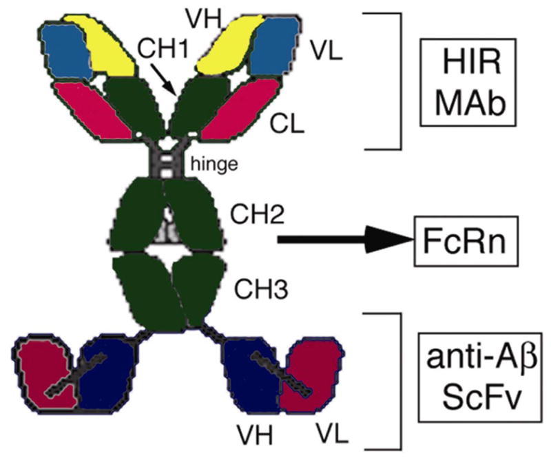
Delivery of monoclonal antibody therapeutics across the blood-brain barrier is an obstacle to the diagnosis or therapy of CNS disease with antibody drugs. The immune therapy of Alzheimer's disease attempts to disaggregate the amyloid plaque of Alzheimer's disease with an anti-Abeta monoclonal antibody. The present work is based on a three-step model of immune therapy of Alzheimer's disease: (1) influx of the anti-Abeta monoclonal antibody across the blood-brain barrier in the blood to brain direction, (2) binding and disaggregation of Abeta fibrils in brain, and (3) efflux of the anti-Abeta monoclonal antibody across the blood-brain barrier in the brain to blood direction. This is accomplished with the genetic engineering of a trifunctional fusion antibody that binds (1) the human insulin receptor, which mediates the influx from blood to brain across the blood-brain barrier, (2) the Abeta fibril to disaggregate amyloid plaque, and (3) the Fc receptor, which mediates the efflux from brain to blood across the blood-brain barrier. This fusion protein is a new antibody-based therapeutic for Alzheimer's disease that is specifically engineered to cross the human blood-brain barrier in both directions.


Figures

Figure 1
Figure 1. Import-export model of Abeta antibody therapeutics
The fusion antibody clears amyloid from brain in AD via 3 sequential steps, and each of these 3 sequential steps uses separate parts of the fusion antibody molecule. Step 1 is the influx of the fusion antibody from blood to brain across the BBB, which is mediated by binding of the fusion antibody to the BBB HIR. Step 2 is binding of the fusion antibody to the amyloid plaque in AD, which promotes disaggregation of the amyloid plaque, and this binding to the plaque is mediated by the anti-AβScFv part of the fusion antibody. Step 3 is the efflux of the fusion antibody from brain to blood across the BBB, which is mediated by binding of the fusion antibody to the BBB FcRn at the CH2-CH3 interface of the Fc region of the fusion antibody.
Figure 2
Figure 2. Antibody fusion protein with 3 functional domains
The first domain, the HIRMAb, binds the BBB HIR to trigger influx across the BBB. The second domain, the CH2/CH3 interface of the Fc region, binds to the BBB FcRn to trigger efflux from brain back to blood. The third domain, the anti-AβScFv fused to the CH3 region, binds to the Aβ amyloid peptide of AD to cause disaggregation of amyloid in brain.
Figure 3
Figure 3. Immunocytochemistry of frozen sections of AD autopsy brain
The brain sections were immune stained with medium conditioned by COS cells that were transfected with pCD- ScFv (a, c), with medium conditioned by COS cells which were exposed only to Lipofectamine 2000 (b), or with the mouse IgG1 isotype control antibody (d). Magnification in panels a and b is the same, and the magnification bar in panel b is 88 um; magnification in panels c and d is the same and the magnification bar in panel d is 35 um.
Figure 4
Figure 4. SDS-PAGE and Western blotting of fusion antibody
(a) SDS-PAGE and Coomasie blue staining shows correct processing of intact fusion antibody by transfected COS cells. (b) Western blotting with an anti-human IgG primary antibody. The HIRMAb and the fusion antibody are comprised of the same light chain, which is 28 kDa. The size of the heavy chain of the HIRMAb is 55 kDa, whereas the size of the heavy chain of fusion antibody is 82 kDa. The heavy chain of the fusion antibody includes the 55 kDa heavy chain of the HIRMAb fused to the 27 kDa anti-AβScFv.
Figure 5
Figure 5. Bi-functional binding of fusion antibody both to Abeta and to the human insulin receptor
(a) The binding of [125I]-murine anti-Aβ MAb to Aβ1–40 is competitively inhibited by either unlabeled murine anti-Aβ MAb or by the fusion antibody. The binding affinity of the fusion antibody is not significantly different from the affinity of the murine anti-Aβ MAb. (b) Binding of the genetically engineered HIRMAb or the fusion antibody to the HIR extracellular domain is measured with an ELISA. There is no binding of human IgG1 to the HIR ECD. The affinity of the fusion antibody for the HIR ECD is >50% of the affinity of the HIRMAb.
Figure 6
Figure 6. Blood to brain transport of fusion antibody in the primate
(a) The [125I]-fusion antibody, and the [3H]-mouse anti-Aβ antibody, were injected intravenously into the adult Rhesus monkey and serum concentrations [% of injected dose (I.D.)/mL] were determined over a 3 hour period. There is no measurable clearance from blood of the [3H]-mouse anti-Aβ antibody during this time period, whereas the fusion antibody is rapidly cleared from blood, owing to uptake via the insulin receptor. (b) Brain volume of distribution (VD) of the [3H]-mouse anti-Aβ antibody and the [125I]-fusion antibody in Rhesus monkey brain at 3 hours after a single intravenous injection. The VD for the homogenate and the post-vascular supernatant are shown. The VD for the [3H]-mouse anti-Aβ antibody, 10 uL/g, is equal to the brain arterial blood volume, which indicates this antibody is not transported across the primate BBB in vivo in the blood to brain direction. The VD for the [125I]-fusion antibody is >100 uL/g in both the brain homogenate and the post-vascular supernatant, which indicates the [125I]-fusion antibody is transported across the BBB.
Figure 7
Figure 7. Global distribution of fusion antibody to primate brain
Brain scans of adult Rhesus monkey at 3 hours after the intravenous administration of the [125I]-fusion antibody demonstrates widespread distribution of the fusion antibody into the primate brain in vivo from blood. The top scan is the most frontal part of brain, and the bottom scan is the most caudal part of brain, and includes the cerebellum.
Figure 8
Figure 8. Brain to blood transport of fusion antibody in the rat
The [125I]-fusion antibody was injected into the cortex under stereotaxic guidance, and the efflux of the fusion antibody from brain across the BBB was measured at 90 minutes after the injection. At this time nearly 60% of the injected fusion antibody has effluxed from brain. This efflux was >90% blocked (p<0.01) by the co-injection of human Fc fragments, which indicates the efflux is mediated by an Fc receptor at the BBB. Data are mean ± S.E. (n=4 rats per point).
Figure 9
Figure 9. Fusion antibody disaggregates Abeta amyloid fibrils in vitro and in vivo
(a) Outline of Aβ plaque disaggregation assay. The Abeta fibrils are sandwiched by an anti-Abeta antibody that binds the carboxyl terminus (CT) of Abeta, and by the fusion antibody, or the murine anti-Abeta MAb, which binds the amino terminus (NT) of Abeta. (b) Disaggregation of Aβ fibrils in vitro by the fusion antibody. Aβ1–40 aggregates were formed over 6 days, followed by incubation with the fusion antibody, with human IgG1 (hIgG1), or with phosphate buffered saline (PBS) for either 1 or 4 hours at 37C. The antibody that binds to the carboxyl terminal region of the Aβ1–40 peptide was plated in 96 well plates. The anti-Aβ ScFv portion of the fusion antibody binds to the amino terminal part of Aβ1–40. A positive ELISA signal is generated only if Abeta fibrils are present. The data show that the fusion antibody binds to Aβ1–40 plaque, and that this binding causes fibril disaggregation over a 4-hour period. (c) Amyloid plaque in brain of double transgenic APPswe/PS1dE9 mice stained with thioflavine-S. (d) The percent of brain occupied by amyloid plaque in either frontal cortex or hippocampus in the transgenic mice was quantitated with confocal microscopy. Amyloid was measured at 48 hours after the intra-cerebral injection of 20 pmol of fusion antibody into the frontal cortex and the hippocampus on one side of the brain. The amyloid burden in brain is expressed as a ratio of the % brain amyloid in the side ipsilateral to the injection relative to the % amyloid content in the contralateral frontal cortex or hippocampus, in the animals injected with either the fusion body, or with saline. Data are mean ± SE (n=3 mice for each treatment group). The decrease in amyloid content in either frontal cortex or hippocampus on the side of the fusion antibody injection, relative to the saline control, is statistically significant (*p<0.01).

References

    1. Schenk D, Barbour R, Dunn W, Gordon G, Grajeda H, Guido T, Hu K, Huang J, Johnson-Wood K, Khan K, Kholodenko D, Lee M, Liao Z, Lieberburg I, Motter R, Mutter L, Soriano F, Shopp G, Vasquez N, Vandevert C, Walker S, Wogulis M, Yednock T, Games D, Seubert P. Immunization with amyloid-beta attenuates Alzheimer-disease-like pathology in the PDAPP mouse. Nature. 1999;400:173–177. - PubMed
    1. Solomon B, Koppel R, Frankel D, Hanan-Aharon E. Disaggregation of Alzheimer beta-amyloid by site-directed mAb. Proc Natl Acad Sci USA. 1997;94:4109–4112. - PMC - PubMed
    1. Solomon B, Koppel R, Hanan E, Katzav T. Monoclonal antibodies inhibit in vitro fibrillar aggregation of the Alzheimer beta-amyloid peptide. Proc Natl Acad Sci USA. 1996;93:452–455. - PMC - PubMed
    1. Lombardo JA, Stern EA, McLellan ME, Kajdasz ST, Hickey GA, Bacskai BJ, Hyman BT. Amyloid-beta antibody treatment leads to rapid normalization of plaque-induced neuritic alterations. J Neurosci. 2003;23:10879–10883. - PMC - PubMed
    1. Wilcock DM, DeCarlo G, Henderson D, Jackson J, Clarke K, Ugen KE, Gordon MN, Morgan D. Intracranially administered anti-A beta antibodies reduce beta-amyloid deposition by mechanisms both independent of and associated with microglial activation. J Neurosci. 2003;23:3745–3751. - PMC - PubMed

Publication types

MeSH terms