Comparative Study
. 2007;9(1):R18.
doi: 10.1186/ar2127.

Effects on osteoclast and osteoblast activities in cultured mouse calvarial bones by synovial fluids from patients with a loose joint prosthesis and from osteoarthritis patients

Affiliations
Comparative Study

Effects on osteoclast and osteoblast activities in cultured mouse calvarial bones by synovial fluids from patients with a loose joint prosthesis and from osteoarthritis patients

Martin K Andersson et al. Arthritis Res Ther. 2007.

Abstract

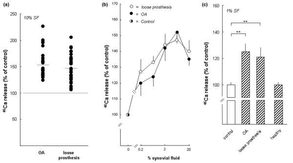
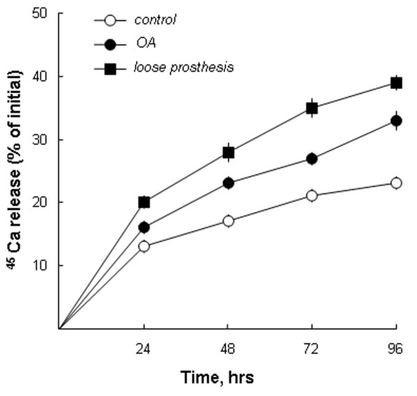
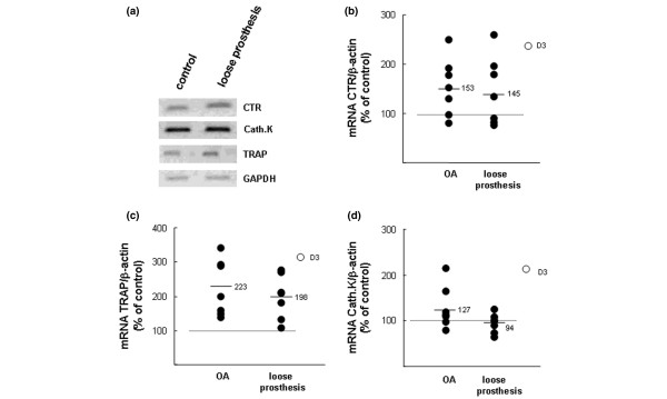
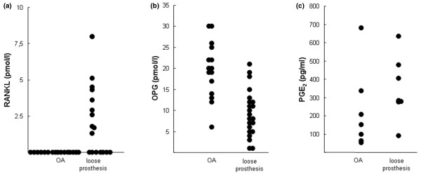
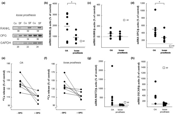
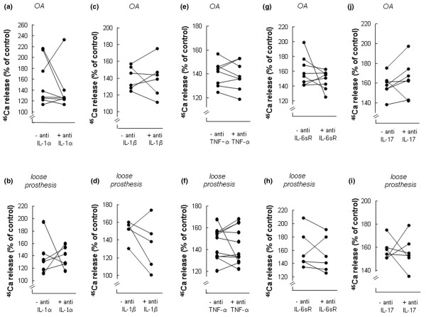
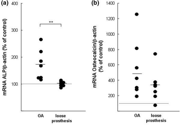
Aseptic loosening of a joint prosthesis is associated with remodelling of bone tissue in the vicinity of the prosthesis. In the present study, we investigated the effects of synovial fluid (SF) from patients with a loose prosthetic component and periprosthetic osteolysis on osteoclast and osteoblast activities in vitro and made comparisons with the effects of SF from patients with osteoarthritis (OA). Bone resorption was assessed by the release of calcium 45 (45Ca) from cultured calvariae. The mRNA expression in calvarial bones of molecules known to be involved in osteoclast and osteoblast differentiation was assessed using semi-quantitative reverse transcription-polymerase chain reaction (PCR) and real-time PCR. SFs from patients with a loose joint prosthesis and patients with OA, but not SFs from healthy subjects, significantly enhanced 45Ca release, effects associated with increased mRNA expression of calcitonin receptor and tartrate-resistant acid phosphatase. The mRNA expression of receptor activator of nuclear factor-kappa-B ligand (rankl) and osteoprotegerin (opg) was enhanced by SFs from both patient categories. The mRNA expressions of nfat2 (nuclear factor of activated T cells 2) and oscar (osteoclast-associated receptor) were enhanced only by SFs from patients with OA, whereas the mRNA expressions of dap12 (DNAX-activating protein 12) and fcrgamma (Fc receptor common gamma subunit) were not affected by either of the two SF types. Bone resorption induced by SFs was inhibited by addition of OPG. Antibodies neutralising interleukin (IL)-1alpha, IL-1beta, soluble IL-6 receptor, IL-17, or tumour necrosis factor-alpha, when added to individual SFs, only occasionally decreased the bone-resorbing activity. The mRNA expression of alkaline phosphatase and osteocalcin was increased by SFs from patients with OA, whereas only osteocalcin mRNA was increased by SFs from patients with a loose prosthesis. Our findings demonstrate the presence of a factor (or factors) stimulating both osteoclast and osteoblast activities in SFs from patients with a loose joint prosthesis and periprosthetic osteolysis as well as in SFs from patients with OA. SF-induced bone resorption was dependent on activation of the RANKL/RANK/OPG pathway. The bone-resorbing activity could not be attributed solely to any of the known pro-inflammatory cytokines, well known to stimulate bone resorption, or to RANKL or prostaglandin E2 in SFs. The data indicate that SFs from patients with a loose prosthesis or with OA stimulate bone resorption and that SFs from patients with OA are more prone to enhance bone formation.


Figures

Figure 1
Figure 1
Stimulation of calcium 45 (45Ca) release from neonatal mouse calvarial bones by synovial fluids (SFs) from patients with osteoarthritis (OA) and patients with a loose prosthesis, but not by SFs from healthy individuals. (a) The effect of SFs from 25 patients with OA and 31 patients with a loose prosthesis. SF from each individual was added to bone culture medium (10%), and each sample was added to five or six bone cultures and incubated for 120 hours. The percentage release of 45Ca induced by the different SFs was compared to that observed in unstimulated control bones (100%). Filled circles represent the mean of the effect on 45Ca release caused by SF from the individual samples. The effect was statistically different (p < 0.05) in 3 of 31 samples from patients with a loose prosthesis and in 25 of 25 samples from patients with OA. (b) The concentration-dependent effect on 45Ca release by SFs from patients with OA and patients with a loose prosthesis. The data are based on 12 different experiments (SFs from 6 patients with OA and 6 with a loose prosthesis) in which SFs from each patient in each category were incubated as described in (a) for 120 hours with five or six calvarial bones and the degree of stimulation was compared to unstimulated bones (100%). Data shown are the cumulative data for six patient samples in each category, and standard error of the mean (SEM) is shown as vertical bars. (c) The data from a comparison between SFs (1%) from patients with OA, patients with a loose prosthesis, and healthy subjects. Each sample was incubated for 120 hours with six or seven bones, and 45Ca release was compared to unstimulated controls (100%). Values are expressed as mean ± SEM. Asterisks denote statistically significant stimulation (p < 0.01).
Figure 2
Figure 2
Synovial fluids cause time-dependent stimulation of calcium 45 (45Ca) release from neonatal mouse calvarial bones. Synovial fluids from one patient with osteoarthritis (OA) and one patient with a loose prosthesis, at concentrations of 10%, were added to six or seven cultured mouse calvarial bones, and the release of 45Ca was compared to that from unstimulated (control) bones. Small amounts of media were withdrawn at the stated time points, and 45Ca release was analysed as described in Materials and methods. The data shown represent the absolute percentage of 45Ca release. Standard error of the mean is shown as vertical bars when the height of the error bar is larger than the radius of the symbol.
Figure 3
Figure 3
Effects of synovial fluids (SFs) from patients with osteoarthritis (OA) or with a loose prosthesis on the mRNA expressions of the calcitonin receptor (CTR), tartrate-resistant acid phosphatase (TRAP), and cathepsin K (Cath.K) in neonatal mouse calvarial bones. (a) Semi-quantitative reverse transcription-polymerase chain reaction (RT-PCR) analysis on samples obtained by incubating five bones in control medium and five in medium containing SF (10%) from a patient with a loose prosthesis. RNA from the five different bones in each group was pooled and used for RT-PCR analysis. (b) Quantitative real-time PCR analysis of calcitonin receptor (CTR) mRNA in mouse calvarial bones stimulated by SFs from either patients with OA OK or patients with a loose prosthesis. (c) Quantitative real-time PCR analysis of tartrate-resistant acid phosphatase (TRAP) mRNA in mouse calvarial bones stimulated by SFs from either patients with OA OK or patients with a loose prosthesis. (d) Quantitative real-time PCR analysis of cathepsin K (Cath.K) mRNA in mouse calvarial bones stimulated by SFs from either patients with OA OK or patients with a loose prosthesis. In (b-d), data were obtained by incubating one calvarial bone for 48 hours with SF (10%) from an OA patient or with SF (10%) from a patient with a loose prosthesis and the effects were compared to those obtained in unstimulated bones (controls = 100%) or in bones stimulated by D3 (10-8 M). Data shown represent the effects by SFs from seven patients with OA and seven with a loose prosthesis. Effects were compared to the means of two unstimulated bones and two bones treated with D3. At the end of the experiments, RNA was extracted and the mRNA expressions were analysed with quantitative real-time PCR. The mRNA expression of the gene of interest was expressed in relation to that of β-actin, used as a housekeeping gene. D3, 1,25(OH)2-vitamin D3.
Figure 4
Figure 4
Concentrations of receptor activator of nuclear factor-kappa-B ligand (RANKL), osteoprotegerin (OPG), and prostaglandin E2 (PGE2) in synovial fluids (SFs) from patients with a loose prosthesis or with osteoarthritis (OA). RANKL (a) and OPG (b) in SFs were analysed with commercially available enzyme-linked immunosorbent assays. (c) PGE2 in SFs was analysed with a commercially available radio-immunoassay.
Figure 5
Figure 5
The importance of the RANKL-RANK-OPG pathway in bone resorption induced by synovial fluids (SF) from patients with osteoarthritis (OA) or with a loose prosthesis. (a) Semi-quantitative reverse transcription-polymerase chain reaction (RT-PCR) analysis on samples obtained by incubating five bones in control medium and five in medium containing SF (10%) from a patient with a loose prosthesis. RNA from the five different bones in each group was pooled and used for RT-PCR analysis. The expressions of the genes of interest were compared to that of GAPDH, and the values below each gel show the number of cycles in the PCRs. (b) Quantitative real-time PCR analysis of rankl mRNA in mouse calvarial bones stimulated by SFs from either patients with OA OK or patients with a loose prosthesis. (c) Quantitative real-time PCR analysis of rank mRNA in mouse calvarial bones stimulated by SFs from either patients with OA or patients with a loose prosthesis. (d) Quantitative real-time PCR analysis of opg mRNA in mouse calvarial bones stimulated by SFs from either patients with OA OK or patients with a loose prosthesis. In (b-d), one calvarial bone was incubated for 48 hours with SF (10%) from an OA patient or with SF (10%) from a patient with a loose prosthesis and the effects were compared to those obtained in unstimulated bones (controls = 100%) or in bones stimulated by D3 (10-8 M). Data shown represent the effects by SFs from seven patients with OA and seven with a loose prosthesis. Effects were compared to the means of two unstimulated bones and two bones treated with D3. The mRNA expression of the gene of interest was expressed in relation to that of β-actin, used as a housekeeping gene. Data shown in (b-d) for the different SFs represent the values obtained in individual bones, and the asterisk denotes a statistically significant (p < 0.05) effect between averages of SFs from patients with OA and those from patients with a loose prosthesis. (e) Addition of OPG to culture medium inhibits calcium 45 (45Ca) release induced by SFs from patients with OA. (f) Addition of OPG to culture medium inhibits 45Ca release induced by SFs from patients with a loose prosthesis. In (e,f), five or six calvarial bones were incubated with SF (10%) from one patient and five or six bones with the same SF and OPG (300 ng/ml). In total, seven patients in each category were tested with and without OPG. At the end of the experiment (96 hours), 45Ca release was analysed and compared to that in unstimulated control bones (100%). OPG significantly (p < 0.05) inhibited the effect of seven of seven SFs from patients with OA and five of seven OKSFs from patients with a loose prosthesis. (g) Quantitative real-time PCR analysis of nfat2 mRNA in mouse calvarial bones stimulated by SFs from either patients with OA or patients with a loose prosthesis. (h) Quantitative real-time PCR analysis of oscar mRNA in mouse calvarial bones stimulated by SFs from either patients with OA or patients with a loose prosthesis. In (g,h), experiments and analysis were performed as described for (b-d) above. Co, control; D3, 1,25(OH)2-vitamin D3; GAPDH, glyceraldehyde-3-phosphate dehydrogenase; NFAT2, nuclear factor of activated T cells 2; OPG, osteoprotegerin; OSCAR, osteoclast-associated receptor; PCR, polymerase chain reaction; RANK, receptor activator of nuclear factor-kappa-B; RANKL, receptor activator of nuclear factor-kappa-B ligand.
Figure 6
Figure 6
Effects of antisera neutralising different cytokines on calcium 45 (45Ca) release from neonatal mouse calvarial bones stimulated by synovial fluids (SFs) from patients with osteoarthritis (OA) or with a loose prosthesis. (a) Effect of antiserum neutralising IL-1α on 45Ca release induced by SFs from patients with OA. (b) Effect of antiserum neutralising IL-1α on 45Ca release induced by SFs from patients with a loose prosthesis. (c) Effect of antiserum neutralising IL-1β on 45Ca release induced by SFs from patients with OA. (d) Effect of antiserum neutralising IL-1β on 45Ca release induced by SFs from patients with a loose prosthesis. (e) Effect of antiserum neutralising TNF-α on 45Ca release induced by SFs from patients with OA. (f) Effect of antiserum neutralising TNF-α on 45Ca release induced by SFs from patients with a loose prosthesis. (g) Effect of antiserum neutralising IL-6sR on 45Ca release induced by SFs from patients with OA. (h) Effect of antiserum neutralising IL-6sR on 45Ca release induced by SFs from patients with a loose prosthesis. (i) Effect of antiserum neutralising IL-17 on 45Ca release induced by SFs from patients with OA. (j) Effect of antiserum neutralising IL-17 on 45Ca release induced by SFs from patients with a+ loose prosthesis. In each experiment, five or six calvarial bones were incubated for 96 hours with SF (10%) from one patient and five or six bones with the same SF and different antisera. At the end of the experiment (96 hours), 45Ca release was analysed and compared to that in unstimulated control bones (100%). IL, interleukin; TNF-α, tumour necrosis factor-alpha.
Figure 7
Figure 7
Effects of synovial fluids (SFs) from patients with osteoarthritis (OA) or with a loose prosthesis on the mRNA expressions of alkaline phosphatase (ALP) and osteocalcin in neonatal mouse calvarial bones. (a) Quantitative real-time polymerase chain reaction (PCR) analysis of alkaline phosphatase mRNA in mouse calvarial bones stimulated by SFs from either patients with OA or patients with a loose prosthesis. (b) Quantitative real-time PCR analysis of osteocalcin mRNA in mouse calvarial bones stimulated by SFs from either patients with OA or patients with a loose prosthesis. Calvarial bones were incubated for 48 hours with SFs (10%) from seven OA patients and with SFs (10%) from seven patients with a loose prosthesis, and the effects were compared to those in two unstimulated bones (controls = 100%). At the end of the experiments, RNA was extracted and the mRNA expressions were analysed with quantitative real-time PCR. The mRNA expression of the gene of interest was expressed in relation to that of β-actin, used as a housekeeping gene. Data shown for the SFs represent the values obtained by the different SFs in individual bones. Asterisks denote a statistically significant (p < 0.01) effect between the averages of SFs from patients with OA and those from patients with a loose prosthesis.

Similar articles

Cited by

References

    1. Atkins RM, Langkamer VG, Perry MJ, Elson CJ, Collins CM. Bone-membrane interface in aseptic loosening of total joint arthroplasties. J Arthroplasty. 1997;12:461–464. doi: 10.1016/S0883-5403(97)90203-5. - DOI - PubMed
    1. Chun L, Yoon J, Song Y, Huie P, Regula D, Goodman S. The characterization of macrophages and osteoclasts in tissues harvested from revised total hip prostheses. J Biomed Mater Res. 1999;48:899–903. doi: 10.1002/(SICI)1097-4636(1999)48:6<899::AID-JBM20>3.0.CO;2-E. - DOI - PubMed
    1. Takagi M, Santavirta S, Ida H, Ishii M, Takei I, Niissalo S, Ogino T, Konttinen YT. High-turnover periprosthetic bone remodeling and immature bone formation around loose cemented total hip joints. J Bone Miner Res. 2001;16:79–88. doi: 10.1359/jbmr.2001.16.1.79. - DOI - PubMed
    1. Schneider U, Breusch SJ, Termath S, Thomsen M, Brocai DR, Niethard FU, Kasperk C. Increased urinary crosslink levels in aseptic loosening of total hip arthroplasty. J Arthroplasty. 1998;13:687–692. doi: 10.1016/S0883-5403(98)80014-4. - DOI - PubMed
    1. Wilkinson JM, Hamer AJ, Rogers A, Stockley I, Eastell R. Bone mineral density and biochemical markers of bone turnover in aseptic loosening after total hip arthroplasty. J Orthop Res. 2003;21:691–696. doi: 10.1016/S0736-0266(02)00237-1. - DOI - PubMed

Publication types