. 2007 Jun;21(8):1844-56.
doi: 10.1096/fj.06-7398com. Epub 2007 Feb 22.

Proopiomelanocortin (POMC), the ACTH/melanocortin precursor, is secreted by human epidermal keratinocytes and melanocytes and stimulates melanogenesis

Affiliations

Proopiomelanocortin (POMC), the ACTH/melanocortin precursor, is secreted by human epidermal keratinocytes and melanocytes and stimulates melanogenesis

Karine Rousseau et al. FASEB J. 2007 Jun.

Abstract

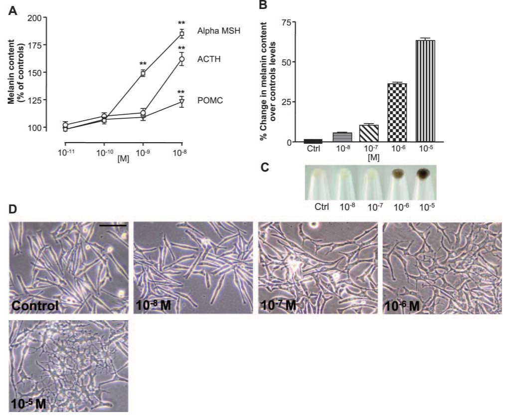
Proopiomelanocortin (POMC) can be processed to ACTH and melanocortin peptides. However, processing is incomplete in some tissues, leading to POMC precursor release from cells. This study examined POMC processing in human skin and the effect of POMC on the melanocortin-1 receptor (MC-1R) and melanocyte regulation. POMC was secreted by both human epidermal keratinocytes (from 5 healthy donors) and matched epidermal melanocytes in culture. Much lower levels of alpha-MSH were secreted and only by the keratinocytes. Neither cell type released ACTH. Cell extracts contained significantly more ACTH than POMC, and alpha-MSH was detected only in keratinocytes. Nevertheless, the POMC processing components, prohormone convertases 1, 2 and regulatory protein 7B2, were detected in melanocytes and keratinocytes. In contrast, hair follicle melanocytes secreted both POMC and alpha-MSH, and this was enhanced in response to corticotrophin-releasing hormone (CRH) acting primarily through the CRH receptor 1. In cells stably transfected with the MC-1R, POMC stimulated cAMP, albeit with a lower potency than ACTH, alpha-MSH, and beta-MSH. POMC also increased melanogenesis and dendricity in human pigment cells. This release of POMC from skin cells and its functional activity at the MC-1R highlight the importance of POMC processing as a key regulatory event in the skin.


Figures

Figure 1
Figure 1
More POMC than α-MSH is secreted by epidermal kertinocytes (A), epidermal melanocytes (B), and hair follicle melanocytes (C). Results are presented as fmol/flask to allow direct comparison of peptides in extracts and media. POMC shaded bars), ACTH (open bars), and α-MSH (hatched bars) were measured in epidermal melanocytes and matched keratinocytes from five normal volunteers after culturing for 24 h in serum-free medium. Levels of peptides in extracts from confluent cultures are compared with those secreted into culture medium (10 ml/flask). Hair follicle melanocytes from four separate subjects were cultured and the secreted levels of POMC and α-MSH were measured at 72 h. Limit of detection for ACTH and α-MSH in culture medium are <24 fmol/flask and in cell extracts are < 7 fmol/flask. Bars represent the mean ± sd (n=3).
Figure 2
Figure 2
A) PC1, PC2, and 7B2 are expressed in epidermal melanocytes in vitro. The POMC processing machinery was variably expressed in cultured epidermal melanocytes (passage 3). Expression correlated positively with differentiation status. Stronger expression was observed in differentiated dendritic melanocytes (arrow); it was weaker in less differentiated (flatter) melanocytes (arrowhead) and exhibited a cytoplasmic distribution pattern. Positive staining was observed with the antibody against gp100 (melanocyte lineage-specific marker). In the negative control, the primary antibody was replaced by preimmune serum from secondary antibody host. Scale bar: 17 µm. B) POMC processing machinery is expressed in epidermal keratinocytes in vitro. The expression of PC1, PC2, and 7B2 was variable in cultured epidermal keratinocytes (passage 2). Expression was higher in keratinocytes with a more differentiated phenotype (arrow) compared with those with a basal cell phenotype (arrowhead). Strong expression was confined to the perinuclear region, where it showed a granular distribution pattern. Positive staining was observed with the keratin-specific antibody, AE3. In the negative control, the primary antibody was replaced by preimmune serum from secondary antibody host. Scale bar: 17 µm
Figure 3
Figure 3
Effects of CRH on POMC release. A) CRH stimulation of POMC and α-MSH secreted into culture medium by human hair follicle melanocytes cultured for 72 h. Mean ± se (n=4 patients); *P < 0.05, **P < 0.01 relative to unstimulated control. B) Time course of the responsiveness of POMC to CRH in cultured human hair follicle melanocytes. Mean ± se (n=4 patients). C) Dose response of CRH agonists d-Pro5CRH, d-Pro4-r-urocortin, and d-Glu20 CRH on the release of POMC by cultured hair follicle melanocytes.
Figure 4
Figure 4
Activation of MC-1R by POMC. A) Gel chromatography profile of the human POMC after purification of POMC secreted into culture medium from the small cell lung carcinoma cell line DMS 79. Fractions were assayed in specific immunoassays for POMC and ACTH. All ACTH results were below the limit of detection. A) Western blot based on an antibody to ACTH 10–18 identifies the purified POMC (lane 3) with no band at the molecular weight of ACTH compared with synthetic ACTH (lane 1) and no recognition of synthetic β-LPH (lane 2). B) ChOK1 cells stably expressing the human MC-1R and a β-galactosidase reporter construct were incubated with increasing concentrations of β-MSH (▲), ACTH (Δ), αMSH (■), POMC (□), and γMSH (●). Ligand binding to the MC-1R stimulates cAMP, which activates the reporter gene to synthesize β-galactosidase. When substrate is added, this generates a color reaction that is measured as optical density (OD) units. Data points represent means of quadruplicate samples ± sd. The curves are representative of 3 independent experiments.
Figure 5
Figure 5
Effects of POMC on melanoma cells. A) POMC, ACTH, and α-MSH bind to the endogenous MC-1R in mouse melanoma cells. Melanin content was increased in response to POMC (▽), ACTH (○), and α-MSH (□) in mouse melanoma cells (S91) cultured for 6 days. Melanin content was determined spectrophotometrically at 475 nm against a synthetic melanin standard. Results were calculated as µg melanin per mg protein and expressed as % of the controls (n=3). B) POMC stimulated melanogenesis in human melanoma cells in culture. Melanin content was determined spectrophotometrically at 475 nm in cells stimulated with POMC at 10−8-10−5 M. Results are expressed as % increase in melanin content over control levels (n=3). C) The corresponding cell pellets revealed the melanogenic effects of POMC at the most potent concentrations. D) POMC stimulated dendricity in cultured human melanoma cells. Cell dendricity was assessed by counting cells with 3 or more dendrites before and after stimulation with POMC at 10−8-10−5 M. Melanoma cells were established in RPMI 1640 medium, FCS was omitted from the culture medium 24 h prior to stimulation and replaced with 0.25% BSA. POMC at 10−5 and 10−6 M were potent inducers of melanoma cell dendricity and subtle effects were observed at 10−7 M. Scale bar: 50 µm.(top right of control)
Figure 6
Figure 6
Effects of POMC on human epidermal melanocytes. AC) The MC-1R is present in cultured epidermal melanocytes and is found at the cell surface (arrowheads). Cells were cultured in FCS/BPE-free medium for 48 h prior to immunostaining, then fixed in 5% paraformaldehyde and the MC-1R was visualized by binding of an anti-rabbit antibody. Melanocyte identity was confirmed by gp100 expression (B). DF) Epidermal melanocyte cultures (passage 4) were established in bFGF/ET-1 supplemented medium. FCS and BPE were omitted from the culture medium 48 h prior to stimulation. A marked increase in cell dendricity was seen 72 h after POMC stimulation at 10−5 and 10−6 M concentrations compared with melanocytes maintained under basal medium conditions. Scale bar: 50 µm. G) Effect of POMC on melanin content in human epidermal melanocytes. Melanin content was determined spectrophotometrically (475 nm) after sodium hydroxide solubilization. H) Visible increases in melanogenesis after 10−5 and 10−6 M POMC stimulation were observed. Results are expressed as a percentage increase in melanin content over control unstimulated levels. I) Effect of POMC on proliferation in human epidermal melanocytes. Cell proliferation was assessed by determining cell counts before and after 10−5 and 10−6 M POMC stimulation. A marked increase in melanocyte proliferation was observed after 72 h stimulation with POMC. Results are expressed as a percentage increase in cell number over control unstimulated levels and experiments were performed in triplicate.

Similar articles

Cited by

References

    1. Slominski A, Wortsman J, Luger T, Paus R, Solomon S. Corticotropin releasing hormone and proopiomelanocortin involvement in the cutaneous response to stress. Physiol. Rev. 2000;80:979–1020. - PubMed
    1. Pritchard LE, Turnbull AV, White A. Proopiomelanocortin processing in the hypothalamus: impact on melanocortin signalling and obesity. J. Endocrinol. 2002;172:411–421. - PubMed
    1. Pritchard LE, Oliver RL, McLoughlin JD, Birtles S, Lawrence CB, Turnbull AV, White A. Proopiomelanocortin-derived peptides in rat cerebrospinal fluid and hypothalamic extracts: evidence that secretion is regulated with respect to energy balance. Endocrinology. 2003;144:760–766. - PubMed
    1. Gibson S, Crosby SR, Stewart MF, Jennings AM, McCall E, White A. Differential release of proopiomelanocortin-derived peptides from the human pituitary: evidence from a panel of two-site immunoradiometric assays. J. Clin. Endocrinol. Metab. 1994;78:835–841. - PubMed
    1. White A, Gibson S. ACTH precursors: biological significance and clinical relevance. Clin. Endocrinol. (Oxford) 1998;48:251–255. - PubMed

Publication types

LinkOut - more resources