. 2007 Jun 1;109(11):4825-931.
doi: 10.1182/blood-2006-08-043810. Epub 2007 Feb 22.

Interferon-producing killer dendritic cells (IKDCs) arise via a unique differentiation pathway from primitive c-kitHiCD62L+ lymphoid progenitors

Affiliations

Interferon-producing killer dendritic cells (IKDCs) arise via a unique differentiation pathway from primitive c-kitHiCD62L+ lymphoid progenitors

Robert S Welner et al. Blood. .

Abstract

Interferon-producing killer dendritic cells (IKDCs) have only recently been described and they share some properties with plasmacytoid dendritic cells (pDCs). We now show that they can arise from some of the same progenitors. However, IKDCs expressed little or no RAG-1, Spi-B, or TLR9, but responded to the TLR9 agonist CpG ODN by production of IFNgamma. The RAG-1(-)pDC2 subset was more similar to IKDCs than RAG-1(+) pDC1s with respect to IFNgamma production. The Id-2 transcriptional inhibitor was essential for production of IKDCs and natural killer (NK) cells, but not pDCs. IKDCs developed from lymphoid progenitors in culture but, unlike pDCs, were not affected by Notch receptor ligation. While IKDCs could be made from estrogen-sensitive progenitors, they may have a slow turnover because their numbers did not rapidly decline in hormone-treated mice. Four categories of progenitors were compared for IKDC-producing ability in transplantation assays. Of these, Lin(-)Sca-1(+)c-Kit(Hi)Thy1.1(-)L-selectin(+) lymphoid progenitors (LSPs) were the best source. While NK cells resemble IKDCs in several respects, they develop from different progenitors. These observations suggest that IKDCs may arise from a unique differentiation pathway, and one that diverges early from those responsible for NK cells, pDCs, and T and B cells.


Figures

Figure 1
Figure 1
IKDCs differ from otherwise similar Ly6C+ pDCs with respect to RAG-1 and patterns of gene expression. (A) The B220+RAG-1/GFP+ population freshly isolated from BM did not include IKDCs. (B) Conversely, BM IKDCs do not express RAG-1 detectable in RAG-1/GFP reporter mice or by RT-PCR. (C) Transcripts for a series of genes were measured in highly purified cells by RT-PCR. (D) Wild-type mice were compared with Id2 gene–targeted animals (Id2−/−) by flow cytometry. Mean ± SEM values are shown for NK1.1+ CD11c NK cells, B220+CD11c+CD19Ly-6C NK1.1+ IKDCs, and B220+CD11c+CD19Ly-6C+NK1.1 pDCs in BM. (E) Rigorously sorted DC subsets were placed in overnight cultures with CpG ODN, and supernatants were tested for the 3 indicated cytokines. The results are representative of 3 independent experiments.
Figure 2
Figure 2
Lymphoid progenitors are a source of IKDCs, and their formation is insensitive to Notch receptor ligation. LSKs and RAG-1+ ELPs were sorted from BM and placed in coculture with either vector (A) or Delta-like-1–transduced (B) OP9 stromal cells for 8 days in the presence of Flk-2/Flt-3 ligand. The indicated flow cytometry gates were used to discriminate B220+CD19CD11c+Ly6C IKDCs, B220+CD19CD11c+Ly6C+ pDCs, and B220CD19CD11c+ cDCs. Total numbers of recovered cells of each type were calculated and expressed in the graphs as yields per input progenitor (mean ± SEM). Asterisks denote significant differences (P < .05) by t test.
Figure 3
Figure 3
IKDCs are not rapidly depleted in estrogen-treated mice, but their functions may be hormone modulated. Mice were given subcutaneous time-release pellets containing placebo or 17β-estradiol. (A) IKDCs, pDC1s, pDC2s, and NK cells in BM were enumerated by flow cytometry and mean ± SEM values are shown (*P < .05). (B) Typical flow cytometry analyses at 2 weeks after treatment are shown. Average frequencies for each of the gated populations are given in the figure. (C) LSKs remaining in BM of mice treated for one week were also sorted and placed in stromal cell cocultures (OP9 + Flk-2/Flt-3 ligand) for an additional week. The results are given as yields of IKDCs per input progenitor. (D) IKDCs were recovered and double sorted from BM of mice treated for one week before being evaluated in overnight CpG-containing cultures for cytokine production. The data are representative of 3 similar experiments.
Figure 4
Figure 4
IKDCs develop efficiently from CD62L+ lymphocyte progenitors. (A) LSPs, ELPs, CMPs, and CLPs were sorted from Thy1.1 congenic RAG-1/GFP reporter mice, and 5 × 10³ cells of each were transferred to sublethally irradiated recipients. Flow cytometry analyses were then performed 16 days after transplantation. Absolute numbers of donor-derived pDC (B220+ CD19CD11c+ Ly6C+), IKDC (B220+ CD19CD11c+ Ly6C NK1.1+), and NK (NK1.1+ CD11c) BM cells per femur are given (* denotes significant differences [P < .05]). (B) LSPs (103/well) were placed in OP9 cocultures with Flk-2/Flt-3 ligand and IL-15. They were subcultured to fresh stromal cell monolayers after one week, and gated B220+CD19CD11c+ Ly6C cells were assessed for NK1.1 expression by flow cytometry at the indicated intervals.
Figure 5
Figure 5
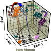
Possible relationship between IKDCs and other lineages in bone marrow. Lymphoid progenitors can be conveniently resolved on the basis of c-Kit, Sca-1, and RAG-1 densities as depicted here. Our new findings suggest that CD62L/L-selectin+ progenitors (LSPs) are the most effective source of IKDCs. In contrast, NK cells are normally produced from c-KitLo RAG-1+ prolymphocytes (including IL-7Rα+ common lymphoid progenitors [CLPs]). Although IKDCs do not express RAG-1, they could be made under experimental conditions from progenitors that express RAG-1 (not illustrated; see Figure 2A). Among other distinctions from pDCs, IKDC development was insensitive to Notch and dependent on the Id-2 transcriptional repressor (not illustrated).

Similar articles

Cited by

References

    1. Pelayo R, Hirose J, Huang J, et al. Derivation of two categories of plasmacytoid dendritic cells in murine bone marrow. Blood. 2005;105:4407–4415. - PMC - PubMed
    1. Chan CW, Crafton E, Fan HN, et al. Interferon-producing killer dendritic cells provide a link between innate and adaptive immunity. Nat Med. 2006;12:207–213. - PubMed
    1. Taieb J, Chaput N, Menard C, et al. A novel dendritic cell subset involved in tumor immunosurveillance. Nat Med. 2006;12:214–219. - PubMed
    1. Baba Y, Pelayo R, Kincade PW. Relationships between hematopoietic stem cells and lymphocyte progenitors. Trends Immunol. 2004;25:645–649. - PubMed
    1. Pelayo R, Welner RS, Nagai Y, Kincade PW. Life before the pre-B cell receptor checkpoint: specification and commitment of primitive lymphoid progenitors in adult bone marrow. Sem Immunol. 2006;18:2–11. - PubMed

Publication types