Comparative Study
. 2007 Mar 21;26(6):1591-601.
doi: 10.1038/sj.emboj.7601588. Epub 2007 Feb 22.

A conserved role for cytoplasmic poly(A)-binding protein 1 (PABPC1) in nonsense-mediated mRNA decay

Affiliations
Comparative Study

A conserved role for cytoplasmic poly(A)-binding protein 1 (PABPC1) in nonsense-mediated mRNA decay

Isabelle Behm-Ansmant et al. EMBO J. .

Abstract

The nonsense-mediated mRNA decay (NMD) pathway degrades mRNAs with premature translation termination codons (PTCs). The mechanisms by which PTCs and natural stop codons are discriminated remain unclear. We show that the position of stops relative to the poly(A) tail (and thus of PABPC1) is a critical determinant for PTC definition in Drosophila melanogaster. Indeed, tethering of PABPC1 downstream of a PTC abolishes NMD. Conversely, natural stops trigger NMD when the length of the 3' UTR is increased. However, many endogenous transcripts with exceptionally long 3' UTRs escape NMD, suggesting that the increase in 3' UTR length has co-evolved with the acquisition of features that suppress NMD. We provide evidence for the existence of 3' UTRs conferring immunity to NMD. We also show that PABPC1 binding is sufficient for PTC recognition, regardless of cleavage or polyadenylation. The role of PABPC1 in NMD must go beyond that of providing positional information for PTC definition, because its depletion suppresses NMD under conditions in which translation efficiency is not affected. These findings reveal a conserved role for PABPC1 in mRNA surveillance.


Figures
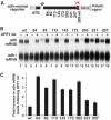
Figure 1
Figure 1
Boundary-dependent NMD in D. melanogaster. (A) Schematic representation of the adh reporters. Black boxes: exons; gray boxes: sequences derived from vector pAc5.1B; red box: V5 epitope; IN: intron. (B, C) S2 cells were transfected with vectors expressing adh-wt or the PTC reporters indicated above the lanes. A truncated version of adh (adhΔ) served as a transfection control. Transfected cells were then divided into two pools and treated with either GFP dsRNA (–) or UPF1 dsRNA (+). Total RNA samples were analyzed by Northern blot (B). The levels of the adh reporters were normalized to the levels of adhΔ mRNA. For each reporter, the normalized values obtained in UPF1-depleted cells were divided by those obtained in control cells (C). Mean values±s.d. of three independent experiments are shown. Kd: knockdown.
Figure 2
Figure 2
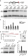
The ability of a PTC to trigger NMD depends on its distance from the 3′ UTR. (A) Schematic representation of the adh reporters. Symbols are as in Figure 1A. The distance of some PTCs to the polyadenylation site, as well as the positions of the insertion and deletion in adh-long and adh-short, respectively, are indicated. (B, C, E) S2 cells depleted of XRN1 were transfected with the reporters indicated above the lanes. RNA samples were analyzed by Northern blot (C, E) using a probe hybridizing to the 3′ UTR of the reporters (as shown in panel A). The levels of the 3′ decay intermediates were normalized to the levels of the full-length transcript (B). Mean values±s.d. of three independent experiments are shown. (D, F) S2 cells expressing the indicated adh reporters were treated with actinomycin D. Total RNA samples were collected at the indicated time points and analyzed by Northern blot. The levels of the adh reporters normalized to the levels of rp49 mRNA are plotted as a function of time. mRNA half-lives are indicated in brackets.
Figure 3
Figure 3
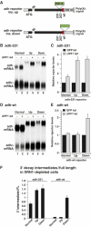
Redefinition of a natural stop as premature by increasing the length of the 3′ UTR. (A) Schematic representation of the adh reporters having an insertion upstream (ins. up) or downstream (ins. down) of the natural stop (green boxes). Symbols are as in Figure 1A. (B–E) S2 cells were transfected with vectors expressing the indicated reporters. Plasmid adhΔ served as a transfection control. Transfected cells were treated with GFP (–) or UPF1 (+) dsRNAs. Panels B and D show representative Northern blots. In panels C and E, the levels of the adh mRNA reporters were normalized to the levels of adhΔ mRNA in three independent experiments. For each reporter, these ratios were set to 1 in control cells treated with GFP dsRNA (black bars). Mean values±s.d. are shown. (F) The levels of the 3′ decay intermediates accumulating in XRN1-depleted cells were normalized to the levels of the full-length transcript. Mean values±s.d. of three independent experiments are shown.
Figure 4
Figure 4
Binding of PABPC1, regardless of cleavage and polyadenylation, is sufficient for PTC definition. (A) Schematic representation of the adh reporters in which the polyadenylation signal of SV40 is replaced by the histone H4 3′ stem–loop (SL) or a self-cleavable hammerhead ribozyme (HhR). Symbols are as in Figure 1A. The insertion of a poly(A) stretch is indicated by a green box. (B–E) S2 cells transfected with the reporters indicated above the panels were treated with GFP (–) or UPF1 (+) dsRNAs. Panels B and E show representative Northern blots. In panels C and D, the levels of the adh reporters were normalized to the levels of adhΔ mRNA in three independent experiments and analyzed as described in Figures 3C and E. (F, G) S2 cells were transfected with the indicated adh reporters. Total protein and RNA samples were analyzed by Western and Northern blots, respectively, and quantitated in three independent experiments.
Figure 5
Figure 5
Tethering of PABPC1 suppresses NMD in a polar manner. (A) Schematic representation of the adh-4BoxB reporters. Symbols are as in Figure 1A. White box: 4BoxB tethering sites. (B, C) S2 cells were transfected with the adh-4BoxB reporters, adhΔ and plasmids encoding the λN-peptide, λN-PABPC1 or untagged PABPC1 as indicated. Total RNA samples were analyzed by Northern blot (B). The levels of the adh reporters were normalized to the levels of adhΔ mRNA. For each reporter, these levels were set to 1 in cells expressing the λN-peptide alone (C, white bars). Mean values±s.d. of three independent experiments are shown. (D) S2 cells expressing Adh-113-4BoxB in the presence of the λN-peptide or λN-PABPC1 were treated with actinomycin D. Samples were collected at the indicated time points and analyzed by Northern blot. The levels of the adh mRNA normalized to the levels of rp49 mRNA are plotted as a function of time. The half-lives of the adh mRNA are indicated. (E–G) S2 cells were transfected with adh-wt-4BoxB or adh-113-4BoxB and plasmids encoding the proteins indicated above the lanes. Samples were analyzed as described in panel C. In panel E, the expression levels of PABPC1 and PABPN1 were analyzed by Western blot. ADH-V5 served as a transfection control.
Figure 6
Figure 6
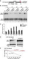
Tethered PABPC1 suppresses NMD. (A) Schematic representation of the CAT-4BoxB reporters. Symbols are as in Figure 5A. (B, C) S2 cells were transfected with vectors expressing CAT-4BoxB reporters and plasmids encoding the proteins indicated above the lanes. Total RNA samples were analyzed by Northern blot (B). In panel C, the levels of the CAT reporters were analyzed as described in Figure 5C. (D, E) S2 cells expressing CAT-126-4BoxB in the presence of the λN-peptide or λN-PABPC1 were treated with actinomycin D. Samples were analyzed as described in Figure 5D.
Figure 7
Figure 7
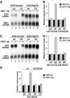
Hsp70 3′ UTR counteracts NMD. (A–D) S2 cells transfected with vectors expressing the reporters indicated above the panels were treated with GFP (–) or UPF1 (+) dsRNAs. Panels A and C show representative Northern blots. The levels of the adh or CAT reporters were normalized to the levels of adhΔ mRNA in three independent experiments (B, D). For each reporter, these normalized values were set to 1 in cells treated with GFP dsRNA (black bars). Mean values±s.d. are shown. (E) Cells were transfected with vectors expressing adh or adh-64 reporters carrying 5BoxB elements inserted in the 3′ UTR, adhΔ and plasmids encoding λN or λN-GW182. Transfected cells were treated with GFP or UPF1 dsRNAs as indicated. Total RNA samples were analyzed by Northern blot (not shown). The levels of the adh reporters were normalized to the levels of the adhΔ transfection control. For each reporter, these levels were set to 1 in control cells treated with GFP dsRNA (black bars). Mean values±s.d. of three independent experiments are shown.
Figure 8
Figure 8
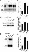
PABPC1 is required for NMD. (A–D) Cells transfected with the reporters indicated above the panels were treated with dsRNAs targeting GFP, UPF1 or PABPC1. Panel A shows a representative Northern blot. The levels of the adh mRNA reporters were normalized to the levels of adhΔ mRNA in three independent experiments. The normalized values obtained for adh-64 were divided by those obtained for adh-wt for each knockdown (to compensate for potential unspecific effects of the depletions), and set to 1 in cells treated with GFP dsRNA. Expression of ADH-V5 from the wild-type reporter was analyzed by Western blot (C). Tubulin served as a loading control. ADH-V5 protein levels were normalized to adh-wt mRNA levels in three independent experiments for each knockdown (D). (E, F) The levels of endogenous ODA mRNA in cells treated with GFP, UPF1 or PABPC1 dsRNAs were analyzed by Northern blot (E), and normalized to the levels of rp49 mRNA in three independent experiments. These ratios were set to 1 in cells treated with GFP dsRNA. Mean values±s.d. from three independent experiments are shown.

References

    1. Adamson TE, Price DH (2003) Cotranscriptional processing of D. melanogaster histone mRNAs. Mol Cell Biol 23: 4046–4055 - PMC - PubMed
    1. Amrani N, Ganesan R, Kervestin S, Mangus DA, Ghosh S, Jacobson A (2004) A faux 3′-UTR promotes aberrant termination and triggers nonsense-mediated mRNA decay. Nature 432: 112–118 - PubMed
    1. Amrani N, Sachs MS, Jacobson A (2006) Early nonsense: mRNA decay solves a translational problem. Nat Rev Mol Cell Biol 7: 415–425 - PubMed
    1. Baker KE, Parker R (2006) Conventional 3′ end formation is not required for NMD substrate recognition in Saccharomyces cerevisiae. RNA 12: 1441–1445 - PMC - PubMed
    1. Behm-Ansmant I, Rehwinkel J, Doerks T, Stark A, Bork P, Izaurralde E (2006) mRNA degradation by miRNAs and GW182 requires both CCR4:NOT deadenylase and DCP1:DCP2 decapping complexes. Genes Dev 20: 1885–1898 - PMC - PubMed

Publication types

LinkOut - more resources