. 2007 Mar 7;26(5):1410-22.
doi: 10.1038/sj.emboj.7601580. Epub 2007 Feb 22.

Ubiquitination of the GTPase Rap1B by the ubiquitin ligase Smurf2 is required for the establishment of neuronal polarity

Affiliations

Ubiquitination of the GTPase Rap1B by the ubiquitin ligase Smurf2 is required for the establishment of neuronal polarity

Jens C Schwamborn et al. EMBO J. .

Retraction in

Abstract

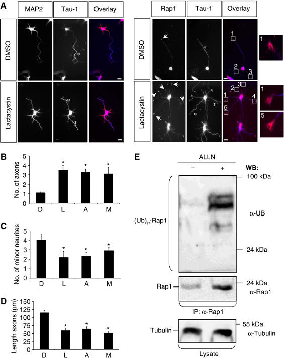
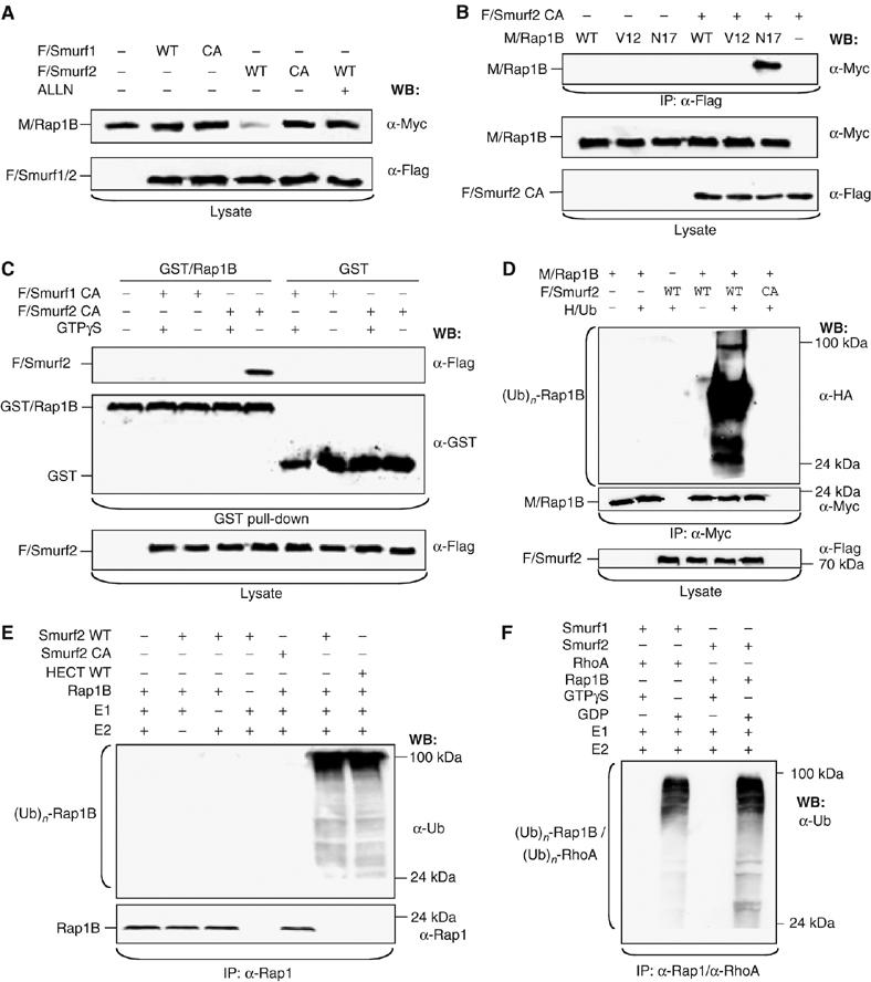
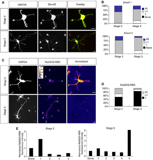
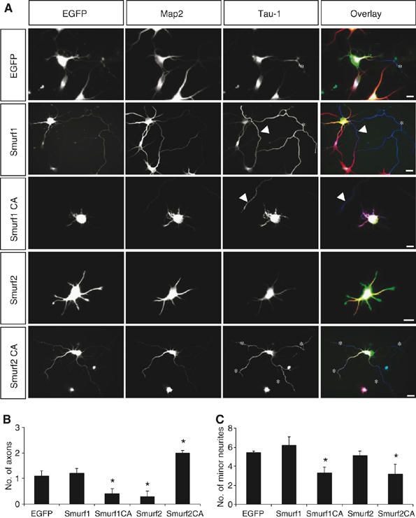
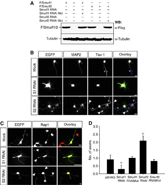
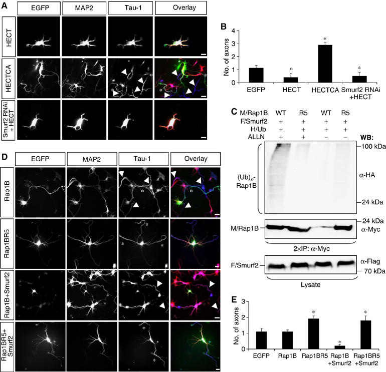
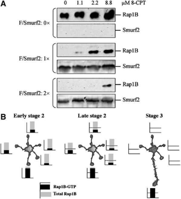
The development of a polarised morphology with multiple dendrites and a single axon is an essential step in the differentiation of neurons. The establishment of neuronal polarity is directed by the sequential activity of the GTPases Rap1B and Cdc42. Rap1B is initially present in all neurites of unpolarised neurons, but becomes restricted to the tip of a single process during the establishment of neuronal polarity where it specifies axonal identity. Here, we show that the ubiquitin ligases Smad ubiquitination regulatory factor-1 (Smurf1) and Smurf2 are essential for neurite growth and neuronal polarity, respectively, and regulate the GTPases Rho and Rap1B in hippocampal neurons. Smurf2 is required for the restriction of Rap1B to a single neurite. Smurf2 ubiquitinates inactive Rap1B and initiates its degradation through the ubiquitin/proteasome pathway (UPS). Degradation of Rap1B restricts it to a single neurite and thereby ensures that neurons extend a single axon.


Figures

Figure 1
Figure 1
Inhibition of the proteasome disrupts neuronal differentiation. (A–D) Hippocampal neurons were cultured in the presence of solvent (DMSO), 1.5 μM clasto-Lactacystin β-Lactone (Lactacystin), 40 μM ALLN, or 1.5 μM MG-132 (all dissolved in DMSO) for 48 h and analysed at 3 d.i.v. (stage 3) by staining with an anti-MAP2 antibody (A; red), the Tau-1 (blue), and an anti-Rap1 (red) antibody. Lactacystin induced the formation of multiple axons (asterisks) whose growth cones were all positive for Rap1B (A, arrows). Insets show higher magnifications of the marked growth cones. Axons identified by Tau-1 immunoreactivity are marked by asterisks. The scale bar is 12 μm. (B–D) The effect of Lactacystin (L), ALLN (A), or MG-132 (M) was analysed by determining the number of axons (B) or minor neurites (C) per cell and the length of axons (E) (means±s.e.m.; *P<0.001 compared to control (DMSO (D); three independent experiments). (E) Hippocampal neurons were incubated overnight with solvent (DMSO, −) or 40 μM ALLN (+). Rap1B was precipitated from cell lysates using an anti-Rap1 antibody (IP) and analysed by Western blot using anti-ubiquitin, anti-Rap1, anti-α-tubulin, and the Tau-1 antibodies (WB). Luminescence signals from Western blots were collected for 1 s (s) and 30 min to detect poly-ubiquitinated Rap1B visible after proteasome inhibition. Staining for tubulin confirmed the loading of comparable amounts of protein. The slight increase in the amount of α-tubulin upon inhibition of the proteasome is consistent with the formation of supernumerary axons.
Figure 2
Figure 2
Smurf2 ubiquitinates Rap1B. (A) HEK 293T cells were transfected with expression vectors for wild -type (WT) or catalytically inactive (CA: Smurf1C699A or Smurf2C716A) Flag-tagged Smurf1 (F/Smurf1) or Smurf2 (F/Smurf2) and myc-tagged Rap1B (M/Rap1B) as indicated. ALLN (40 μM) was added 16 h before cells were lysed as indicated and protein expression in cell lysates analysed by Western blot using anti-myc and anti-Flag antibodies. (B) HEK 293T cells were transfected with expression vectors for Flag-Smurf2C716A and WT or mutant myc-Rap1B (constitutively active: V12; dominant-negative: N17). Smurf2C716A was precipitated from cell lysates using the anti-Flag antibody (IP) and Rap1B detected by Western blot (WB) using the anti-myc antibody. The expression of equivalent amounts of protein was confirmed by Western blot. (C) HEK 293T cells were transfected with expression vectors for Flag-Smurf1C699A or Flag-Smurf2C716A and the cell lysates incubated with bacterially expressed GST or GST-Rap1B coupled to glutathione-Sepharose and preloaded with 0.7 mM GTPγS (+) as indicated. Bound Smurf2 (top panel) was detected in Western blots using an anti-Flag antibody. The expression of equivalent amounts of Smurf2 and GST-Rap1B or GST was confirmed by Western blot. (D) HEK 293T cells were transfected with vectors for HA-tagged ubiquitin (HA/Ub), myc-Rap1B (M/Rap1B), and WT or catalytically inactive Flag-Smurf2 (CA), incubated overnight with 40μM ALLN, and Rap1B was immunoprecipitated with an anti-myc antibody. Ubiquitin-conjugated Rap1B ((Ub)n-Rap1B) and Rap1B were detected with anti-HA and anti-myc antibodies, respectively. Flag-Smurf2 and myc-Rap1B expression was confirmed by Western blot of cell lysates using anti-Flag and anti-myc antibodies, respectively. (E) Bacterially expressed and purified Rap1B and WT or catalytically inactive Smurf2 (CA) or the HECT domain of Smurf2 were used for an in vitro ubiquitination assay after combining the indicated proteins. Recombinant ubiquitin, ubiquitin-activating enzyme (E1), and UbcH5c (E2) were added to the reaction mixture as indicated. After performing the in vitro ubiquitination, Rap1B was immunoprecipitated from the samples (IP) using an anti-Rap1 antibody. Poly-ubiquitinated Rap1B ((Ub)n-Rap1B) and Rap1B were detected by Western blot using anti-ubiquitin and anti-Rap1B antibodies. (F) Bacterially expressed and purified Rap1B, RhoA, Smurf1, and Smurf2 were used for an in vitro ubiquitination assay after combining the indicated proteins. Rap1B and RhoA were preloaded with 0.7 mM GDP or 0.7 mM GTPγS. Recombinant ubiquitin, ubiquitin-activating enzyme (E1), and UbcH5 (E2) were added to the reaction mixture as indicated. After performing the in vitro ubiquitination, Rap1B and RhoA were immunoprecipitated from the samples (IP) using an anti-Rap1 or an anti-RhoA antibody. Ubiquitinated GTPases ((Ub)n-Rap1B/(Ub)n-RhoA) were detected by Western blot using an anti-ubiquitin antibody.
Figure 3
Figure 3
Distribution of Smurf1 and Smurf2 and active Rap1B during neuronal differentiation. (A, B) Hippocampal neurons were fixed at stage 2 or 3 and stained with anti-Smurf1 (not shown) or anti-Smurf2 antibodies (A, red). Neurons were analysed by confocal microscopy. Projections of z-stacks that contained the complete cell are shown. The immunofluorescence signals were normalised for cell volume by labelling with CMFDA (A, green). The percentage of cells that contain Smurf1 (B, top) or Smurf2 (B, bottom) in all, several (>1; the axon and one or more minor neurites in stage 3), one (1; the axon in stage 3), or none of the neurites is shown. Arrows indicate staining at the tips of neurites. At stage 2, 71±5% of the cells contained Smurf2 in more than one but not all neurites, 23±3% in all neurites. 5±2% of the neurons showed Smurf2 in a single and 1±1% in none of the neurites (n=75; three experiments). At stage 3, Smurf2 was restricted to the axon in 17±3% of the cells (1). 7±1% contained Smurf2 in all growth cones and 75±5% in the axon and one or more minor neurites in addition (>1; n=150; three independent experiments). For each growth cone, the normalised fluorescence intensity was calculated as the ratio of the fluorescence intensities of Smurf1 or Smurf2 and CMFDA in the growth cone. A growth cone was scored as positive for Smurf1 or Smurf2 if the value for the relative fluorescence intensity was at least three times higher than the value for the background. Scale bars are 12 μm. (C–E) Hippocampal neurons were fixed at stage 2 or 3, incubated with bacterially expressed GST-RalGDS-RBD that specifically binds Rap1B-GTP, and bound GST-RalGDS-RBD detected in situ with an anti-GST antibody. The immunofluorescence signals were normalised for cell volume by labelling with CMFDA. The intensity of fluorescence (RalGDS-RBD) and the normalised fluorescence intensity (normalised) were colour-coded. The normalised fluorescence intensity was calculated as the ratio of the fluorescence intensities of RalGDS-RBD and CMFDA in the growth cone. Blue indicates weak staining, white strong labelling. The distribution of RalGDS-RBD binding sites, indicating active Rap1B, was analysed by determining the percentage of cells that showed binding in all, several (>1; the axon and one or more minor neurites in stage 3), or one (1; the axon in stage 3) of the neurites (D). (E) The normalised immunofluorescence intensity (arbitrary units) in the growth cones marked in (C) is shown. A growth cone was scored as positive for active Rap1B when the normalised fluorescence intensity was at least double the value of that in the soma.
Figure 4
Figure 4
Expression of Smurf1 and Smurf2 disrupts neuronal differentiation. (A–C) Hippocampal neurons were transfected 2 h after plating with expression vectors for EGFP or EGFP and Smurf1, Smurf1C699A, Smurf2, or Smurf2C716A (A, green). Transfected cells were analysed at 3 d.i.v. by staining with the Tau-1 (blue; asterisks) and anti-MAP2 antibodies (red). The development of neuronal polarity was analysed by counting the number of axons (B) and minor neurites (C) per cell (means±s.e.m.; *P<0.001 compared to EGFP; five independent experiments). Axons extended by untransfected cells are marked by arrowheads. Scale bars are 12 μm.
Figure 5
Figure 5
Smurf2 knockdown induces supernumerary axons. (A) HEK 293T cells were transfected with Flag-tagged Smurf1 (F/Smurf1) or Smurf2 (F/Smurf2) and expression vectors for shRNAs directed against Smurf1 (S1 RNAi) or Smurf2 (S2 RNAi) as indicated. As control, Flag-Smurf1 or Flag-Smurf2 was cotransfected with expression vectors for inactive shRNAs (Mut). Smurf1 and Smurf2 expression was determined by Western blots of cell lysates using an anti-Flag antibody. The loading of equivalent amounts of protein was confirmed by using an anti-α-tubulin antibody. (B, D) Hippocampal neurons were transfected with pSHAG-1 (Mock) or expression vectors for shRNAs directed against Smurf1 (S1 RNAi) or Smurf2 (S2 RNAi) and EGFP (B, green). As control, constructs for inactive shRNAs (Mut) were used. (B) Transfected cells were analysed at 3 d.i.v. (stage 3) by staining with the Tau-1 (blue) and anti-MAP2 antibodies (red). Arrowheads mark the soma or axon of untransfected neurons. The development of neuronal polarity was analysed by counting the number of axons (D) and minor neurites (Supplementary Figure S4C) per cell (mean±s.e.m.; *P<0.001 compared to mock; n=3 independent experiments). (C) To analyse the effect of a Smurf1 or Smurf2 knockdown, neurons were stained with an anti-Rap1 antibody. In control transfections, 82±7% of the neurons (n=53; three experiments) contained Rap1B in a single neurite (arrow). After knockdown of Smurf1, 71±6% of the neurons still showed Rap1B in a single neurite (arrow, n=47). Rap1B was present in all supernumerary axons after knockdown of Smurf2 (arrows). Rap1 immunoreactivty in the soma or in axons of untransfected cells is marked by arrowheads in (B) and (C). Scale bars are 12 μm.
Figure 6
Figure 6
Mutation of a single lysine residue blocks Smurf2-dependent inhibition of Rap1B. (A, B) Hippocampal neurons were transfected 2 h after plating with expression vectors for EGFP-Smurf2-HECT (HECT), -Smurf2-HECT C716A (HECTCA), or EGFP-Smurf2-HECT and an shRNA directed against Smurf2 (A, green). Scale bars are 12 μm. (C) HEK 293T cells were transfected with vectors for HA-tagged ubiquitin (HA/Ub), myc-Rap1B (M/Rap1B, WT) or myc-Rap1BR5 (R5), and Flag-Smurf2 (F/Smurf2), incubated overnight with 40 μM ALLN, and Rap1B immunoprecipitated with an anti-myc antibody. Ubiquitin-conjugated Rap1B ((Ub)n-Rap1B) and Rap1B were detected with anti-HA and anti-myc antibodies, respectively. Flag-Smurf2 and myc-Rap1B expression was confirmed by Western blot of cell lysates using anti-Flag and anti-myc antibodies. (D, E) Hippocampal neurons were transfected 2 h after plating with expression vectors for Rap1B, Rap1BR5, Rap1B and EGFP-Smurf2, or Rap1BR5 and EGFP-Smurf2 (D, green). Transfected cells were analysed at 3 d.i.v. by staining with the Tau-1 (blue; asterisks) and anti-MAP2 antibodies (red). (B, E) The development of neuronal polarity was analysed by determining the number of axons per cell (means±s.e.m.; *P<0.001 compared to EGFP; n=3 independent experiments). Axons extended by untransfected cells are marked by arrowheads in (A) and (D).
Figure 7
Figure 7
The concentration of Smurf2 determines the extent of Rap1B activation required to protect it from degradation. (A) HEK 293T cells were cotransfected with a constant amount of an expression vector for myc-tagged Rap1B (M/Rap1B) and increasing amounts of a vector for Flag-tagged Smurf2 (F/Smurf2) as indicated. 8-CPT was added at the indicated concentrations to activate endogenous Epac 16 h before cell lysates were analysed by Western blot using anti-myc and anti-Flag antibodies. (B) In early stage 2 neurons, comparable amounts of Rap1B protein are found in all neurites (Schwamborn and Püschel, 2004). However, one neurite contains a higher amount of active Rap1B than the other neurites, reflecting intrinsic differences between these neurites (Bradke and Dotti, 1997; Da Silva et al, 2005; de Anda et al, 2005). In late stage 2 neurons, Smurf2 is transported into all neurites and initiates the degradation of Rap1B in those where Rap1B activity is below the threshold defined by the local concentration of Smurf2. The degradation of Rap1B removes it from all but a single neurite, which becomes the axon. Active Rap1B together with Smurf1 reduces Rho activity to allow the rapid extension of the axon (Yamada et al, 2005).

Similar articles

Cited by

References

    1. Bos JL, de Rooij J, Reedquist KA (2001) Rap1 signalling: adhering to new models. Nat Rev Mol Cell Biol 2: 369–377 - PubMed
    1. Bradke F, Dotti CG (1997) Neuronal polarity: vectorial cytoplasmic flow precedes axon formation. Neuron 19: 1175–1186 - PubMed
    1. Bradke F, Dotti CG (2000) Establishment of neuronal polarity: lessons from cultured hippocampal neurons. Curr Opin Neurobiol 10: 574–581 - PubMed
    1. Campbell DS, Holt CE (2001) Chemotropic responses of retinal growth cones mediated by rapid local protein synthesis and degradation. Neuron 32: 1013–1026 - PubMed
    1. Campbell DS, Holt CE (2003) Apoptotic pathway and MAPKs differentially regulate chemotropic responses of retinal growth cones. Neuron 37: 939–952 - PubMed

Publication types

MeSH terms

LinkOut - more resources