. 2007 Mar 7;26(5):1423-33.
doi: 10.1038/sj.emboj.7601570. Epub 2007 Feb 22.

Trogocytosis-based generation of suppressive NK cells

Affiliations

Trogocytosis-based generation of suppressive NK cells

Julien Caumartin et al. EMBO J. .

Abstract

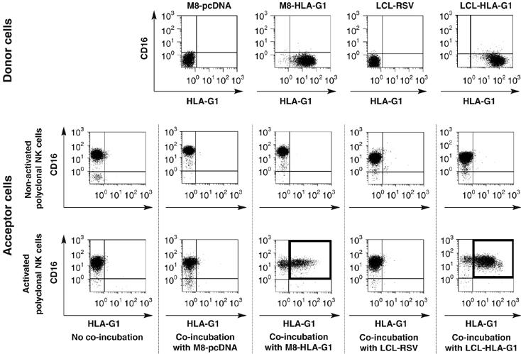
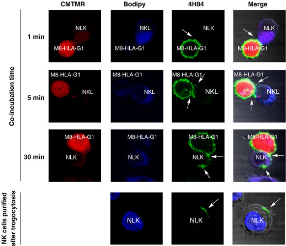
Trogocytosis is a fast uptake of membranes and associated molecules from one cell by another. Trogocytosis between natural killer (NK) cells and tumors is already described, but the functional relevance of NK-tumor targets material exchange is unclear. We investigated whether the immunosuppressive molecule HLA-G that is commonly expressed by tumors in vivo and known to block NK cytolytic function, could be transferred from tumor cells to NK cells, and if this transfer had functional consequences. We show that activated NK cells acquire HLA-G1 from tumor cells, and that upon this acquisition, NK cells stop proliferating, are no longer cytotoxic, and behave as suppressor cells. Such cells can inhibit other NK cells' cytotoxic function and protect NK-sensitive tumor cells from cytolysis. These data are the first demonstration that trogocytosis of HLA-G1 can be a major mechanism of immune escape that acts through effector cells made to act as suppressor cells locally, temporarily, but efficiently. The broader consequences of membrane sharing between immune and non-immune cells on the function of effectors and the outcome of immune responses are discussed.


Figures

Figure 1
Figure 1
HLA-G1 is found on activated NKL cells after coincubation with HLA-G1-positive melanoma cells. Non-activated and IL-2-activated NKL cells were coincubated for 1 h with adherent HLA-G1-negative M8-pcDNA cells and HLA-G1-positive M8-HLA-G1 cells. After coincubation, non-adherent NKL cells were recovered and their HLA-G1 cell-surface expression was evaluated. NK cells were distinguished from potential M8 contamination by size and CD45 expression. CD45 versus HLA-G1 expression is shown for M8 donor cells and for activated and non-activated NKL cells before and after a 1 h coincubation with M8 cells and isolation. Results shown are from one representative experiment out of five.
Figure 2
Figure 2
HLA-G1 is found on activated polyclonal NK cells (pNK) after coincubation with HLA-G1 tumor cells. Non-activated and IL-2-activated pNK cells were coincubated for 1 h with HLA-G1-negative M8-pcDNA or LCL-RSV cells, and HLA-G1-positive M8-HLA-G1 or LCL-HLA-G1 cells. After coincubation, pNK cells were recovered and their HLA-G1 cell-surface expression was evaluated. pNK cells were distinguished from potential donor cell contamination by size and CD45 and CD16 expression. CD16 versus HLA-G1 expression is shown for donor cells and for CD45-positive activated and non-activated pNK cells before and after coincubation with donor cells. Results shown are from one representative experiment out of eight.
Figure 3
Figure 3
Parameters of HLA-G1 trogocytosis mechanism. (A) NK cell-surface HLA-G1 is acquired from tumor cells and not endogenously produced: activated NKL cells were incubated or not with M8-HLA-G1 cells and their HLA-G1 cell-surface expression and corresponding transcription of endogenous HLA-G1 were investigated. Plots: cell-surface expression of HLA-G1 (native or recombinant) by flow cytometry. Gels: transcription levels of native (not transfected) HLA-G by RT–PCR. JEG-3 cells were used as positive controls and M8-HLA-G1 cells were used as negative controls for native HLA-G transcription. β-Actin was used as an internal standard. Data are representative of three independent experiments. (B) Acquisition of HLA-G1 by NKL cells is cell-to-cell contact dependent and not due to HLA-G1 shedding. NKL cells and M8-HLA-G1 cells were coincubated for 30 min together (no transwell) or separated by a semipermeable membrane (transwell) before analysis of NKL HLA-G1 cell-surface expression. Data are mean±s.d. of 10 independent experiments. Coincubation with M8-HLA-G1-GFP: M8 cells transfected with HLA-G1 fused to EGFP at its intracellular part were used as HLA-G1 donors. After 1 h of coincubation, the presence of HLA-G1-EGFP was detected on NKL cells by flow cytometry. (C) HLA-G1 does not require interaction with its receptors to transfer from target cells to NK cells. HLA-G1 interactions with its receptors on NKL cells were prevented by masking HLA-G1 or ILT2 with blocking antibodies before coincubation. Transfer of HLA-G1 to NKL cells was analyzed by flow cytometry. Data are mean±s.d. of three independent experiments. (D) HLA-G1 acquisition kinetics by activated NKL and pNK cells. Activated NKL or activated pNK cells were coincubated with M8-HLA-G1 cells for the indicated times and NK-HLA-G1 expression was determined by flow cytometry. The results are mean±s.d. of five independent experiments. (E) Lifetime of acquired HLA-G1 at the surface of activated NKL cells. NKL cells that had acquired HLA-G1 from M8-HLA-G1 cells were purified, put back in culture, and their cell-surface HLA-G1 levels were analyzed at the indicated times. Data are representative of seven independent experiments.
Figure 4
Figure 4
Visualization of HLA-G1 transfer from M8-HLA-G1 cells to NKL cells by confocal microscopy. Red: CMTMR cytoplasmic labeling of M8-HLA-G1 cells; blue: BODIPY cytoplasmic labeling of NKL cells; green: HLA-G labeling by anti-HLA-G antibody 4H84. Pictures were taken at the indicated time after the beginning of coincubation. Arrows indicate areas of interest. M8-HLA-G1/NKL conjugates (top lines) and isolated NKL cells (bottom line) are shown. Data are representative of three independent experiments.
Figure 5
Figure 5
Acquisition of HLA-G1 by NK cells stops their proliferation. (A) NKL, NKL-HLA-G1acq−, and NKL-HLA-G1acq+ cells, and (B) pNK, pNK-HLA-G1acq−, and pNK-HLA-G1acq+ cells were generated in the presence of the indicated antibodies, isolated, and put back in culture. Proliferation was evaluated for the 12-h period before the indicated times. Data are representative of seven independent experiments.
Figure 6
Figure 6
HLA-G1 acquisition makes activated NK cells non-cytotoxic. The capability to lyse M8-pcDNA target cells was investigated by 51Cr release assay for (A) NKL-HLA-G1acq− and NKL-HLA-G1acq+ cells, and (B) pNK-HLA-G1acq− and pNK-HLA-G1acq+ taken immediately or 24 h after HLA-G1 acquisition (NKL only). When indicated, isotypic control or blocking mAb was added before trogocytosis and was kept present throughout the experiment. Results shown for NKL cells are mean±s.d. of three independent experiments, and those for pNK are representative of three experiments.
Figure 7
Figure 7
NK-HLA-G1acq+ cells are immunosuppressive. (A) NKL-HLA-G1acq− and NKL-HLA-G1acq+ cells, and (B) pNK-HLA-G1acq− and pNK-HLA-G1acq+ cells taken immediately or 24 h after HLA-G1 acquisition (NKL only), were added as third-party cells to cytotoxic assay between autologous activated NK and M8-pcDNA cells. E:T:S, effector:target:suppressor ratio. When indicated, isotypic control or blocking mAb was added before trogocytosis and kept present throughout the experiment. Results shown for NKL cells are mean±s.d. of three independent experiments, and those for pNK are representative of three experiments.

References

    1. Bahri R, Hirsch F, Josse A, Rouas-Freiss N, Bidere N, Vasquez A, Carosella ED, Charpentier B, Durrbach A (2006) Soluble HLA-G inhibits cell cycle progression in human alloreactive T lymphocytes. J Immunol 176: 1331–1339 - PubMed
    1. Carlin LM, Eleme K, McCann FE, Davis DM (2001) Intercellular transfer and supramolecular organization of human leukocyte antigen C at inhibitory natural killer cell immune synapses. J Exp Med 194: 1507–1517 - PMC - PubMed
    1. Carosella ED, Moreau P, Le Maoult J, Le Discorde M, Dausset J, Rouas-Freiss N (2003) HLA-G molecules: from maternal–fetal tolerance to tissue acceptance. Adv Immunol 81: 199–252 - PubMed
    1. Game DS, Rogers NJ, Lechler RI (2005) Acquisition of HLA-DR and costimulatory molecules by T cells from allogeneic antigen presenting cells. Am J Transplant 7: 1614–1625 - PubMed
    1. Horuzsko A, Lenfant F, Munn DH, Mellor AL (2001) Maturation of antigen-presenting cells is compromised in HLA-G transgenic mice. Int Immunol 13: 385–394 - PubMed

MeSH terms