Review
. 2007 Mar;7(3):192-201.
doi: 10.1038/nrc2073.

Candidate mechanisms for chemotherapy-induced cognitive changes

Affiliations
Review

Candidate mechanisms for chemotherapy-induced cognitive changes

Tim A Ahles et al. Nat Rev Cancer. 2007 Mar.

Abstract

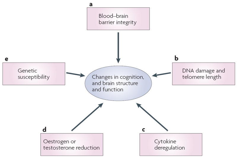
The mechanism(s) for chemotherapy-induced cognitive changes are largely unknown; however, several candidate mechanisms have been identified. We suggest that shared genetic risk factors for the development of cancer and cognitive problems, including low-efficiency efflux pumps, deficits in DNA-repair mechanisms and/or a deregulated immune response, coupled with the effect of chemotherapy on these systems, might contribute to cognitive decline in patients after chemotherapy. Furthermore, the genetically modulated reduction of capacity for neural repair and neurotransmitter activity, as well as reduced antioxidant capacity associated with treatment-induced reduction in oestrogen and testosterone levels, might interact with these mechanisms and/or have independent effects on cognitive function.


Conflict of interest statement

Competing interests statement

The authors declare no competing financial interests.

Figures

Figure 1
Figure 1. Neuroimaging methods relevant to the assessment of cognitive changes
a ∣ Structural magnetic resonance imaging (MRI) provides a high resolution picture of normal neuroanatomic details and atrophy (T1-weighted scans), and visible pathology such as microvascular and inflammatory lesions (T2-weighted scans; fluid-attenuated inversion recovery (FLAIR) scans). Semi-automated methods can be used to segment or classify the structural images into the main tissue compartments, including grey and white matter, cerebrospinal fluid and hyperintense lesions, which reflect microvascular changes or areas of demyelination. Software is available to quantify the volume and other characteristics of each tissue type. b ∣ Diffusion tensor imaging (DTI) is a recently developed technique that can be used to assess pathological changes in grey matter (increased mean diffusivity) and the loss of integrity of white matter fibre bundles (decreased fractional anisotropy). Tractography software enables the identification of directional fibre bundles such as subregions of the corpus callosum (shown). c ∣ Functional MRI (fMRI) uses blood-oxygen-level dependent (BOLD) contrast or perfusion measurements to assess the functional activation of cortical and subcortical regions during the performance of cognitive or sensorimotor tasks in the scanner. Bilateral frontal and parietal activation can be seen, representing the mean activation observed in a group of healthy individuals performing a working memory task. d ∣ Positron emission tomography (PET) using the fluorodeoxyglucose (FDG) radiotracer provides a measure of neuronal metabolism. Other applications of molecular imaging methods such as PET provide data on cerebral blood flow or specific neurotransmitter–receptor systems. Most of these methods have not yet been examined in systematic, prospective studies of chemotherapy-induced or cancer-associated cognitive changes, but these approaches hold promise for identifying the neural bases of such changes.
Figure 2
Figure 2. Candidate mechanisms
Several candidate mechanisms for chemotherapy-associated changes in cognitive function and brain structure and function are proposed. a ∣ Although common chemotherapeutic agents do not readily cross the blood–brain barrier, recent data from animal studies suggest that even small doses of chemotherapy can cause cell death and reduce cell division in structures relevant for cognition. b ∣ Chemotherapy has been associated with DNA damage and telomere shortening, both of which have been implicated in neural degeneration and the development of neurodegenerative disorders with cognitive components (for example, Alzheimers disease and mild cognitive impairment). c ∣ Chemotherapy-induced cognitive changes might be associated with neurotoxic effects of inflammation and cytokine deregulation. d ∣ Oestrogen and testosterone levels can be reduced secondary to chemotherapy and as a result of hormonal treatments for cancer. Reduction in hormonal levels has been associated with cognitive decline. e ∣ Genetic variability in blood–brain barrier transporters, DNA-repair mechanisms, rate of telomere shortening, cytokine regulation, neuronal repair and plasticity, and neurotransmission could all increase vulnerability to cognitive changes associated with chemotherapy.

References

    1. Silberfarb PM. Chemotherapy and cognitive defects in cancer patients. Annu Rev Med. 1983;34:35–46. - PubMed
    1. Wieneke MH, Dienst ER. Neuropsychological assessment of cognitive functioning following chemotherapy for breast cancer. Psychooncology. 1995;4:61–66.
    1. van Dam FS, et al. Impairment of cognitive function in women receiving adjuvant treatment for high-risk breast cancer: high-dose versus standard-dose chemotherapy. J Natl Cancer Inst. 1998;90:210–218. comment. - PubMed
    1. Schagen SB, et al. Cognitive deficits after postoperative adjuvant chemotherapy for breast carcinoma. Cancer. 1999;85:640–650. - PubMed
    1. Brezden CB, et al. Cognitive function in breast cancer patients receiving adjuvant chemotherapy. J Clin Oncol. 2000;18:2695–2701. - PubMed

Publication types

Substances