

Review
doi: 10.1038/nrm2125.
Matrix metalloproteinases and the regulation of tissue remodelling
Affiliations
- PMID: 17318226
- PMCID: PMC2760082
- DOI: 10.1038/nrm2125
Item in Clipboard
Review
Matrix metalloproteinases and the regulation of tissue remodelling
Nat Rev Mol Cell Biol.
2007 Mar.
Abstract
Matrix metalloproteinases (MMPs) were discovered because of their role in amphibian metamorphosis, yet they have attracted more attention because of their roles in disease. Despite intensive scrutiny in vitro, in cell culture and in animal models, the normal physiological roles of these extracellular proteases have been elusive. Recent studies in mice and flies point to essential roles of MMPs as mediators of change and physical adaptation in tissues, whether developmentally regulated, environmentally induced or disease associated.
Conflict of interest statement
The authors declare no competing financial interests.
Figures
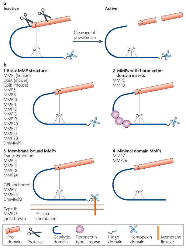
a | Matrix metalloproteinases (MMPs) are expressed as pro-proteins. A conserved Cys residue in the pro-domain coordinates the zinc ion, which would otherwise be used for catalysis. The pro-domain is removed by a combination of a cleavage in the domain and a cleavage between the pro-domain and the catalytic domain. b | Most MMPs share a conserved domain structure of pro-domain, catalytic domain, hinge region and hemopexin domain (1). All MMPs are synthesized with a signal peptide, which is cleaved during transport through the secretory pathway. MMP2 and MMP9 have three fibronectin type II repeats in their catalytic domains (2). Membrane type MMPs (MT-MMPs) are linked to the plasma membrane either by a transmembrane domain or by a glycosylphosphatidylinositol (GPI) linkage, attached to the hemopexin domain (3). Minimal MMPs lack the hinge and hemopexin domains (4). MMP21 has a truncated hinge domain. Drosophila melanogaster DmMMP2 has an insertion of 214 amino acids into its hinge domain. MMP23 (not shown) has a non-conserved N-terminal domain that consists of an immunoglobulin IgC2 domain and a ShKT domain; it is unclear if MMP23 contains a Cys residue switch.
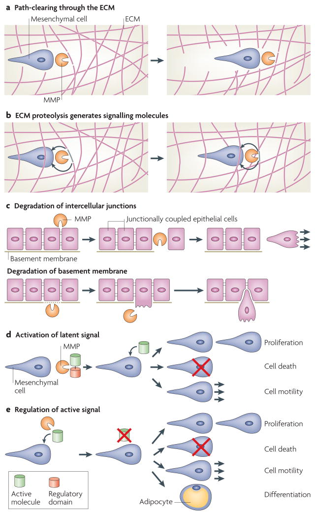
Recent work has dramatically expanded our understanding of the possible substrates that can be subject to matrix metalloproteinase (MMP) cleavage, and has similarly expanded our awareness of the means by which MMPs can affect cell behaviour. a | MMPs can cleave components of the extracellular matrix (ECM), resulting in increased space for cell or tissue movement. b | Alternatively, MMP proteolysis can generate specific cleavage products that then signal in an autocrine or paracrine manner (for example, cleavage of collagen IV α3 chain by MMP9 yields tumstatin, an anti-angiogenic peptide that functions by binding to the αvβ3 integrin127). c | MMPs can also directly regulate epithelial tissue architecture through cleavage of intercellular junctions or the basement membrane. d | MMPs can activate or modify the action of latent signalling molecules, resulting in diverse cellular consequences. For example, cleavage of vascular endothelial growth factor (VEGF) by MMPs changes angiogenic outcome by modifying the binding and diffusion properties of VEGF. e | MMPs can deactivate or modify the action of active signalling molecules, resulting in changes in proliferation, cell death, differentiation or cell motility. For example, MMP2 cleavage of stromal-cell derived factor-1 (SDF1, also known as CXC-motif ligand-12 (CXCL12)) results in its inactivation.
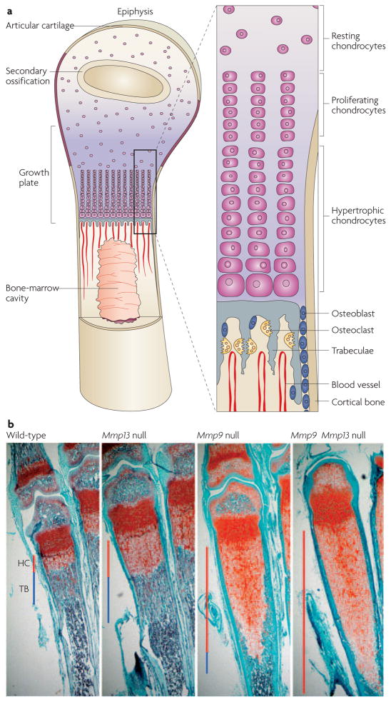
a | Long bones in mice and humans develop through the process of endochondral ossification, in which a cartilage template forms first and then is resorbed and replaced by mineralized bone. This process requires extensive matrix remodelling and invasion of new blood vessels. The schematic is adapted from REF. © (2000) Elsevier, and REF. © (1999) Macmillan Magazines Ltd. b | Matrix metalloproteinase-9 (Mmp9)- and Mmp13-null femurs display greatly expanded hypertrophic cartilage zones (HC; red line) and altered trabecular bone (TB; blue line). Despite this expansion, Mmp9- and Mmp13-null phenotypes eventually resolve, resulting in good bone formation. The Mmp9 Mmp13 double mutant has an even greater expansion of hypertrophic cartilage, and significantly and persistently shorter long bones. Images courtesy of D. Stickens, D. Behonick and N. Ortega, University of California, San Francisco, USA.
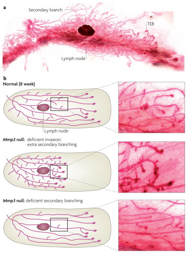
a | The length of the ductal network of the mammary gland is formed through the invasion and bifurcation of terminal end buds (TEBs). New branches initiate off of these primary ducts, in a process known as secondary branching. b | Mmp2- and Mmp3-null animals have reciprocal phenotypes; Mmp2-null mammary glands have deficient primary invasion and excess secondary branching, whereas Mmp3-null mammary glands have normal invasion and deficient secondary branching. These findings show that different proteases are used to accomplish these tissue-invasion processes. Images courtesy of A.J.E. and J. Trumbull, University of California, San Francisco, USA.
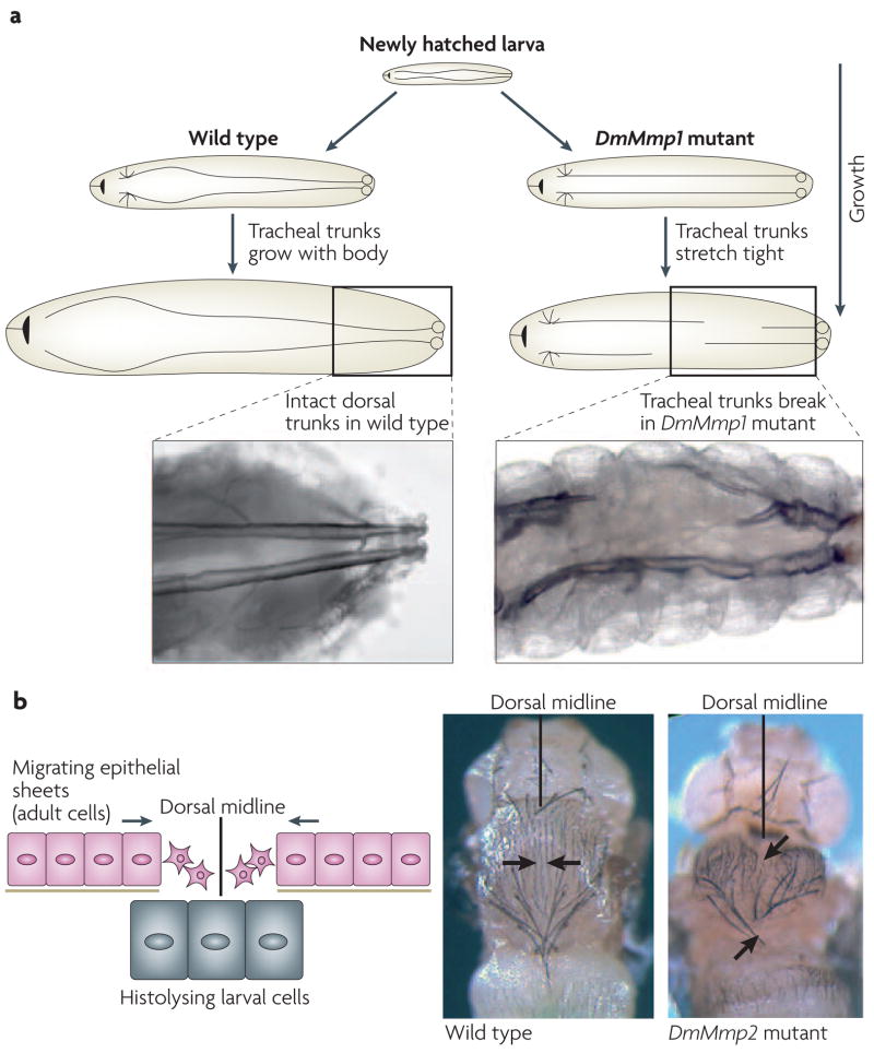
a | Drosophila melanogaster tracheal development requires the dorsal tracheal trunks to extend with the growth of the larva. In DmMmp1-mutant flies, the tracheal trunks stretch tight and finally break, resulting in larval lethality. b | During D. melanogaster metamorphosis, larval tissue is histolysed and adult dorsal epithelium migrates to the midline and fuses. In DmMmp2 mutants, the midline closure of the epithelium fails. Images adapted from REF. © (2003) Elsevier.
References
-
- Brinckerhoff CE, Matrisian LM. Matrix metalloproteinases: a tail of a frog that became a prince. Nature Rev Mol Cell Biol. 2002;3:207–214. - PubMed
-
- Birkedal-Hansen H, et al. Matrix metalloproteinases: a review. Crit Rev Oral Biol Med. 1993;4:197–250. - PubMed
-
- Welgus HG, Kobayashi DK, Jeffrey JJ. The collagen substrate specificity of rat uterus collagenase. J Biol Chem. 1983;258:14162–14165. - PubMed
-
- Egeblad M, Werb Z. New functions for the matrix metalloproteinases in cancer progression. Nature Rev Cancer. 2002;2:161–174. - PubMed
Publication types
MeSH terms
Substances
Grants and funding
LinkOut - more resources
Full Text Sources
Other Literature Sources
