. 2007 Mar;117(3):784-93.
doi: 10.1172/JCI30443. Epub 2007 Feb 22.

Impaired neutrophil activity and increased susceptibility to bacterial infection in mice lacking glucose-6-phosphatase-beta

Affiliations

Impaired neutrophil activity and increased susceptibility to bacterial infection in mice lacking glucose-6-phosphatase-beta

Yuk Yin Cheung et al. J Clin Invest. 2007 Mar.

Abstract

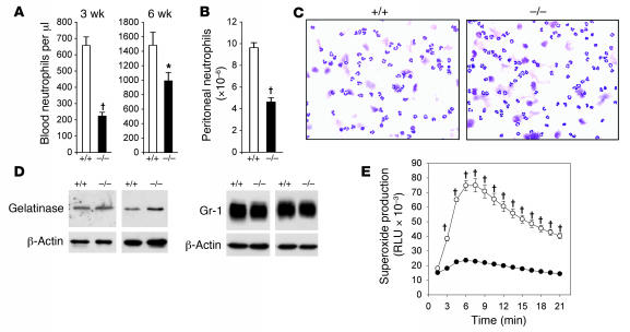
Neutropenia and neutrophil dysfunction are common in many diseases, although their etiology is often unclear. Previous views held that there was a single ER enzyme, glucose-6-phosphatase-alpha (G6Pase-alpha), whose activity--limited to the liver, kidney, and intestine--was solely responsible for the final stages of gluconeogenesis and glycogenolysis, in which glucose-6-phosphate (G6P) is hydrolyzed to glucose for release to the blood. Recently, we characterized a second G6Pase activity, that of G6Pase-beta (also known as G6PC), which is also capable of hydrolyzing G6P to glucose but is ubiquitously expressed and not implicated in interprandial blood glucose homeostasis. We now report that the absence of G6Pase-beta led to neutropenia; defects in neutrophil respiratory burst, chemotaxis, and calcium flux; and increased susceptibility to bacterial infection. Consistent with this, G6Pase-beta-deficient (G6pc3-/-) mice with experimental peritonitis exhibited increased expression of the glucose-regulated proteins upregulated during ER stress in their neutrophils and bone marrow, and the G6pc3-/- neutrophils exhibited an enhanced rate of apoptosis. Our results define a molecular pathway to neutropenia and neutrophil dysfunction of previously unknown etiology, providing a potential model for the treatment of these conditions.


Figures

Figure 1
Figure 1. Generation of G6pc3–/– mice.
(A) Targeting strategy. The wild-type allele is presented along with the targeting vector and the anticipated outcome of the recombination (targeted allele). Introns are denoted as lines, exons as boxes. Gene targeting resulted in the replacement of exons III–V with a loxP-flanked (arrowheads) neo cassette containing a diagnostic EcoRV site. The locations of the diagnostic 5′ and 3′ probes used in Southern blot analysis are shown. B, BamHI; N, NotI; R, EcoRI; RV, EcoRV; Sd, SanDI. (B) Southern blot analysis of genomic DNA from wild-type (+/+) and targeted ES (+/–) clones digested with EcoRV. (C) PCR analysis of genomic DNA of F1 intercross progeny. A wild-type locus–specific primer pair, EIVs/EVas, amplified a 440-bp fragment in the wild-type and heterozygous animals and 2 targeted locus–specific primer pairs, Neo1s/Neo2as and Neo2s/EVIas, amplified 616- and 722-bp fragments, respectively, in the heterozygous and homozygous (–/–) animals. (D) Western blot analysis of brain, muscle, and testis microsomal protein preparations (50 μg of each tissue) using an antibody against human G6Pase-β (48).
Figure 2
Figure 2. Analysis of the serum profiles and expression of G6Pase-β and G6PT mRNA.
(A) Serum concentrations of glucose, cholesterol, triglyceride, uric acid, and lactic acid in wild-type and G6pc3–/– mice. Fourteen mice of each genotype were used. Values represent mean ± SEM. (B) Northern blot analysis of human G6Pase-β and G6PT mRNA expression in human neutrophils. β-Actin was used as a control. Each lane contained 3 μg poly (A)+ RNA. (C) Quantification of murine G6Pase-β and G6PT mRNA by real-time RT-PCR in 6-week-old wild-type mice. Leukocytes were isolated from the whole blood, and neutrophils were isolated from thioglycollate-induced peritoneum. Expression levels are shown relative to that of G6Pase-β or G6PT transcript in the brain, which was arbitrarily assigned as 100%.
Figure 3
Figure 3. Analysis of myeloid functions.
(A) Neutrophil counts in 3-week-old (n = 8) and 6-week-old (n = 12) wild-type and G6pc3–/– mice. (B) Total peritoneal neutrophil counts in 6- to 7-week-old wild-type (n = 15) and G6pc3–/– (n = 16) mice challenged with thioglycollate. (C) Hema 3–stained cytospins of peritoneal neutrophils obtained from 7-week-old wild-type and G6pc3–/– mice. (D) Western blot analysis of protein extracts of peritoneal neutrophils (50 μg/lane) from 6- to 7-week-old wild-type and G6pc3–/– mice using antibodies against gelatinase, Gr-1, or β-actin. (E) Neutrophil respiratory burst activity. Results represent triplicate determinations using peritoneal neutrophils from 6- to 7-week-old wild-type (open circles) or G6pc3–/– (filled circles) mice. RLU, relative light unit. Values are mean ± SEM. *P < 0.03; P < 0.0005 versus wild-type.
Figure 4
Figure 4. Impaired chemotaxis and calcium flux of G6pc3–/– neutrophils in response to fMLP, KC, and MIP-2.
Neutrophils were isolated from thioglycollate-induced peritoneum of 6- to 7-week-old mice. (A) Concentration-dependent chemotaxis. Open circles, wild-type; filled circles, G6pc3–/–. Values represent mean ± SEM of quadruplet determinations. *P < 0.03, P < 0.005 versus wild-type. (B) Ca2+ flux in response to 10–6 M fMLP, KC, or MIP-2. Representative experiments are shown. Open circles, wild-type; open and filled triangles, both G6pc3–/–.
Figure 5
Figure 5. Susceptibility of G6pc3–/– mice to bacteria infection.
(A) Survival curves and (B) viable blood-born bacteria at 5 hours after infection for wild-type (open circles) and G6pc3–/– (filled circles) mice (n = 24 per group) challenged with E. coli at 4 × 10⁷ CFU/30 g body weight. Results are from 2 independent experiments. P < 0.0001 for survival curves, log-rank test.
Figure 6
Figure 6. Analysis of hematopoiesis.
(A) Western blot analysis of bone marrow aspirates (50 μg/lane) from 7-week-old wild-type and G6pc3–/– mice using antibodies against gelatinase, Gr-1, or β-actin. (B) Myeloid progenitor cells in the femur plus tibia of wild-type and G6pc3–/– mice at 3 and 7 weeks of age. CFUs were determined following stimulation of bone marrow cells with G-CSF, GM-CSF, or M-CSF. Results are mean ± SEM from 4 separate experiments, in which each mouse was assessed individually. ΧP < 0.001 versus wild-type. (C) Hema 3–stained cytospins of granulocyte CFUs obtained from 3-week-old wild-type and G6pc3–/– mouse bone marrow cells following 9 days’ stimulation with G-CSF.
Figure 7
Figure 7. Real-time PCR and Western blot analyses of GRP gene expression.
Peritoneal neutrophils and bone marrow aspirates were isolated from 6- to 7-week-old wild-type and G6pc3–/– mice during thioglycollate-elicited peritonitis. (A and B) Real-time PCR analysis. The expression levels of the GRP78 and GRP170 transcripts were normalized to β-actin RNA and then scaled relative to the transcript in a wild-type littermate, arbitrarily assigned as 1. (A) Peritoneal neutrophils (n = 10 and 13 for wild-type and G6pc3–/–, respectively). (B) Bone marrow (n = 14 and 18 for wild-type and G6pc3–/–, respectively). Values represent mean ± SEM. ΧP < 0.001. (C and D) Immunoblot analysis of protein extracts of neutrophils (50 μg/lane; C) and bone marrow (D) using antibodies against GRP78, GRP170, or β-actin.
Figure 8
Figure 8. Analysis of neutrophil apoptosis.
Peritoneal neutrophils were isolated from 6- to 7-week-old wild-type and G6pc3–/– mice during thioglycollate-elicited peritonitis. (A) Immunofluorescence of annexin V surface staining of peritoneal neutrophils (green fluorescence) and DAPI staining of nuclei (blue fluorescence). Note the colocalization of annexin V staining with DAPI staining in the merged image. Original magnification, ×200. (B) Expression of the caspase-3 transcript in wild-type and G6pc3–/– mice (n = 12 per group) quantified by real-time PCR. The expression levels of the caspase-3 transcript were normalized to β-actin RNA and then scaled relative to the transcript in a wild-type littermate, arbitrarily assigned as 1. Values represent mean ± SEM. #P = 0.016. (C) Immunoblot analysis of protein extracts of neutrophils (50 μg/lane) using antibodies against caspase-3 or β-actin. (D) DEVD-cleaving activity of active caspase-3 in protein extracts of peritoneal neutrophils from wild-type and G6pc3–/– mice (n = 3 per group). Activities were normalized to that of wild-type caspase-3. Values represent mean ± SEM. ΧP < 0.001.

Similar articles

Cited by

References

    1. Chou J.Y., Matern D., Mansfield B.C., Chen Y.T. Type I glycogen storage diseases: disorders of the glucose-6-phosphatase complex. Curr. Mol. Med. 2002;2:121–143. - PubMed
    1. Chou, J.Y., and Mansfield, B.C. 2003. Glucose-6-phosphate transporter: the key to glycogen storage disease type Ib. In Membrane transporter diseases. S. Broer and C.A. Wagner, editors. Springer. New York, New York, USA. 191–205.
    1. Beaudet A.L., Anderson D.C., Michels V.V., Arion W.J., Lange A.J. Neutropenia and impaired neutrophil migration in type 1B glycogen storage disease. J. Pediatr. 1980;97:906–910. - PubMed
    1. Gitzelmann R., Bosshard N.U. Defective neutrophil and monocyte functions in glycogen storage disease type 1b: a literature review. Eur. J. Pediatr. 1993;152(Suppl.):S33–S38. - PubMed
    1. Rothbaum R., et al. Shwachman-Diamond syndrome: report from an international conference. J. Pediatr. 2002;141:266–270. - PubMed

Publication types

MeSH terms